Nose
Historical document, translated for reference. It reflects medical knowledge of the 1920s–30s and is not medical advice.
Summary
This article from the first edition of the Great Medical Encyclopedia covers the comparative anatomy, embryology, anatomy, physiology, pathology, and surgery of the nose, detailing its evolutionary development from aquatic vertebrates to mammals.
Encyclopedia article (1928–1936)
NOSE. Contents: I. Comparative Anatomy and Embryology . . 577 II. Anatomy ................... 581 III. Physiology .................. 590 IV. Pathology .................. 591 V. General Operative Surgery of the Nose ..... 609 F. Comparative Anatomy and Embryology. The nose (nasus), the organ of smell in vertebrates, develops as a paired thickening of the ectoderm at the anterior end of the head (olfactory placodes); these plates sink deeper, forming olfactory pits, and then more or less complexly structured olfactory cavities, lined for the most part by sensory epithelium. The olfactory epithelium consists of tall, almost fibrous primary sensory cells (i.e., each extending directly into a nerve fiber), provided with bundles of sensory hairs, and no less long supporting epithelial cells, among which they are usually evenly scattered (in higher fishes and amphibians sometimes concentrated in the form of special buds). The olfactory sacs are protected on the outside by a skeletal capsule that fuses with the anterior part of the braincase at the site of the olfactory nerve openings. The surface of the epithelium of the olfactory sacs increases through the formation of a more or less complex system of folds, into which supporting skeletal outgrowths or conchae from the walls of the olfactory capsule may protrude. In fishes, numerous folds of the olfactory epithelium are usually arranged radially with respect to the longitudinal axis of the organ. Already in fishes, adaptations are observed to facilitate the exchange of water in the olfactory sac. In cartilaginous fishes, the nostrils, located on the lower side of the snout in front of the mouth, are connected to the latter by means of grooves covered by skin folds. This connection with the oral cavity obviously acquires some importance when testing food already in the mouth. Furthermore, thanks to the development of the nasal passage, the movement of the animal creates a continuous current of water, contributing to its rapid exchange in the olfactory sac itself. Through the fusion of the skin fold over the groove, the latter turns in most vertebrates into a channel connecting the anterior, inlet opening with the posterior, outlet one. Thus, in higher fishes there are two pairs of nostrils, usually located on the dorsal side of the head. In land vertebrates, the pair of nostrils closest to the mouth has moved into the oral cavity, forming the choanae, while the other pair remains on the dorsal surface of the head in the form of external nostrils. The establishment of a connection between the nostrils and the oral cavity made it possible to use the indicated nasal passage as a respiratory tract. This largely determined the further evolution of the organ in land vertebrates. The olfactory sac is already subdivided in amphibians into the olfactory region proper, lined with sensory epithelium, and the respiratory region, lined with ordinary epithelium. In reptiles, the respiratory region lies below the olfactory region and is further subdivided into three sections: the vestibule, lined with squamous epithelium; the middle section, lined with cylindrical ciliated epithelium and connected with the olfactory region; and finally the posterior section, opening through the choanae into the oral cavity. With the development of the secondary palate in higher reptiles (especially crocodiles) and in mammals, the upper part of the oral cavity into which the choanae open becomes isolated, and thus long nasopharyngeal channels develop, opening much further behind by the so-called secondary choanae. The progressive development of the olfactory region led in land vertebrates, as in fishes, to the formation of a complex system of folds, appearing here in the form of more or less twisted horizontal outgrowths. These outgrowths are supported by processes of the skeletal walls of the nasal cavity, in which independent ossifications, termed nasal conchae, can also develop. The very simply structured conchae of reptiles and birds achieve considerable complexity in mammals. In the vestibule, the maxillary (maxillo-turbinale) and nasal (naso-turbinale) conchae develop, obviously serving to warm and humidify the inhaled air (and associated with the bones of the same name). In the olfactory section, a more or less significant number of ethmoid conchae (ethmoturbinalia) lined with olfactory epithelium develop, usually arranged parallel to one another. Some of these conchae extend deeper toward the nasal septum (endoturbinalia), while others lie at the outer wall (ectoturbinalia). In animals with a well-developed sense of smell, the number of conchae can be very large (5--8 internal ones) and at the same time they are very complexly twisted. In primates, the number of internal conchae is reduced to two, and the external ones disappear entirely. In many mammals, a projection bearing the nostrils develops on the face, the external nose, supported by nasal bones and cartilages and sometimes extending into a more or less long muscular proboscis (shrews, pigs, tapirs, and especially elephants). By isolation from the olfactory cavity, land vertebrates develop another special, also olfactory section that independently opens into the oral cavity, the so-called Jacobson's organ.
I. Schmalhausen. Embryology of the Nose. From the end of the 3rd embryonic week, olfactory fields are laid down on both sides in the human embryo beneath the telencephalon and anterior to the optic vesicles in the form of a thickening of the covering epithelium. The olfactory fields are transformed into olfactory pits by the ingrowth and deepening of the epithelium. At the beginning of its development, the opening of the oral depression is a wide pentagonal hole surrounded by five prominences. The upper unpaired prominence represents the frontal process, and of the remaining four, the two upper ones are called the maxillary processes, and the two lower ones the mandibular processes. Simultaneously, a so-called median frontal process and two lateral nasal processes differentiate on the frontal process. Further, the pit plunges deep in the form of an olfactory sac, bounded besides the nasal processes also laterally by the adjoining maxillary process of the first visceral fold. The latter also encompasses the olfactory sac from below; the role of the partition between it and the median nasal process is played by a thin and small pharyngeal membrane (membrana bucco-nasalis) lying in the continuation of the nasal groove, which is part of the partition between the olfactory sac and the primary oral cavity. This membrane is subsequently ruptured by the deepening olfactory pits, which open at their posterior ends into the primary oral cavity by means of small openings called primary choanae. The maxillary processes grow significantly and gradually fuse both with the lateral nasal processes, giving rise to the subsequently disappearing nasolacrimal groove, and with the median frontal process, forming together with the latter the upper margin of the oral opening. Subsequently, on the inner side of both maxillary processes, prominences begin to form—palatine shelves—growing toward each other and gradually dividing the primary oral cavity into the upper section, or nasal cavity, where the primary choanae open, and the lower section, the oral cavity proper. During the 2nd month of embryonic life, the formation of the face and nose takes place. The latter arises initially in the form of a transverse, so-called nasal ridge, formed on the median frontal process and representing the rudiment of the tip of the nose; the openings of the olfactory pits are transformed into the external nasal openings, and the lateral nasal processes give rise to the wings of the nose. Simultaneously, on the inner side of the median frontal process, a so-called nasal septum arises perpendicular to the palatine processes (Fig. 1), growing gradually
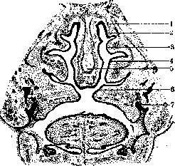
Figure 1. Section through the nasal cavity of an embryo: 1, 2, and 3—ethmoturbinalia; 4—inferior concha; 5—cartilaginous nasal septum; 6—palatine processes; 7—tooth germ.
inward and dividing the nasal cavity forming above the palatine processes into two halves. The edges of the palatine processes gradually fuse with the lower edge of the nasal septum and with each other, which leads to the formation of the definitive palate. The posterior edges of the palatine processes form small growths that subsequently give rise to the uvula, while the openings remaining above them and leading into the nasal cavities represent the secondary choanae. Conchiform formations appearing on the outer wall of the nasal cavity are of great importance for its formation. Here one can distinguish the os maxillo-turbinale, naso-turbinale, and several ethmo-turbinalia; from these, the inferior concha and the ethmoid labyrinth are formed. From the epithelium of the olfactory field, and later from the epithelium of the pit and sac, through a special differentiation, the epithelium of the partis olfactoriae of the mucous membrane of the nose with its olfactory and supporting cells develops. Folds arising on the lateral surfaces of the nasal cavities subsequently form the nasal conchae, of which there are four in the newborn. Accessory cavities of the nose appear due to the fact that the mucous membrane of the nasal cavity, increasing in surface area, penetrates into the thickness of the walls of the cavity. B. Anatomy. The nose (nasus) is divided into the external nose and the nasal cavity with accessory cavities. The shape of the nose varies within significant limits not only in connection with racial features of the structure of the facial skeleton, but also depending on age, during the growth period in the same subject. It is impossible to indicate the shape of the nose in all possible variants. Martin, for the purpose of anthropological study, divided (Fig. 2) all forms into 15 groups. Each group is characterized by 5 main features: 1) the shape of the nasal bridge, 2) its length, 3) the location of the root of the nose, 4) the shape of the tip of the nose, and 5) the direction of the base of the nose. Figure 3. Various nostril shapes. The lateral surfaces of the nose are convex downwards, bounded by a clearly expressed groove (sulcus naso-labialis), mobile, and form the wings of the nose (alae nasi), the lower free edge of which forms the nostrils (nares) (Fig. 3). The latter are separated from each other by the mobile part of the nasal septum (septum mobile nasi). The external nose consists of bony, cartilaginous (hyaline cartilage), and soft parts. The bony section in the upper part is formed by the nasal processes of the frontal bones (processus nasales ossis frontalis) and the nasal bones (ossa nasalia). Downward and laterally, the frontal process of the upper jaw (processus frontalis ossis maxillae) adjoins the nasal bones. The nasal bones fill the space between the frontal processes of the upper jaws and participate in the formation of the nasal bridge; their upper edge connects with the frontal bone, the outer edge with the frontal process of the upper jaw, the inner edge with the corresponding bone of the other side, and the lower free edge forms the upper boundary of the piriform aperture (apertura piriformis). The cartilaginous skeleton of the nose is a continuation of the bony skeleton and is firmly fused around the circumference of the piriform aperture with the latter. The lateral (median) wall of the external nose on each side is formed by a cartilaginous plate of irregular triangular shape (cartilago triangularis) (Fig. 4). The upper edge of this cartilage extends somewhat backward, under the nasal bones and the frontal process of the upper jaw, attaching to them by means of dense connective tissue. The inner edge in the region of the nasal bridge connects with the anterior edge of the nasal septum cartilage. The anteroinferior angle of the triangular cartilage reaches the large cartilage of the ala. Scheme of various nose shapes (according to Martin) (to Fig. 2). Shape of nasal bridge | Length of nasal bridge | Location of nasal root | Tip of nose | Direction of base of nose. Concise table of shapes. The external nose (nasus externus) has the shape of an irregular triangular pyramid, the base of which coincides with the facial skeleton, and the apex constitutes the tip of the nose. The upper, narrow end of the nose, starting from the forehead, is called the root of the nose (radix nasi); downwards and anteriorly from it, the nasal bridge (dorsum nasi) extends obliquely, passing into the tip of the nose (apex nasi). The large cartilage of the ala of the nose (cartilago alaris major), also paired, is particularly thin and has a variegated shape. It consists of two plates, of which one, wider, called the outer crus, forms the ala of the nose, and the other, inner, is located next to the same crus of the other side and is part of the mobile septum of the nose, being loosely connected both with it and with the septum cartilage of the nose. The small alar cartilages of the nose (cartilagines alares minores) are small irregular pieces of cartilage found in varying numbers in the posterior part of each of the alae of the nose. Small additional cartilaginous plates in the amount of one or two, located between the triangular cartilage and the large cartilage of the ala of the nose, are called sesamoid cartilages (cartilagines sesamoideae nasi). The part of the nasal ala in the posteroinferior section contains no cartilage and is formed only by a doubling of the skin. The cartilaginous section of the external nose also includes the quadrangular cartilage of the nasal septum (cartilago quadrangularis septi nasi), which is an irregular
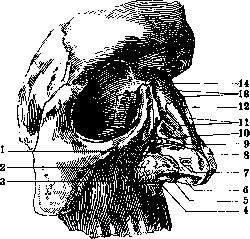
Figure 4. Dissected nasal cartilages: 1 - cartilago alaris minor; 2 - tela subcutanea cutis; 3 - maxilla; 4 - spina nasalis ant.; 5 - cartilago septi nasi; 6 and 7 - crus mediale et laterale cartilaginis alaris majoris dext.; 8 - cartilago alaris major sinist.; 9 - cartilago sesamoidea nasi; 10 - incisura nasalis maxillae; 11 - cartilag. nasi lat. (triangul.); 12 - cartilago septi nasi; 13 - ossa nasalia; 14 - sutura internasalis.
quadrilateral cartilage plate, which forms the lower part of the nasal septum and is wedged between the perpendicular plate of the ethmoid bone, while its posteroinferior margin lies in the groove of the vomer and the anterior section of the nasal crest of the maxilla. The anteroinferior margin of the cartilage forms the upper boundary of the mobile nasal septum in front, descends somewhat lower posteriorly, and is attached here to the anterior nasal spine (spina nasalis anterior). Under the name of the vomeronasal cartilage of Jacobson (cartilago vomero-nasalis Jacobsoni), a small cartilaginous strip is described, which directly adjoins both sides of the nasal septum cartilage, immediately below the anterior nasal spine. The muscles of the external nose in man are vestigial in character and have no practical significance. Among the muscular bundles of greater or lesser significance, one may note m. levator alae nasi et labii superioris, a superficially situated muscle that elevates the wing of the nose and the upper lip; it originates from the frontal process of the maxilla and attaches to the posterior margin of the wing of the nose, partly passing into the skin of the upper lip. Two other muscles: 1) m. nasalis and 2) m. depressor septi nasi lead to the narrowing of the nostril. The integument of the nose is very thin and is connected to the underlying parts above by means of loose, fat-poor connective tissue, while on the wings of the nose it is closely connected by means of elastic connective tissue with the underlying muscle layer. The skin of the nose is studded with numerous sebaceous glands, which, especially in the posterior section of the nasal wings, are exceptionally large and whose orifices are visible to the naked eye. In the skin of the nose, apart from hair follicles and fine hairs, there are also sweat glands. In the region of the nostrils, the skin turns inward into the nose and, in the region of the fold of the inner surface of the wings, called the nasal threshold (limen nasi), gradually passes into the mucous membrane of the nasal cavity. In the initial part, directly at the entrance to the nose, the nasal cavity is lined first by skin, which curls inward and is provided with hairs and sebaceous glands. The hairs located here are called vibrissae and can reach a considerable length. This is followed by an intermediate zone, which further passes into the actual respiratory mucous membrane of the nose. Vessels and nerves of the nose. The external nose is very richly supplied with vessels. The arteries originate 1) from a. dorsalis nasi, the terminal branch of a. ophthalmicae, which runs above lig. palpebrale mediale to the skin of the nasal root and its dorsum, and 2) from branches of a. maxillaris externa running toward the medial angle of the eye (a. angularis, seu a. naso-frontalis) and to the wing of the nose (rami alares). These arteries directly transition into the rami alares of the other side; further, they anastomose with aa. septi narium, which branch off from a. labialis and run upward to the nasal septum. The external arterial network is mostly located under the skin, and furthermore directly connects with the arteries of the nasal cavity (in the vestibulum). The veins of the external nose, branching similarly to the arteries but not accompanying the latter everywhere, empty with several branches into v. nasalis anterior (profunda). The lymphatic vessels of the nose empty into the large lymphatic trunks of the face, which in turn run to the lymph nodes of the submandibular region. Nerves. The sensory nerves originate from n. ethmoidalis and n. infraorbitalis, the motor nerves to the muscles of the nose from n. facialis. The nasal cavity (internal nose) is located between the cranial cavity, the eye orbits, and the oral cavity. Anteriorly, it opens with nostrils located on the lower surface of the external nose and bordered only by skin, which have a very diverse shape and are arranged somewhat obliquely. Posteriorly, the nasal cavity communicates with the upper section of the pharynx by means of two adjacently located, oval-shaped posterior nasal apertures called choanae. By a median nasal septum (septum nasi), which usually deviates partially to one side or the other, the entire cavity is divided into two identical halves. The upper and posterior part of the septum is bony (septum nasi osseum), the anterior is formed by the quadrilateral cartilage (see
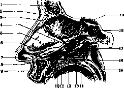
Figure 5. Nasal cavity without mucous membrane: 1 - sinus frontalis; 2 - spina frontalis; 3 - os nasale; 4 - processus sphenoidalis septi cartilaginei; 5 - cartilago septi nasi; 6 - cartilago vomero-nasalis (jacobsoni); 7 - cartilago alaris major (crus mediale); 8 - spina nasalis ant.; 9 - labium sup.; 10 - canalis incisivus; 11 - palatum durum; 12 - crista nasalis; 13 - sutura palatina transversa; 14 - pars horizontalis ossis palatini; 15 - palatum molle; 16 - spina nasalis post.; 17 - vomer; 18 - sinus sphenoidalis; 19 - lamina perpendicularis ossis ethmoidalis.
above); adjoining it anteriorly and inferiorly is the membranous septum (septum membranaceum, or septum mobile nasi). The nasal cavity has four walls forming it: internal, external, upper, and lower (figures 5 and 6).
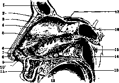
Figure 6. Nasal cavity with mucous membrane: 1 - sinus frontalis; 2 - spina frontalis; 3 - os nasale; 4 - membrana mucosa nasi; 5 - cartilago nasi lat.; 6 - cartilago alaris major; 7 - vestibulum nasi; 8 - apex nasi; 9 - septum mobile nasi; 10 - margo nasi; 11 - labium superius; 12 - palatum durum; 13 - palatum molle; 14 - choana; 15 - ostium pharyngeum tubae auditivae; 16 - sinus sphenoidalis; 17 - septum nasi.
The internal wall is formed by the nasal septum, the bony part of which in the posterosuperior section is formed by the perpendicular plate of the ethmoid bone (lamina perpendicularis ossis ethmoidalis), and in the posteroinferior section by an independent bone of the nasal septum, the vomer (vomer). In the lower part of the septum, closer to the front, at the border with the vomer, there is the opening of the vomeronasal (Jacobson's) organ; in man it is undeveloped, a few millimeters in length, whereas in animals it has the shape of an elongated pouch lined with olfactory epithelium. Posteriorly and beneath the tubule of Jacobson's organ, on the floor of the nasal cavity, there is often a small opening leading into a tubule called the incisive canal (ductus incisivus); the latter usually ends blindly, although it may open with a very thin unpaired opening on the incisive papilla of the hard palate. This canal represents a rudiment of Stensen's canal, which is well developed in many mammals. The external or lateral wall of the nasal cavity appears the most complex. Its bony skeleton includes the nasal bone, then the medial surface of the body of the maxilla with the frontal process, further posteriorly adjoins the lacrimal bone, followed by the cavity system of the ethmoid bone and
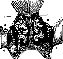
Figure 7. Frontal section of the head behind the crista galli: 1 - upper meatus; 2 and 6 - sinus lat.; 3 - recessus praelacrimalis; 4 - ostium maxillare; 5 - canalis infraorbitalis. Finally, the greater part of the posterior half of the outer wall is formed by the perpendicular part of the palatine bone and the internal plate of the pterygoid process of the sphenoid bone. On the bony part of the outer wall are located three nasal conchae: the inferior, middle, and superior (conchae nasales inferior, media et superior). The free space located between the nasal septum and the nasal conchae on the one hand and between the nasal vault and the nasal floor on the other constitutes the so-called common nasal meatus (meatus nasi communis). Besides this, under each of the nasal conchae there are separate nasal meatuses: 1) between the inferior concha and the floor of the nasal cavity lies the inferior nasal meatus (meatus nasi inferior), 2) between the middle concha and the lateral wall of the nose lies the middle nasal meatus (meatus nasi medius), and above the middle concha is located the superior nasal meatus (meatus nasi superior). The rearmost part of the nasal cavity (Fig. 7), behind the posterior ends of the middle and inferior conchae, which directly adjoins the choanae, is called the nasopharyngeal meatus (meatus nasopharyngeus). The inferior nasal concha is an independent bone (os turbinale), attached to the crista turbinata. The middle and superior conchae are parts of the ethmoid labyrinth. Under the vault of the inferior concha in the anterior third of the passage, at a distance of 1 cm from the anterior end of the concha, is the opening of the nasolacrimal canal (canalis nasolacrimalis). The width of the inferior nasal passage depends on the size of the concha and the position of the nasal septum. Above the inferior concha is the middle one, which extends neither as far anteriorly nor posteriorly as the inferior; its free anterior vertical edge meets the lower horizontal edge at a right angle. Frequently, one of the cells of the ethmoid labyrinth develops in the bone itself forming the anterior part of the middle concha, the latter increasing significantly in size and forming the so-called concha bullosa. In the middle nasal meatus open almost all the paranasal sinuses, namely: 1) the maxillary sinus, 2) the frontal sinus, and 3) the anterior cells of the ethmoid labyrinth. Because of this relation to the paranasal sinuses, the middle nasal meatus clinically represents the most important part of the lateral wall of the nose. If on a bony or soft-tissue preparation, after removing or bending the middle concha upward, one examines the lateral wall of the nasal passage, one notices first of all on it a passage running from anterior and upper to posterior and lower, slightly convex anteriorly, 2-3 mm wide, which is called from its shape the hiatus semilunaris. This passage is bounded anteriorly and posteriorly by the process of the ethmoid bone (processus uncinatus), and upward by one of the cells of the ethmoid labyrinth (bulla ethmoidalis). The semilunar hiatus in its posterior part widens funnel-wise into a special depression, a sort of funnel (infundibulum ethmoidale). At the bottom of this funnel, near the posterior end of the hiatus semilunaris, is the ostium maxillare—the entrance opening of the maxillary sinus. If one traces the semilunar hiatus with a probe anteriorly and upward, one enters mostly the ductus nasofrontalis and through it into the frontal sinus, more rarely into the opening of some other cell of the ethmoid labyrinth. If the end of the probe is directed along the floor of the semilunar hiatus, it enters the opening of the maxillary sinus. On the anterior and posterior walls of the semilunar hiatus or near it, several anterior cells of the ethmoid labyrinth usually open. From the large opening connecting the nasal cavity with the maxillary sinus on the bony preparation, only the ostium maxillare located in the depths of the infundibulum remains noticeable on the preparation with soft parts; the rest of the opening is closed by the adjacent mucous membranes of the lateral wall of the nose and the inner wall of the maxillary sinus. By means of delicate bony processes extending from the processus uncinatus posteriorly and inferiorly, the large opening is divided into several smaller openings; both lower ones are closed by a membrane. The bony openings are called the anterior and posterior fontanelle. In the posterior fontanelle, in almost 10% of cases, a second opening communicating with the maxillary cavity is encountered—the ostium maxillare accessorium.
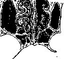
The superior concha, the smallest of the nasal conchae, is a weakly expressed bony protrusion of the ethmoid labyrinth in the region above the middle concha. Below the superior concha is located the superior nasal meatus, in the region of which the posterior cells of the ethmoid labyrinth open. The opening of the sphenoid sinus opens above the superior concha. In newborns, the superior concha, or rather the posterior end of the superior concha, appears divided by a longitudinal cleft, and ...
appears as if a separate new concha, which in such cases is called the fourth nasal concha (concha suprema). The lower wall of the nose (nasal floor) is formed mainly by the palatine process of the maxilla and posteriorly by the horizontal plate of the palatine bone. The floor of the nasal cavity is slightly concave in both the frontal and sagittal planes. The upper wall of the nasal cavity, or vault, is formed by the horizontally situated cribriform plate of the ethmoid bone (lamina cribrosa ossis ethmoidalis), through the openings of which branches of the olfactory nerve pass from the cranial cavity into the nasal cavity. Posteriorly, the nasal cavity communicates with the nasopharyngeal space through the choanae. The latter are bounded medially by the vomer, laterally by the pterygoid process (processus pterygoideus) of the sphenoid bone, superiorly by the sphenoid sinus (sinus sphenoidalis), and inferiorly by the horizontal plate of the palatine bone. The choanae are separated laterally by means of the nasopharyngeal sulcus (sulcus naso-pharyngeus) from the nasal part of the pharynx, into which they transition. Histological structure of the mucous membrane of the nasal cavity. In the mucous membrane of the nasal cavity, with the exception of a small space in the nasal vestibule, two regions are distinguished: the respiratory region (pars respiratoria) and the olfactory region (pars olfactoria). The nasal vestibule is lined initially by skin, which turns inward into the nose. The olfactory region extends over the surface of the superior concha, part of the middle concha, and the corresponding part of the nasal septum. The entire remaining part of the nasal cavity between the vestibule and the olfactory region belongs to the respiratory region. 1. The mucosa of the respiratory region (membrana Schneideriana) is covered with a pseudostratified ciliated columnar epithelium, the hairs of which perform movements inward toward the choanae. The mucosa is tightly fused with the periosteum and perichondrium and differs in various parts of the cavity only in thickness, which in the inferior nasal conchae reaches up to 4 mm (Figure 8). Beneath the epithelium in the tunica propria lie branching tubulo-alveolar glands of a mixed character. In the mucosa, alongside the columnar cells, there are also special goblet cells, the process of mucus formation in which is particularly intense during inflammations. The main feature of the mucosa in the respiratory region (regio respiratoria) is the presence of dense venous networks in the submucosal layer, forming within the nose cavernous tissue, or erectile bodies of the nasal mucous membrane. This cavernous tissue is especially developed on the medial surface and edge of the inferior concha, on the edge of the middle concha, and on the posterior end of the middle concha. It consists of a deeper, coarser venous network and a superficially located, finer venous network. The walls of the vessels are distinguished by an abundant content of muscle and elastic fibers. Owing to the extreme swelling capacity of the cavernous tissue, the nasal mucous membrane swells very easily and collapses under the influence of mechanical, thermal, or also psychic influences. The filling and emptying of the vessels occur under the influence of irritations originating from the fibers of the trigeminal nerve coming from the sphenopalatine ganglion (ganglion spheno-palatinum). In the anterior part of the septum, or on the floor of the nose, according to the studies of Kiesselbach, there is a network of dilated blood vessels from which nosebleeds very frequently occur as a result of mild injuries (locus Kiesselbachi). Extending to all the accessory sinuses, the mucosa lines their walls in the form of a thin (up to 0.02 mm), poorly glandular lining. The color of the mucous membrane is reddish, with varying degrees of intensity. 2. The mucosa of the olfactory region is already macroscopically distinguished by its yellowish-brown tint from the rest of the mucosa. It consists of the olfactory epithelium, in which there are two kinds of cells: 1) olfactory cells and 2) supporting cells, between the basal parts of which there is yet a third form of cells—replacement cells. The olfactory cells have a long, thread-like shape, with a thickening approximately in the middle, in which the round nucleus is located. At the free peripheral end of this cell, there is a tuft of non-motile hairs that protrude above the surface of the mucosa. Inferiorly, the olfactory cells elongate into a process that directly continues into the axis cylinder of a nerve fiber. Surrounding the olfactory cells are supporting cells, which are simple columnar epithelial cells. In the region of the olfactory epithelium are Bowman's glands, which secrete mucus and serous fluid (Simanovsky). Vessels of the nasal cavity. The main vessel supplying the nasal cavity with arterial blood is the sphenopalatine artery (a. spheno-palatina), branching off from the internal maxillary artery (a. maxillaris interna). It enters the nasal cavity through the sphenopalatine foramen (foramen spheno-palatinum), where it divides into the posterior nasal artery (a. nasalis posterior) and the nasopalatine artery (a. naso-palatina). The posterior nasal artery in turn divides into two branches, of which the lateral posterior nasal artery (a. nasalis posterior lateralis) spreads over most of the lateral wall of the nose, while the posterior septal nasal artery (art. nasalis posterior septi) goes to the nasal septum. The upper part of the lateral wall of the nose receives blood in addition from the anterior and posterior ethmoidal arteries (a. ethmoidalis anterior and posterior), which originate from the ophthalmic artery (a. ophthalmica). Furthermore, there are numerous anastomoses between the vessels of the lateral wall of the nose and the external maxillary artery (a. maxillaris ext.) and the ascending palatine artery (a. palatina ascendens). The nasal septum is supplied with blood, aside from the posterior septal nasal artery, mainly through the nasopalatine artery with its various branches. The outflow of venous blood occurs through very numerous vessels, mainly into the anterior facial vein (v. facialis anterior) and the ophthalmic vein (v. ophthalmica). However, a large number of small venous vessels of the nose and superior accessory sinuses pour their blood directly into the superior sagittal sinus (sinus longitudinalis). The lymphatic vessels of the nose represent a deeper and more superficially located, very dense network. Of no small importance is the circumstance that, as studies have shown (Axel Key, Retzius), the lymphatic pathways of the nose communicate with the subdural and subarachnoid spaces and can be injected from these spaces. The sensory nerves of the nasal cavity originate from the 1st and 2nd branches of the trigeminal nerve. The anterior ethmoidal nerve (n. ethmoidalis ant.), branching off from the 1st branch of the trigeminal nerve and extending downward from the nasal vault, innervates the medial and lateral walls of the vestibule. Posterior nasal nerves (pp. nasales posteriores) emerge from the sphenopalatine ganglion and, heading anteriorly, branch out on the lateral wall and septum of the nose. The most strongly developed branch running along the nasal septum is named the nasopalatine nerve (n. naso-palatinus Scarpae); it passes through the nasopalatine canal (canalis naso-palatinus) to the anterior part of the palatal mucous membrane. The branches of the olfactory nerve penetrate into the interior of the nose from the olfactory bulb (bulbus olfactorius) through the openings of the cribriform plate (lamina cribrosa) of the ethmoid bone and are distributed in the olfactory region (regio olfactoria), i.e., on the medial surface of the superior concha, as well as on the very uppermost parts of the septum. The cleft located between the lateral and medial surfaces of the olfactory region is named the olfactory slit (rima olfactoria). III. Physiology. The nasal cavity performs respiratory, olfactory, and protective functions. The air current, entering through the nasal apertures, rises upward toward the nasal vault and passes in its main mass through the middle nasal meatus, after which it descends in an arch posteriorly and inferiorly, heading through the choanae into the nasopharyngeal cavity. Thus, a more prolonged contact of the air with the mucous membrane is achieved, as well as a slowdown of its current encountering on its way the irregularities of the concha of the lateral wall of the nose, which creates favorable conditions for the protective function of the nose. Passing through the nasal cavity, the air is warmed and moistened. Warming (or cooling) is easily achieved by the blood filling the cavernous tissue of the nasal mucosa. Foreign bodies suspended in the air, such as dust, bacteria, etc., in their larger parts are retained already in the nostrils thanks to the filter made of hairs placed here. With the further movement of the air current, the tortuosity of the nasal passages and the moisture of the walls of the latter create conditions favorable for the settling of a significant part (up to 50%) of the dust contained in the air on the walls of the nasal cavity. Dust particles, thanks to the action of the cilia of the ciliated epithelium, enter the nasopharynx. The air is moistened almost to full saturation at the expense of nasal mucus. The mucous membrane of the nose secretes about 500 g of moisture within 24 hours. Bacteria that have entered the nose with the air and have not been removed along with dust particles are largely neutralized and killed by the action of nasal mucus. But even in a normal state, the nasal cavity always has a certain amount of bacteria (concerning the microflora of the nose, see Human microflora), which do not always have a harmful effect on the mucosa of the nasal cavity thanks to the acquired local immunity (Besredka). The nasal cavity also neutralizes to a significant degree gaseous (smoke) and chemically harmful substances. The nasal and nasopharyngeal cavities serve as resonators for the voice, the air in which amplifies the sound of the voice and gives it timbre and individual sonorously resonant quality.
When the nose is obstructed, clear changes in the voice are observed; it loses its sonority and acquires a muffled shade, which is called closed nasality (rhinolalia clausa). If, due to sagging of the soft palate (its paralysis) during phonation, the nose remains open on the nasopharyngeal side, speech sounds acquire a nasal shade of a different character, which is called open nasality (rhinolalia aperta). If nasal breathing is impossible due to pathological changes, it is replaced by mouth breathing. It has been established by experience and experiment that during nasal breathing, due to the negative pressure produced in the underlying respiratory tract, better lung ventilation is achieved with a greater absorption of atmospheric oxygen than during mouth breathing. A whole series of the above-listed protective functions performed by the mucosa of the nasal cavity during mouth breathing is excluded and performed to a significantly lesser degree than during nasal breathing. The protective capacity of the upper respiratory tract during mouth breathing is depleted more quickly, which leads to various lesions of the upper respiratory tract, primarily the pharynx, which in such cases often serve as gateways for many infectious diseases of the body. The olfactory function of the nose—see Olfaction. IV. Pathology. Diseases of the nose occur very frequently and cause a whole series of subjective disorders and phenomena on the part of distant organs, sometimes diseases of the eyes, as well as the brain, but most often the ears. In all diseases of the nose, the nasal mucosa is mainly and primarily affected. This is explained not only by the fact that the nasal mucous membrane, due to the participation of the latter in the act of respiration, is directly subjected to the influence of various harmful agents entering from the outside with the inhaled air, but also by the fact that upon sudden severe cooling of the skin or individual parts of it, a strong rush of blood occurs to the mucous membranes and especially to the nasal mucosa, which, together with other harmful influences, leads to its prolonged disorders. Deformities of the nose. Among various congenital deformities of the nose, one can point to the rarely observed doubling of the external nose, the infrequently encountered cleft of the tip of the nose, so that both nostrils are separated by a groove (called a "pug's nose"). Somewhat more common are defects of the nasal bones. Developmental defects of the turbinates, concerning their size and shape, are also far from common. Of much greater interest to the physician are congenital and acquired adhesions, as well as abnormalities of the nasal septum. In adhesions, a distinction is made between synechiae, i.e., the formation of bridges or thin connections between individual parts of the nose, and atresias, more or less completely closing the nasal lumen. Adhesions can be cartilaginous, bony, or connective tissue. Synechiae only occasionally are congenital, but in the majority of cases, like atresias, are the consequence of ulcerating processes in the nose on the basis of acute infections such as smallpox, diphtheria, typhus, which leads to subsequent cicatricial narrowing or complete obliteration, e.g., of the entrance to the nose. Causes causing deformations in the nasal cavity and nasal orifices may also include lupus, syphilis, rhinoscleroma, less frequently trauma, as well as cauterization in the nose, accidental or for therapeutic purposes, with caustic chemical substances or galvanocautery. The nature of such obliterations is usually connective tissue, less frequently bony. The diagnosis is easily established by inspection and probing; attention must be paid to the free state of the choanae and nasopharynx. Adhesions located in the posterior part, near the choanae, are often of a bony character. In prophylactic terms, during galvanocautery and blood-letting operations in the nasal cavity, it is necessary to avoid performing one or another operation simultaneously on two mutually opposing parts; this particularly applies to galvanocautery. If there is injury to the mucosa on both sides simultaneously, it is necessary to take measures by repeatedly separating the appearing adhesions with a buttoned probe after cocaine application or by wearing rubber, metal, or plastic plates (drainages) to prevent the formation of cicatricial adhesions. Treatment consists in bloody cleavage of synechiae with the use of plates or excision of scars and adhesions with the subsequent use of thick-walled rubber drainages. As for abnormalities on the part of the external sections of the nose, among such can be noted the sinking of the nasal wings and those deformations of the nose which most often are the consequence of syphilitic lesions of the bones of the nasal skeleton or trauma (saddle nose and lorgnette nose). Irregularities of the nasal septum can be expressed by curvatures (deviationes) and thickenings with the formation of spurs and crests (spinae, cristae), and a combination of these changes is also possible (Fig. 9). In adults, the nasal septum is rarely a regular plate established in the median plane, but is more often curved to one side or the other. Curvatures of the septum can develop in all sections of the latter, although the posterior bony section is affected significantly less often. The septum may be curved entirely to one side or in its upper part it is ex
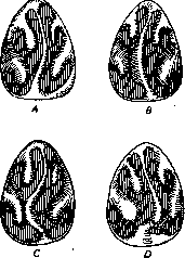
Figure 9. Deformations of the nasal septum. Curvatures: A - mild; B - with swollen mucous membrane; C - S-shaped; D - angular with swelling of the mucous membrane.
bent to one side, and in the lower part to the other, forming an S-shape on the frontal section. Sometimes the upper part is curved at an angle in relation to the lower, forming a fracture-like curvature, or the septum falls away to one side in the form of steps or a terrace. Thickenings in the form of spurs and crests are usually located on the convex part of the septum, predominantly at the junction of the cartilage with the upper edge of the vomer. They occur both in the posterior and in the anterior section of the nasal septum, in some cases located on the smooth straight part of the septum, and in others (more often) developing in the form of an isolated thickening—a crest—along the entire length of the nasal septum. As for the etiology of nasal septum curvatures, at present the majority of authors adhere to the view that changes in shape are the consequence of growth anomalies—uneven growth of various sections of the nasal septum and its bony frame; trauma and rickets also play a role here. A symptom of septum curvatures with the formation of crests and spurs on it is insufficient patency of one or the other side. Sometimes projections on the septum, especially if they impinge on the opposing turbinates, serve as a cause for the development of reflex neuroses in various areas of the body. The unfavorable effect of nasal narrowing on the Eustachian tube and middle ear should also be noted. Diagnosis is made using anterior rhinoscopy and usually presents no difficulties. With a simple curvature, a depression will be observed on one side of the septum, and a convexity on the opposite side. It is also not difficult to recognize S-shaped forms of curvature, as well as dislocations of the septum and its partial and general thickenings, formed symmetrically on both sides. Treatment of septum curvatures can be carried out by the operation of submucous resection of the nasal septum. Partial resection of the nasal septum was proposed by Hartmann and Petersen. This method was subsequently improved (Krieg, Bönninghaus, Cholewa, Zarniko, and Killian). Based on modern observations, the operation of submucous resection of the nasal septum is indicated in the following cases (according to Denker): 1) With insufficient air passage due to a curvature of the septum, even if only on one side. 2) With a straight septum, but a very narrow nasal lumen and abnormal thickness of the septum. 3) With unsatisfactory outflow conditions in cases of suppuration of the accessory sinuses. 4) As a preparatory operation during endonasal opening of the ethmoid bone, resp. the operation on the frontal sinus according to Killian. 5) With frequently recurring catarrhs of the pharynx and diseases of the Eustachian tube (see Nasal catarrh). 6) With the development of reflex neuroses (see below).—Crests and spurs without simultaneous curvature of the septum are rare. Therefore, their removal generally coincides with the operation for curvature. For this purpose, at present, submucous resection of the septum is performed. The principle of the operation is that between the two leaves of the mucous membrane, which are preserved intact as much as possible, through an incision of the mucous membrane on one side, the cartilaginous and bony septum is carefully exposed, after which they are resected from behind and above, preserving (which is especially important) the strip on the nasal dorsum. After removal of parts of the skeleton of the nasal septum, both leaves of the mucous membrane are applied to each other again. Instead of the curved osteocartilaginous nasal septum, a straight, mostly membranous septum is obtained.—Much less frequently than irregularities of the septum, developmental anomalies on the lateral wall of the nose are observed: vesicular swellings on the middle turbinate or less frequently on the bulla ethmoidalis, which can bulge inward strongly, pushing the septum to the other side, and thus reduce the patency of the nose for air. Such swellings are subject to removal by means of a cutting snare or conchotome.—Nose injuries. Both external and internal injuries of the nose are the result of a fall on the nose, a blow to it (so-called boxer's nose), etc., as a result of which contusions, damage to soft parts, simple and complex fractures of the cartilaginous and bony skeleton of the nose occur. The nasal bones are most often damaged, less often the frontal processes of the upper jaw, and least of all the lateral walls of the nose, which are damaged simultaneously only in very severe injuries caused by firearms. In many cases, damage to the nasal septum occurs simultaneously in the form of its curvature, dislocation, fracture, or break. Curvatures predominantly form on the cartilaginous septum. Dislocations also most frequently occur on the anterior edge of the cartilage and at the border with the upper edge of the vomer. Fractures are rarely observed in the bony septum, more often in the cartilaginous one. Very often during injuries, hemorrhage occurs under the periosteum or perichondrium, forming a hematoma, which often leads to an abscess of the nasal septum. Bleeding accompanying nasal injuries is sometimes insignificant, while in other cases blood loss even threatens the patient's life. In addition, pain and nasal obstruction are observed.—Diagnosis in severe injuries accompanied by a fracture of the nasal framework in the first hours does not present particular difficulties. Displacement of the fractured parts, especially if dealing with the nasal bones, local pain on pressure, and sometimes crepitus lead to the correct diagnosis. Unfortunately, patients arrive some time after the trauma, when, due to severe swelling of the soft parts, the shape and general appearance of the nose are already strongly altered. By inspection and probing of the nasal cavity, and palpation of its external parts, they try to determine the nature of the injury. An X-ray in the frontal or transverse diameter is sometimes useful for clarifying the damage to the bony parts of the nose.—Treatment in the initial moments of injury should consist in stopping the bleeding, after which they try to reduce the fragments and fix them in the proper position. In most cases, this has to be done under general anesthesia. Regarding the treatment of hematoma and abscess, see below. Inflammatory diseases. A. Acute. Rhinitis—see Nasal catarrh. a) Acute perichondritis, hematoma, and abscess of the nasal septum. Acute purulent inflammation of the perichondrium of the nasal septum can develop in erysipelas, caries of the roots of the incisors, or metastatically in acute infectious diseases. Much more frequently, external injuries of the nose lead to a hematoma under the mucous membrane, which under the influence of secondary infection almost always turns into an abscess. The latter is accompanied by an elevated temperature with significantly expressed subjective symptoms, headaches, and nasal obstruction. Often the external part of the nose turns out to be swollen, red, and sensitive to pressure on the tip of the nose. Upon rhinoscopy, the hematoma and abscess appear as a noticeable fluctuating swelling located on both sides of the nasal septum near the nasal opening. In case of doubt in diagnosis, one can resort to puncturing the tumor with a syringe.—Treatment consists in a wide opening of the hematoma or abscess and tamponade of the sac in case of pus. b) Furuncles of the nose are often accompanied by generalized furunculosis and are the result of scratching in the nose with the introduction of staphylococcal infection into the sebaceous and hair follicles located in the area of the entrance to the nose. During the development of the furuncle, there is greater or lesser pain in the integuments of the nose; the latter swell, sometimes involving neighboring parts of the face. The skin on the tip or ala of the nose is sharply red, tense, and very sensitive to pressure. Upon inspection of the inner surface of the entrance to the nose, a cone-shaped infiltration, redness, and severe pain upon touch are noticeable in a limited area, especially upon the formation of limited pustules with breakdown in the center. Sometimes an abscess forms, and before its opening the temperature can be significantly elevated.—Treatment is conservative; it consists in the application of indifferent ointments or inserting into the nose every 3–4 hours a gauze tampon moistened with Burow's solution. When an abscess forms—an incision with the insertion of a gauze strip. With prolonged furunculosis, phototherapy, protein therapy, and autohemotherapy are used. c) Erysipelas of the nose. Erysipelatous inflammation of the nasal cavity usually develops secondarily due to the spread of the erysipelatous process from the pharynx or skin. Primary erysipelas of the nose is rarely observed. The disease is usually accompanied by a strong rise in temperature, severe disorder of the general condition, and severe headaches. Upon rhinoscopic examination, the mucous membrane turns out to be strongly red and very painful when touched; sometimes vesicles appear on the mucous membrane. It should be noted that such patients often have empyema of the accessory sinuses, which explains the frequently recurring, so-called habitual erysipelas. The starting point of erysipelas of the pharynx and nose can be a chronically inflamed pharyngeal tonsil.—The diagnosis of erysipelas usually presents no difficulties, because it is preceded by facial erysipelas, or is accompanied by it. In the course of erysipelas, it is sometimes necessary to note its transition from the nose to the entire face, scalp, chest, and back.
In the latter cases, erysipelas spreads up and down the trunk and may therefore last for several weeks and exhaust the patient. Complications of erysipelas result from the further spread and metastasizing of the inflammation. When the inflammation spreads deep, parotitis or phlegmon of the orbital cellular tissue may develop, which can lead to suppuration of the eyeball and meningitis. When erysipelas spreads along the mucous membrane of the nasal cavity to the pharynx and larynx, an acute inflammatory narrowing of the larynx develops, requiring tracheotomy and often ending unfavorably. - Treatment. Preventive measures are of primary importance above all—treatment of empyemas of the nasal accessory sinuses, elimination of abrasions and excoriations around the nose, through the scratching of which infection is introduced, leading to erysipelas. In treating developed erysipelas, compresses moistened with a 10% solution of aluminum acetate can be applied day and night (changing frequently) to the affected areas and surrounding healthy parts. At the same time, small tampons moistened in this solution are inserted into the nose, which are changed 5-6 times a day. Others use a 5-10% ichthymal ointment in treating erysipelas, covering the affected areas with it (see Erysipelas). B. Chronic inflammatory diseases of the nasal cavity. a) Among other diseases of the septum, ulcus septi perforans should be noted, which develops in connection with rhinitis sicca anterior and represents a perforation on the cartilaginous part, round or oval in shape, up to one centimeter in diameter. This disease owes its origin to an epithelial defect due to nose-picking with a fingernail; as a result of infection by staphylococci and streptococci, a limited ulcer forms on the septum, which gradually, from both sides, penetrating into the depth, leads to perforation. A perforating ulcer of the septum is especially often observed in workers who by virtue of their profession are forced to inhale air containing chemical or mechanical irritating and corrosive substances (cement and accumulator factories, industries associated with the extraction and processing of chromium salts), as well as in cocaine addicts (see Cocainism). - In differential diagnosis, it is necessary to exclude tuberculosis and syphilis. The tuberculous ulcer is always surrounded by granulating edges, whereas the syphilitic ulcer is not limited to the cartilaginous part, but more often passes onto the bony nasal septum as well. - Treatment should be aimed at strengthening preventive measures in the given industry, which should lead to the removal of harmful impurities from the air. The patient must be forbidden to pick the nose with a finger to prevent the introduction of infection. When a perforation has formed, the edges of the perforation are carefully cleaned and a 2% yellow mercuric ointment is applied. The healing process proceeds very slowly. b) Sycosis of the nostrils (sycosis vestibuli nasi)—an inflammatory-suppurative disease of the hair follicles of the hairs located in the nasal vestibule. Sycosis of the nostrils often exists simultaneously with sycosis of the beard. The causes of sycosis are staphylococcal infection either penetrating from the side of suppuratively affected nasal accessory sinuses or introduced from the outside by contaminated fingers. - Treatment. After softening the crusts with some ointment (Unguentum diachylon), it is necessary to remove with tweezers all hairs surrounded by pustules. The skin is disinfected with soapy alcohol or mercuric chloride (1 : 1,000). If an inflammatory reaction occurs, compresses of Burow's solution and the following ointment are prescribed: Bismuthi subnitratis 1.0, Vaselini albi 10.0. After cure, the patient must remain under a physician's observation for a long time, since relapses of the disease are very frequent. c) Eczema of the nasal vestibule (eczema vestibuli nasi) is most often the consequence of irritation caused by the purulent discharge of diseased accessory sinuses (see Accessory nasal sinuses). In many cases, chronic rhinitis with abundant mucus discharge can serve as the cause of the formation and prolonged course of eczema of the nasal vestibule. Eczema of the nasal vestibule may also exist as one of the manifestations of generalized eczema. In cases of acute forms of eczema of the nasal vestibule, the latter is characterized by redness, swelling, the formation of excoriations, and sometimes vesicles on the skin. Sometimes crusts form, and beneath them painful fissures, especially in the recessus apicis nasi and in the posterior angle of the nasal vestibule. In chronically proceeding cases, crusts form especially on the inner surface of the nasal ala, being firmly adhered to the skin. Through the formed fissures, pathogenic microorganisms can penetrate, repeatedly causing facial erysipelas, boils, and sycosis of the nasal vestibule, and sometimes septic diseases. - The prognosis in acute eczema is always favorable; in chronic cases, recovery is also achieved with proper treatment. - Treatment must first of all be aimed at eliminating the causes that caused the disease. Local treatment begins with the removal of crusts, which is best achieved by introducing a cotton tampon impregnated with some indifferent ointment, olive oil, or almond oil. In chronic cases, where sometimes such treatment is not sufficient, mercury ointments from Hydrargyri praecipitati rubri or oxydati flavi 0.2, Vaselini 15.0, or Unguentum diachylon Hebrae are also used. Fissures are lightly smeared with a 5% solution of silver nitrate or iodine and potassium iodide in glycerin. After the eczema is cured, ointments must still be applied for some time to avoid relapses (until the skin acquires normal color and elasticity). d) Syphilis of the nose is encountered in the form of primary sclerosis, secondary, and tertiary forms. Primary sclerosis in the nose is encountered much more rarely than in the oral cavity and on the lips, but by no means as rarely as was believed. Thus, out of 3,676 cases of primary syphilis of the upper respiratory tract and digestive tract, Sendziak found primary sclerosis in the nose in 118 cases. The hard chancre is localized mainly at the nasal vestibule, on the nasal alae, and on the cutaneous part of the nasal septum, i.e., in those places that are most exposed to infection thanks to traumatization by the fingernail. The infiltrate appearing in this case is limited, of dense consistency, with thickened roller-like edges, ulcerating from the surface over time. Deeper in the nasal cavity, a true ulcer can form, surrounded by a dense infiltrate. With the onset of the secondary period, the ulcer heals. The occipital and submandibular lymph nodes are the first regional glands that swell in cases of localization of primary sclerosis in the nose and are for the most part painless to the touch. - Secondary syphilis is encountered in the form of erythema and syphilitic papule. Erythema is manifested by phenomena of catarrh, redness and swelling of the mucous membrane, as well as the discharge of a bloody-serous or mucous secretion. The specific character of the catarrh must be especially suspected in congenitally syphilitic newborns and infants. Syphilitic catarrh (coryza syphilitica neonatorum) can be distinguished from simple rhinitis only in the subsequent course on the basis of the more protracted and stubborn nature of the disease. In such cases, sucking may be difficult for children due to nasal obstruction, which disrupts the child's nutrition. Papular syphilis, broad condyloma usually develop at the nasal vestibule, more rarely in the nasal cavity itself. As a result of the breakdown of papules and under the influence of constant irritation by the secretion, fissures and small ulcers form at the nasal vestibule.
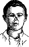
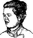
Fig. 10. Syphilitic noses.
The tertiary form of syphilis is observed most frequently. Anatomically, one deals with the formation of syphilitic infiltrates, the so-called gummatous tumors (syphilomas) and the products of their breakdown. If a syphilitic infiltrate has formed in the mucous membrane, a deep ulcer with sharply defined margins and a lardaceous base is formed, which may subsequently spread to the bone and cartilage. If a gumma is primarily localized in the bone or periosteum, bone necrosis occurs as a result of nutritional disorders, leading to the formation of a sequestrum. Tertiary syphilis can affect all the walls of the nose, but the most favored site is the bony part of the nasal septum; upon the breakdown of the gumma, a large perforation in the septum results. The floor of the nasal cavity may also undergo destruction, in which case the latter communicates with the oral cavity. As a result of necrosis of the cribriform plate of the ethmoid bone, intracranial complications may develop. Subjective symptoms in tertiary syphilis of the nose consist of nasal obstruction due to tissue swelling and stagnation of secretions drying into crusts. Patients often complain of severe pain in the nose, in the forehead, and the orbital region. Sense of smell is often diminished. In bone necrosis, a strong odor emanates from the nose. Characteristic changes in neglected cases appear in the shape of the nose; they result from the destruction of the nasal septum and nasal bones combined with cicatricial shrinkage of the connective tissue. The shape of the nose arising in this manner is termed a saddle-shaped, bulldog-like nose, or flat nose (platyrrhinia) (Fig. 10). It should be borne in mind, however, that platyrrhinia may also be a congenital condition of both specific (in congenital syphilis) and non-syphilitic character, for example, in achondroplasia (see). The course of syphilis is almost always progressive. If the process is not arrested by means of timely specific treatment, extremely severe destruction occurs inside the nose, which may even lead to a fatal outcome due to breakthrough through the cribriform plate into the cranial cavity or due to the destruction of the venous sinuses. Diagnosis. Primary sclerosis is localized chiefly at the entrance to the nose, and is often recognized only after establishing the swelling of the occipital and submandibular lymph glands. One has to differentiate between a furuncle of the nasal vestibule and the nasal septum. In a furuncle, there are limited pustules with a central breakdown and without an infiltrate in the periphery, which is not the case in syphilis. Secondary manifestations are mostly recognized on the basis of the simultaneous appearance of papules on the lips, in the oral cavity, and around the anus. In newborn and infant congenital syphilitics, poor general condition, the emaciated appearance of the child, and flabby, wrinkled skin lead one to suspect syphilis. The diagnosis of tertiary syphilis can sometimes present difficulties, especially in the initial stage of gummatous infiltration, if at the same time there are no other signs of syphilis on the body. In such cases, the Wassermann reaction, sometimes a trial treatment, or a biopsy helps to clarify the disease. In the developed stage of tertiary manifestations, when ulceration, perforation of the septum, and sequestration have already occurred, the diagnosis is usually not difficult to set (differential diagnosis from tuberculosis of the nose—see below). The prognosis, if the disease is diagnosed in the initial stage, is generally favorable. Treatment of syphilis must be primarily general (see Syphilis, treatment). Cases of tertiary forms very often respond very well to the combined administration of iodine and mercury in the form of Ricord's solution (Sol. Kalii jodati 10.0 : 300.0 Hydrarg. bijodati 0.2, three times a day a tablespoon with milk). Deformities of the external nose (saddle nose) are best corrected by means of injections of plastic paraffin (Stein's mixture) or by endonasal transplantation of bone plates taken from the tibia or costal cartilage (see Plastic operations, Rhinoplasty). 60S d) Tuberculosis of the nose. Etiology. In the majority of cases, cracks in the mucous membrane probably serve as the site of entry for the infection, especially with scratching by the fingernails in the initial part of the nasal cavity. On the other hand, with the existence of a tuberculous focus in the body (lungs, larynx, joints), bacilli can enter the nose via lymphatic and blood vessels. Tuberculosis in the nose appears in the form of an infiltrate or an ulcer; if the infiltrate takes on a tumor-like form, we are dealing with a tuberculoma. In addition to the nasal septum, tuberculous changes are found on the inferior and, more rarely, on the middle turbinate. The breakdown of tuberculous tubercles on the surface of the mucous membrane leads to the formation of ulcerations. Symptoms and course. Subjective symptoms in the initial stage are barely noticeable; in the subsequent course, as the infiltrates enlarge, a more or less complete nasal blockage occurs. After the breakdown of the infiltrates and the formation of ulcers, a purulent, sometimes blood-stained discharge appears, which may simultaneously lead to the formation of crusts. Diagnosis of a tuberculous disease of the nasal cavity is not particularly difficult in those cases when other tuberculous foci are simultaneously present in the organism or lupus is detected on the skin. To distinguish from syphilis, it must be borne in mind that tuberculosis usually affects the cartilaginous nasal septum, whereas tertiary syphilis affects the bony septum as well. Syphilitic diseases, as a result of bone necrosis, are accompanied by a strong odor and pain in the nose region, which do not occur in tuberculous lesions of the nose. In addition, the Wassermann reaction and, in children, the Pirquet reaction together with a general examination of the patient can provide final certainty in the diagnosis. Tumor-like tuberculous infiltrates may also be confused with a sarcomatous neoplasm; in such cases, the diagnosis is established on the basis of a biopsy. Prognosis in those cases where the formation developed primarily in the nose, while the other organs are healthy, cannot be considered unfavorable, although it is difficult to prevent recurrences. If, however, tuberculosis of the nose is added to manifest tuberculosis of the lungs or larynx, the hopes for a complete cure are extremely doubtful, although even here energetic measures often succeed in achieving significant improvement or a temporary arrest of the process. In treating tuberculosis of the nose, as with other forms of tuberculosis, main attention must be paid to the general treatment of the patient. Local infiltrates and tumors must be removed with a cold or galvanocauteric snare, diathermy, or cutting instruments. Flat infiltrates and ulcerations are treated by thorough curettage followed by cauterization with 80% lactic acid. In localized lesions of the nasal septum, a partial resection of it within the limits of the affected area can be performed. (Diphtheria, rhinoscleroma—see corresponding articles; lupus of the nose—see Tuberculous diseases of bones; rhinoliths—see corresponding article). e) Glanders (malleus). In individuals dealing with glanders-infected horses, glanders develops, although rarely, due to the entry of horse nasal secretion containing glanders bacilli into cracks of the skin and mucous membrane. The acute form of the disease is expressed by an elevated temperature with phenomena of general infection. On the swollen mucous membrane of the nose, vesicles and small yellowish nodules are formed, which also frequently spread to the erysipelatously inflamed skin of the face. In the subsequent course, ulcerations form along with the discharge of a viscous, later blood-stained secretion. The process may also spread to the bone, destroy the nasal septum, and mostly leads to death with phenomena of pyemia. The chronic form of glanders begins more gradually, without general symptoms; the mucous membrane appears red and moderately swollen, small ulcers form, and the discharge of secretion is not as abundant as in the acute form. At the very beginning of the disease, small abscesses usually appear on the skin. Frequently the process is localized simultaneously in the pharynx, oral cavity, and larynx, and may spread to the stomach and intestines. In diagnosis, confusion with tuberculous and syphilitic processes is possible; mostly, errors are prevented by the anamnestic data and the finding of the glanders bacillus in the nasal mucus. Prognosis in the acute form is unfavorable, in the majority of cases the disease leads to death; in chronic cases the prognosis is better, however, complete recovery is not frequently observed here either. Treatment is symptomatic, aimed at keeping the nose clean, since no specific remedy against glanders exists. In one case, a good result was obtained by Hoffmann from injections of atoxyl. In nasal obstruction, galvanocautery of ulcers and swellings is sometimes resorted to in order to restore nasal patency. f) Leprosy (lepra). In leprosy, the nose is also affected in the majority of cases. Some authors believe that the initial site of the disease caused by the leprosy bacillus is often the nose. Inside the nose, especially near the entrance to the nose, nodular infiltrates are formed, which later, as a result of breakdown, form ulcers and, as in syphilis, cause the destruction of the framework of the nose, and ultimately, as a result of cicatricial shrinkage, lead to severe disfigurement of the nose and the entire face (facies leonina).
Tubercular infiltration usually spreads also to the external nose and surrounding parts, especially the upper lip. The secretion discharged from the nose is usually foul-smelling and often forms extremely dense crusts. Not infrequently, in leprosy, there is anesthesia of the mucous membrane and skin of the nose. The diagnosis of leprous disease is usually not difficult to establish on the basis of clinical phenomena; if characteristic changes on the face and in the nose are not yet noticeable, if anesthesia of the skin and mucous membrane is also absent, then it is necessary to examine the nasal secretion for Hansen's leprosy bacillus. The prognosis is unconditionally unfavorable. Treatment, in view of the incurability of the disease, must be limited to palliative measures, especially cleansing and keeping the nose clean. If abundant crust formation has occurred, they are removed in the same way as indicated for ozenas. The incurability and the danger of infection associated with the disease justify severe measures in the form of complete isolation of leprosy patients. Tumors, a) Of the benign tumors of the nose, the so-called nasal (mucous) polyps are most frequently encountered, which histologically resemble edematous fibromas. As for their etiology, they are products of chronic inflammation of the nasal mucosa, which is also indicated by the abundance of plasma cells in these polyps. In hypertrophic rhinitis (see Cold), hyperplasia of the mucosa occurs, from which polypoid hypertrophies develop, and from the latter, partly due to the influence of mechanical causes (the movement of the stream of inhaled and exhaled air), stalked nasal polyps develop. Often nasal polyps also form as a result of irritation of the mucous membrane by pus constantly flowing from the accessory cavities during chronic inflammations of them. Diseases of the cells of the ethmoid labyrinth of a hyperplastic or purulent nature most often are the cause of the formation of nasal polyps. Polyps have a gray, sometimes yellowish-red gelatinous appearance and a smooth surface. Their size and number vary greatly. Often the entire nasal cavity is stuffed with numerous small polyps, but sometimes a single gigantic polyp can fill the entire cavity. Polyps can form in any part of the nose, but most often they originate from the middle concha and the edges of the hiatus semilunaris. Usually, polyps adapt their shape to the surrounding parts during development, but with strong development they are able to exert such strong pressure on the walls of the nose that the nose swells. Histologically, nasal polyps consist of a wide-meshed, fine-fibrous connective tissue mass containing numerous round and spindle-shaped plasma cells, as well as cavities filled with serous fluid, sometimes connecting with each other into cysts (vesicular polyps). The surface of the mucous membrane of the polyps is covered partly by ciliated columnar epithelium, partly by stratified squamous epithelium. Sometimes polyps abound in glands (adenomatous polyps), granulation tissue (granulation tissue polyps), vessels (angiomatous polyps). The diagnosis of nasal polyps usually presents no particular difficulties, and if the polyps do not reach the nasal orifice, it is established by anterior rhinoscopy and probing. Most often polyps form in both halves of the nose, if they exist in only one, then there is more often to deal with suppuration of the accessory cavities of the nose. The prognosis of nasal polyps is favorable. Treatment is only operative; it consists in removing the polyp under local anesthesia with a cold snare. After thorough swabbing with a 10-20% solution of cocaine with adrenaline and illumination with a mirror, the snare is inserted into the nose, placed on the polyp, advanced to the place of its attachment, and tightened. Many authors recommend, when tightening the stalk of the polyp into the snare, not to cut it off, but to tear it off, then the affected ethmoid cell will also be opened at the same time. Small polyps sitting secretly in the anterior nasal passage are removed with Hartmann's conchotome or cutting forceps. If new relapses constantly appear, it is often necessary to resection part of the labyrinth of the ethmoid bone by means of nasal scissors and a snare or conchotome. Existing suppurations of the accessory cavities, it goes without saying, must be subjected to careful treatment. As a special type of polyp, it is necessary to pay attention to the bleeding polyp of the nasal septum, which departs on a wide stalk from the anterior cartilaginous part of the nasal septum. It ranges in size from a pea to a walnut, its surface is either smooth or lobulated and prone to bleeding. Histologically, it has the same structure as concha hyperplasia; it differs only in its abundant blood content. To avoid bleeding, it is better to remove it with a galvanocautery snare. Of other benign tumors, a soft papilloma sometimes develops in the nose, resembling cauliflower with its uneven surface. The nasal cavity often serves as a site for the development of fibromas, chondromas, and osteomas as well. Fibroma for the most part has the nasopharynx as its starting point and is described under the name of nasopharyngeal fibroma, or fibroma of the skull base (see Pharynx and Nasopharyngeal space). In the nasal cavity, there are also myxomas, lymphangiomas, dermoids, osteomas, and chondromas. Although in patho-anatomical respects they are classified as benign formations, clinically when they develop in the nose they have a malignant character in their course and therefore, with greater or lesser significant development, are subject to the same radical removal as malignant tumors. Osteomas in the nasal cavity are very rare, they develop during the period of final development of the skeleton and are most often encountered at the age from 17 to 25 years, less often up to 30 years. The initial origin of nasal osteoma according to Arnold's theory should be recognized as the embryonic cartilage of the ethmoid labyrinth. Osteoma is a tumor of dense, bony consistency, non-bleeding, painless, and immobile. In their growth, osteomas first and foremost cause facial disfigurement, displacement of the eyeball, and less disturb the function of the nose. Osteomas that have separated from their attachment site are called "dead" osteomas. They are always provided with a bony stalk, which apparently atrophies under the influence of pressure. The prognosis for nasal osteomas is doubtful already for the simple reason that sooner or later they threaten the eye, can grow into the cranial cavity, and consequently are a disease dangerous in its consequences. Treatment of osteomas is only surgical: wide opening of the nasal cavity and its accessory cavities with complete removal of the tumor with its starting origin. Rhinosporidiosis (observed exclusively in hot countries) is a tumor of an infectious character, of irregular shape, located in the lower parts of the nasal cavity singly or multiply on a greater or lesser wide base, resembles a raspberry, reddish in color, covered on the surface with tiny white dots-sporangia. The main symptom is nasal obstruction, sometimes increased serous discharge, there are no bleedings. A correct diagnosis can be made only microscopically. The course is from several weeks to 1-2 decades. Treatment consists in surgical removal of the tumor along with the area of healthy mucous membrane adjacent to it. Multiple tumors of rhinosporidiosis very often recur. b) Malignant neoplasms of the nose. Of malignant neoplasms in the nose, sarcomas (angiosarcomas, round-cell, spindle-cell, melanosarcomas, lymphosarcomas) are often observed, further carcinomas are encountered, more often squamous-cell. They can originate from all walls of the nasal cavity, but most often the starting place is the upper jaw and the region of the ethmoid labyrinth. Symptoms are extremely diverse; patients complain of nasal obstruction and often also of nosebleeds. In the further development, a foul nasal discharge usually appears and the sense of smell on the affected side is lost. Neuralgic pains in the region of the trigeminal nerve are frequent. With the spread of the tumor to surrounding parts, displacement of the eyeball and its protrusion occur, further lesions of the optic nerve develop (amblyopia, amaurosis), of the oculomotor nerve (limitation of mobility of the eyeball), and finally intracranial complications. Protrusions in the region of the root of the nose, in the cheek and palate region are often observed. Upon rhinoscopic examination in the initial stage, a tumor is determined which in its appearance differs from the usual benign tumors - polyp, fibroma. Its surface is greater or lesser bumpy and bleeds easily when examined with a probe. In the further stage, the nasal cavity is usually completely filled with the mass of the tumor, which often ulcerates and secretes a dirty, foul-smelling secretion. Clinically, nasal sarcoma manifests itself by persistent and frequent nosebleeds, and then by the presence of a tumor causing difficulty in nasal breathing. Diagnosis. If the tumor is not yet particularly large, then it is possible to confuse it with a syphilitic or tuberculous process. Confusion with syphilis is sometimes prevented by the result of the Wassermann reaction, trial treatment with potassium iodide, and salvarsan.
To distinguish from tuberculosis, it is necessary to perform a histological examination of an excised piece of the tumor. In any case, a malignant neoplasm should be suspected if an elderly subject presents with an easily bleeding tumor with an uneven surface in the nose. In later stages, in cancer, a sharp picture of a disintegrating tumor is obtained with signs characteristic of a cancerous tumor. If a younger subject has the nasal cavity blocked by a tumor prone to bleeding, there is reason to suspect a sarcoma. In either case, a biopsy must be performed immediately. - Prognosis in malignant diseases of the nose is unfavorable; if, however, the orbit and cranial cavity are not affected, the possibility of cure by surgical means is not ruled out. The treatment of malignant tumors developing in the nose is still mainly surgical. Recently, the Russian and foreign literature has described many cases of reduction and disappearance of tumors located in the nasal cavity under the influence of X-rays, radium, and diathermy. In view of the deep position of such tumors, special types of treatment are very difficult to apply in the nasal cavity, especially since the result from this treatment is not always satisfactory. To remove malignant nasal tumors, many different surgical methods have been proposed. Methods providing good access to the tumor include total or partial resection of the upper jaw (with its numerous modifications). Nasal methods include those operations in which the entire nose is incised and turned back, or a temporary or permanent resection of the entire upper region of the nose is performed. Most of these operations are so bloody that it is recommended to perform a prophylactic tracheotomy with the introduction of a tamponing cannula or Kuhn intubation. Their second drawback is that the skin of the face is incised and scars remain with more or less noticeable disfigurement. - To avoid all these disadvantages, Denker proposed his surgical method for removing malignant tumors from the nose transmaxillary. This method consists in the fact that to the usual opening of the maxillary sinus, as is done in radical operations on it, are added the removal of the apertura piriformis (the entire facial wall of the maxillary sinus) with the capture of the lower part of the nasal bone, the frontal process of the upper jaw up to the border of the lower and inner edge of the orbit. Then the complete removal of the nasal wall of the maxillary cavity is performed, after which sufficient access to the nasal cavity is created, necessary for the removal of the malignant tumor. Since this operation is not accompanied by profuse bleeding, the need for a precautionary ligation of the carotid artery and preliminary tracheotomy disappears. Moore's method consists in making a skin incision that begins from the middle of the eyebrow, goes through it along the lateral wall of the nose downwards, and ends on the upper lip at the level of the attachment of the nasal wing. Then the soft parts are separated from the nasal bone and the frontal process of the upper jaw to the lacrimal bone. Then the frontal process of the upper jaw, the nasal and lacrimal bones, and a part of the lamina papyracea of the ethmoid bone are resected. In this way, wide access to the ethmoid bone is created, and after removing the cells of the latter, one can operate in the region of the sphenoid sinus. Plant and animal parasites in the nose. Among plant parasites in the nose, mold fungi (various species of Aspergillus) and Oidium albicans are found. Ascarids can penetrate the nose from the digestive tract. The introduction of pinworm eggs is also possible through the nostril (by hands and with inhaled dust). A number of cases of finding eggs and larvae of pinworms in the nasal cavity have been described, where there are quite favorable conditions for the development of the egg. In addition, in rare cases, leeches and earwigs were found in the nose. Furthermore, especially in purulent nasal discharge, insects and their larvae were found. - Treatment. In plant parasites in the nasal cavity (rhinomycosis), it is necessary to remove the mycelium with tweezers or by washing, and the remnants that do not yield to removal must be killed by repeated smearing with 5% menthol oil or by blowing in antiseptic powders. In the presence of thrush, it is advised to smear with a 1-3% silver nitrate solution. Larger insects that have entered the nose must be removed by blowing the nose or with an instrument. Insect larvae sit so tightly on the walls of the nose that they can only be removed after being killed with chloroform water (chloroform and water in half). For prophylactic purposes, patients with purulent nasal discharge must be forbidden to sleep in the open air in summer to prevent the crawling of insect larvae. Reflex neuroses associated with the nasal cavity (vasomotor rhinitis, hay fever, nasal asthma). The general characteristic of this group of diseases is the intensification to a pathological state of normal reflexes caused by irritation of the sensitive nerves of the nose (trigeminal nerve) or the olfactory nerve. At the same time, the moments causing reflex phenomena are either purely functional disorders or irritating substances entering the nose along with the inhaled air. - Irritations acting on other parts of the body (skin, optic nerve, stomach nerves, etc.) can cause disorders in the nose. Jurasz and Kuttner divide reflex diseases of nasal origin into 3 groups: 1) reflex neuroses in which excitation originates from the nose and the reflex is transmitted to organs lying outside the nose; 2) reflex neuroses in which excitation is located in the nasal cavity and the reflex ends right there; 3) reflex neuroses in which irritation takes place in other organs and the reflex is transmitted to the nasal cavity. Among reflex neuroses, nasal cough has long been known, but of greatest interest in view of its frequency is nasal asthma. In predisposed individuals, an asthma attack occurs if mucosal hypertrophy, nasal polyps, nasal septum deviations, or protrusions on it strongly narrow the nasal passage, so that the septum and turbinate are more or less closely touching each other. Foreign bodies, plant and animal parasites can, under certain conditions, cause asthma attacks by reflex irritation. Thermal and chemical irritations of the nasal mucosa can act in the same way. Quite often, vasomotor disorders of reflex origin are observed in the nose, such as: sudden swelling and subsidence of the cavernous tissue of the inferior turbinate, abundant discharge from the nose with redness and swelling of the conjunctiva of the eyeballs and eyelids - a condition commonly called vasomotor (nervous) rhinitis. In addition to the listed disorders, reflex neuroses of nasal origin include hay fever, sneezing spasm, dysmenorrhea, hystero-epileptic seizures, heart neuroses, spasm of the glottis, erythema of the external nose, and skin rashes similar to herpes. A reflex seizure can be caused by touching a probe to certain places of the nasal mucosa, which, however, are different in different people; on the other hand, sometimes a seizure can be aborted if these places are smeared with cocaine. Most often, the reflex points are the front part of the inferior and middle turbinate and the so-called tuberculum septi, i.e., the thickened area of the mucosa lying opposite the front end of the middle turbinate. In the diagnosis of reflex neuroses of nasal origin, one must, on the one hand, examine the general condition for the presence of neuropathic predisposition and determine whether the patient suffers from any other reflex neuroses. Rhinoscopy establishes whether there are any changes in the nose that can cause reflex phenomena. The presence of these changes does not yet prove that they serve as the cause of reflex neurosis. If moving a nasal polyp or touching a probe to a reflex point directly causes a seizure, this circumstance speaks with greater probability that one is dealing with a reflex neurosis of nasal origin. The diagnosis is substantially confirmed if it is possible to abort the attack by smearing the named points of the nasal mucosa with cocaine. If the probe examination and the cocaine experiment give a negative result, the existence of nasal reflex neurosis is doubtful, although it cannot be excluded with certainty. - Sometimes reflex neuroses of the nose can manifest themselves in severe attacks of convulsive sneezing (sneezing spasm), which can be so severe that they become agonizing for the patient. In addition to general treatment (bromine preparations) and the elimination of detected pathological conditions in the nose during an attack, spraying a solution of cocaine with adrenaline can be used here, which sometimes stops the seizure. The same treatment can be used for nasal cough. The existing reflex relations between the nose and the genital apparatus are not yet fully elucidated. The relationship between epilepsy and changes in the nose is also far from elucidated.
Eye disorders sometimes also occur on a reflex basis in the form of photophobia, amblyopia, asthenopia, scintillating scotoma, and blepharospasm. Neuralgic pains in the orbit and in the frontal region along the course of the trigeminal nerve sometimes arise by reflex action. These cases also sometimes yield a favorable effect after careful treatment of the changes present in the nose. Bronchial asthma of nasal origin. Voltolini in 1871 was the first to point out the connection between asthma and nasal polyps. Following him, Frankel and Hack (B. Frankel, Hack) pointed out the reflex origin of the asthmatic attack from a cold. All the changes in the nasal mucosa mentioned above can serve as the irritation capable of causing an asthma attack. A cold appears, combined with sneezing and nasal discharge, followed by suffocation and predominantly expiratory noise and whistling in the chest, audible from a great distance. The tightness gradually decreases, mucus appears, and after a shorter or longer time the attack passes, giving way to a completely free period. In the treatment of bronchial asthma of nasal origin, it is first of all necessary to eliminate the causative factors in the nose, remove polyps and foreign bodies, and surgically eliminate ridges, deviations, and hyperplasias. Locally here too, during an attack, it is useful to try spraying the nose with a 2-5% cocaine solution or insufflating anesthesin. For general treatment, see Bronchial asthma.
V. General operative surgery of the nose. During numerous operations in the nasal cavity performed for therapeutic purposes, it is impossible to disinfect the operative field in the way it is done on the skin. In these cases, one has to be limited to cleaning the nasal cavity from pus and mucus by lightly wiping with sterile cotton. Experience shows that wounds in the nasal cavity heal well even without thorough disinfection of the mucosa, but under the indispensable observance of general surgical rules, the use of carefully sterilized material, instruments, etc. The hands of the operating surgeon must be disinfected according to the general rules of surgery. The view is absolutely incorrect that since the operation has to be performed in an area not free from microbes, there is no need for careful observance of all the rules of surgical asepsis and antisepsis. In most cases, operations not only on soft parts, but also on bone parts in the nasal cavity are performed under local anesthesia. For this purpose, a 10-20% cocaine solution is used, and in order to bleed the mucosa more and simultaneously slow down the absorption of cocaine, 1-2 drops of adrenaline (1:1,000) per 1 cm3 of cocaine solution are added. Usually 2-3 brushings of the mucosa are sufficient for operations on soft parts; for more extensive operations on the ethmoid labyrinth and the nasal septum, it is better to insert a strip of gauze moistened in a cocaine solution into the operation site for a short time (15-20 minutes). For operations on the nasal septum, many prefer submucoid injection of Schleich's solution and 0.5-1% novocain solution with adrenaline. General anesthesia for operations in the nasal area is used rarely, almost exclusively in malignant tumors, radical operations of the frontal sinus, and also in some cases of foreign body removal in young children. Surgical operations for diseases of the nose are performed using electrolysis, galvanocautery, diathermy, and the blood method. Electrolysis at the present time for the removal of various nasal tumors is more often replaced by diathermy. Galvanocautery is used much more often than the first two methods. In using galvanocautery, it is necessary to observe the following rules: 1) the cautery must not be heated to white heat, since only heating to red heat has a hemostatic effect, and 2) during cauterization, it is necessary to ensure that two opposing parts of the nose are not cauterized simultaneously in order to avoid subsequent adhesions between both surfaces subjected to cauterization. For bloody operations in the nose, we have a whole series of various instruments, which in their shape and appearance are adapted to the conditions of the location of various formations inside the nose. The main difficulty when working with instruments is to avoid, resp. reduce or stop bleeding. Even the mere introduction of cocaine with adrenaline causes a rather significant contraction of vessels and reduces bleeding during the operation. It goes without saying that it is necessary to first verify whether the patient comes from a bleeding family, and it is also necessary to be careful when operating in kidney diseases, anemia, chlorosis, leukemia, Werlhof's disease.
Related articles
Mentioned in
Cite this page
“Nose.” Soviet Medical Encyclopedia. English translation of Bolshaya Meditsinskaya Entsiklopediya, 1st ed. (Moscow, 1928–1936), ed. N. A. Semashko. https://sovietmedicalencyclopedia.pages.dev/article/nose/