Rhinoplasty
Historical document, translated for reference. It reflects medical knowledge of the 1920s–30s and is not medical advice.
Summary
This article from the 1928–1936 Soviet medical encyclopedia provides a historical overview of rhinoplasty, tracing its development from ancient Indian techniques to the surgical advancements of the 19th and early 20th centuries. It details the evolution of flap surgery, including the Indian, Italian, and brachial methods, and the subsequent development of techniques for creating structural support for the nose.
Encyclopedia article (1928–1936)
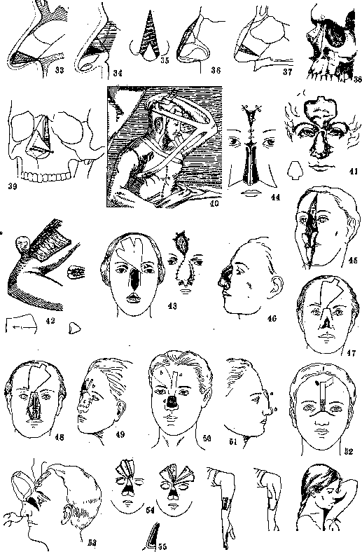
RHINOPLASTY. At the time when nasal plastic surgery was the sole content of plastic surgery, old surgeons (Tagliacozzi, Graefe, Carpue, Dieffenbach, and others) viewed rhinoplasty as the art of restoring the nose exclusively by replacing the defect with skin flaps from the forehead or arm; in the modern understanding, rhinoplasty represents a branch of plastic surgery that studies not only methods for restoring an absent nose and its parts by replacing defects with living tissues, but also methods for correcting congenital and acquired irregularities of the shape of the nose through surgical means. The breadth and complexity of plastic operations on the nose are determined in one case by the degree of deviation from the norm, and in another by the size of the defect of the tissues forming the projection of the nose in front of the piriform aperture. In the history of rhinoplasty, three eras are distinguished: the first era begins approximately 1,000 years before our era in India: a flap from the forehead, from the cheek, a flap from the buttock; precise cutting of flaps. The second era—in Europe from 1450 to 1760: a skin flap from the arm, from the cheek, improvement of the forehead flap, surgical shaping. The third era—from 1760 to the present time: formation of a lining and support for the skin flap, formation of a bony support and mucosa; further development of skin flaps. The first indications of the existence of rhinoplasty we encounter in the most ancient Indian book (Susruta's Ayurweda); there is an exact description of the operation, which was performed by the caste of priests (according to other sources, potters) for the restoration of a cut-off nose (cutting off the nose was a common phenomenon of barbaric times as punishment for misdemeanors). Joseph provides an exact description of this operation: "The physician took a leaf from a plant the size of the nose to be formed, placed it on the forehead or cheek, and according to the size of the leaf cut a piece of skin so that it still hung on a pedicle, quickly freshened the edges of the nose and fitted it to the wound and secured the flap in the proper manner with dressings, below he secured in it two inserted tubes, directed them upward and sprinkled (the wound) with sandalwood." English physicians Findlay and Crusso (James Findlay and Thomas Crusso) provide almost the same description of the operation seen by them in Bombay in 1494; here the shape of the nose is already prepared from a wax plate at the site of the defect, the plate is flattened and transferred to the forehead, where the shape of the flap is cut out along the edge, a skin pedicle is left for nourishment between the eyes, at the root of the nose. According to the accounts of contemporaries, these noses did not differ in any way from normal noses. The English military surgeon Lucas, having learned from Indian operators, operated many times using this method. The Indian method of operation remained unknown in Europe until the middle of the 15th century. The second era of rhinoplasty begins from 1450 in Italy, where this operation was applied by the Sicilian surgeon Branca from Catania. Whether he began to apply this method independently or it was brought from India has not been established with certainty. Branca restored the nose, ears, and lips with flaps from the face. Branca's son, Antonio Branca, was the first to begin taking a flap for the formation of the nose from the arm and is the author of the so-called Italian method. Joseph calls this method brachial rhinoplasty. This operation began to be widely applied by the students of A. Branca in Calabria and by a number of surgeons from the Boiani family. A scientific description of this operation is given by the professor of anatomy in Bologna, Tagliacozzi, in the work "De Chirurgia curtorum per insitionem," published in Venice in 1597. The operation is divided into 6 moments. After careful measurement on the anterior surface of the arm, a skin flap is outlined with two parallel incisions and detached from the underlying tissues, a piece of linen is placed under it, which is changed daily for 4 days; after 14 days the peripheral pedicle is cut and lies freely for another 14 days, passing through the stages of childhood (pueritia), youth (adolescentia), and old age (senectus); when the flap begins to shrink, it is transplanted onto the freshened defect of the nose and the arm is held by a complex bandage (Fig. 40) for 20 days, after which the flap is cut. After 14 days, the wings and septum were formed. After the death of Tagliacozzi, very few followers remained, and this operation was forgotten in Europe for almost 200 years. Only in 1814 did the English surgeon Carpue first in Europe begin to operate according to the Indian method, and he secured the flap to the defect with sutures (Figure 41). In Germany, Graefe (1816) restored the nose according to the method of Tagliacozzi, having previously formed a skin nose on the arm (Fig. 42), but then he began to operate in one stage. He cut the flap on 3 sides and immediately sutured it to the defect; thus, he shortened the period of the operation from 3 months to 1 month. A contemporary of Graefe, Serre, abandoned the brachial method and began to take a flap from the cheek. Graefe, in his monumental work, gives a critique of the methods contemporary to him. He considers the disfiguring scars on the forehead, the frequent complication of erysipelatous inflammation, and the growth of hair on the flap to be the disadvantages of the Indian method, since the flap was taken with a large reserve of skin, capturing the hairy part. He considers the difference in the color of the skin of the arm and face and the difficulty of securing the arm to the head, the difficulty for the patient to endure this bandage for a long time, to be the disadvantages of the Italian method. The Italian and Indian methods, after Carpue and Graefe, began to be widely applied in Europe by surgeons of the pre-antiseptic period with various improvements in the technique of cutting the flap in terms of shape, location (e.g., double flap from the forehead, flap from the lateral surface of the forehead), more convenient rotation, cutting of details, etc. All these methods served and still serve as the basic methods for cutting skin flaps in modern rhinoplasty with the addition of new improvements to them to eliminate the essential disadvantages of single-layer flaps—shrinking and change of shape due to granulation of the exposed surfaces on the inner side. Dieffenbach, the founder of plastic surgery and creator of new methods of plastic surgery in various fields, laid the foundation for a new era of rhinoplasty, having given the idea of doubling the flap in the lower part of the nose, in the region of the wings and septum, by turning in specially cut lobes of the flap for the wings inward. Blasius, to avoid necrosis, pre-folded the flaps for the wings on the forehead. Langenbeck cut a flap from the forehead for the wings and for the septum with three lobes at the ends for doubling; furthermore, he placed the flap obliquely on the forehead (Fig. 43). Since the turning in of the wings did not save the initial shape of the nose from collapsing and shrinking, the idea of placing a stronger lining under the surface layer continued to develop. Szymanowski proposed cutting two long flaps for the support of the nose, located side by side in the middle of the forehead, turning them over and connecting them with their exposed surfaces, and sewing them into the middle of the defect; thus, a septum and a tensioned support for the flap were formed (Fig. 44). He formed the surface layer from lateral triangular flaps from the cheek. Dieffenbach also proposed splitting the formed skin bridge of the nose and sewing in a long flap from the forehead with the skin inward; after the engraftment of this flap, the edges of the initial incision on the bridge were sutured. In 1859, Langenbeck proposed using a bone-periosteal flap from the forehead and obtained more satisfactory results in terms of creating a denser support for the nose, thereby laying the foundation for the formation of a hard framework of the nose. In 1861, Ollier used the remnants of the left nasal bone and part of the frontal process together with the detached skin flap from the forehead and the lateral parts of the piriform aperture to form a hard support for the nose. Figures 45 and 46 give an idea of this operation, which served for the development of further stages of restoration of the bony framework of the nose. Langenbeck, to implement this idea, sawed out two plates from the lateral parts of the nose, leaving them connected at the bottom to the lateral parts of the jaw. Placed vertically, they formed the arch of the framework. At the same time, the collapsed nasal bones were lifted with an elevator. A frontal skin flap, sometimes together with the periosteum, was placed over the formed bony framework. Bardenheuer (1898) operated using a method similar to Ollier's method, but with two flaps, whereby for the inner lining a flap was taken from the root of the nose with the base at the defect together with the nasal bones and part of the bony septum, while the outer flap was taken from the forehead (Fig. 47 and 48). The idea of using a bone plate to form a support for the skin flap also belongs to Langenbeck, but it was implemented by Franz König in 1886. The operation was applied for a severely collapsed nose. At the site of the greatest collapse, a transverse incision was made, reaching from the sides to the base of the nose; the end of the nose, pulled upward, was pulled downward, after which a rather wide defect was obtained in the middle part of the nose. On the forehead, two flaps are cut, one narrow in the middle of the forehead, which is taken with the underlying bone plate and turned over with the skin inward and sewn into the defect. It forms a support for the other flap, which is wider, taken from the lateral side of the forehead. This flap forms the upper layer of the nose.
König's method served as a prototype for methods of osteoplastic operations for the formation of the bony base of the nose. A drawback of König's method is the formation of skin folds at the turn of the flaps, creating skin pockets that retain the discharge from granulating surfaces. This drawback is eliminated by a modified operation proposed by P. Dyakonov in 1891. He also dissected the skin transversely at the site of the greatest depression, sutured a flap with a bone plate into the defect in the same way, skin side inward, and sutured it under the detached end of the lower segment; in order to prevent a skin fold from remaining, he excised a thin layer of epithelium at the site of the flap's fold so that the freshened surfaces would grow together—the transplanted internal flap was covered by tightening the edges of the defect (Figs. 52 and 53). König's operation, like Dyakonov's, can also serve as a method for total rhinoplasty. Helferich took a flap from the cheek for the internal lining and covered it from above with a skin-bone flap from the forehead. Further development of König's operation proceeds in two directions: the formation of the bony bridge of the nose and the formation of a roof-shaped framework of the nose. Hacker, together with a flap cut from the forehead, takes a long strip of bone from the cortical layer of the forehead, cuts it closer to the peripheral end of the flap, and bends it at an angle. The plate, bent at an angle, forms the bony profile of the nose (Figs. 54 and 55). Israel, for the formation of a bony support for the nose, proposed taking a skin-periosteal-bone flap on a long skin pedicle from the lateral surfaces of the ulna (Figs. 56, 57, and 58). Incidentally, this is one of the most difficult positions of the arm to tolerate during fixation. Schimmelbusch applied König's idea for total rhinoplasty by forming a triangular skin-bone flap in the middle of the forehead (Figs. 61, 62, and 63). The flap, taken in its entirety with a bone plate, is kept wrapped in gauze for 4-8 weeks. When it becomes covered with granulations on the bone side and loose bone fragments are sequestered, the granulations are scraped off and the exposed surface is covered with epithelium. After the epithelium has taken, the bone is sawed through in the middle, and the entire flap, folded in the shape of a roof with the epithelium inward, is sutured to the freshened opening. The defect on the forehead is closed by sliding large lateral flaps, bounded by arcuate incisions in the temple region. The operation presents great difficulties, mainly due to the need to take an even plate along the entire length of the flap; complications are also possible during the long healing period. Lexer (1910) improved Schimmelbusch's operation in that he separated the bone plate on the forehead only up to the middle of the flap, then folded it together with the covering skin inward, thus creating a duplication of skin in which the bone plate was placed; from the middle part of the plate on the inner side, a strip of skin on a pedicle was cut out at the edge to form the septum, and the bone was sawed through. The flap was bent in the middle, rotated on the pedicle by 180°, and its lateral edges, after freshening, were sutured to the freshened lateral edges of the defect. After healing, in the region of the rather thick bridge of the nose, it is necessary to excise the subcutaneous tissue to shape the profile of the nose (Figs. 59, 60, and 64). Besides skin-bone flaps from the forehead and shoulder, Hardie (1875) was the first to attempt to form a nose with a solid bony support from a finger in a boy. He freshened the skin of the index finger of the left hand and sutured it into the upper corner of the defect. The skin was dissected in the longitudinal direction on the palmar side and its edges sutured to the edges of the defect. After 15 weeks, the finger was detached in the middle of the second phalanx. The operation was unsuccessful, because the phalanx was pulled into the piriform aperture by scars. In 1902, Vreden implemented this idea in a more perfect form, using the ring finger of the left hand for total rhinoplasty; the bridge of the nose was formed from the first and second phalanges, and the main septum from the third. Volkovich (1908) proposed his own method of forming a nose from a finger of the left hand and operated on 2 cases with a good result. Despite the originality of the method and the ingenious use of the three phalanges for the framework of the nose, this operation does not have wide application due to the mutilation of the finger. Furthermore, the operation does not provide the long-term cosmetic effect expected from it. Simultaneously with the development and complication of the Indian and Italian methods, new ways were being developed to obtain skin and bone material for plastic surgery of the nose. Thus, Serre proposed using flaps from the lateral side of the nose to close the defect, the so-called old Indian or French method. Hueter, Bourow, and Helferich proposed cutting two wide flaps from the cheek: one for the internal layer, the other for the external layer. A combination of a cheek and forehead flap was proposed by Thiersch, and a combination of a skin flap from the arm and from the forehead by Küster. In 1889, Hacker recommended taking a flap from the chest or back and suturing it to the exposed surface of the upper limb, and after it has taken, transferring it with the arm to the face. Steinthal published a method of transplanting a flap from the chest to the arm, and from the arm to the face—a wandering flap (Figs. 65 and 66). In 1913, Hollander used for rhinoplasty a flap cut in the region of the sternum with a base on a pendulous female breast and, together with the breast pulled upward, brought it closer to the nasal defect. Rosenstein, in the same year, proposed fixing a long flap from the chest to the chin and then using the other end for plastic surgery of the nose. Most recently, the tubular flap proposed by Filatov makes it possible to take skin material not only from the arm but also from the most distant places by sequentially moving the pedicles. Furthermore, the platform formed at the end can be provided with bone plates for the formation of the skeleton of the nose. Besides flaps on a feeding pedicle, free skin grafting according to the Thiersch-Reverdin, Wolfe-Krause, and Davis methods is also used in rhinoplasty, for example, to cover exposed surfaces after taking a flap or to line the internal surface of flaps facing the nasal cavity. A huge shift in the development of rhinoplasty in terms of simplifying technique and plastic possibilities was produced by free transplantation of bone and cartilage. Israel (1896) was the first to use free bone transplantation in a case of nasal depression. The bone plate was taken from the lower leg. To introduce the transplant, a longitudinal incision was made along the bridge of the nose at the site of the greatest depression. A bone plate was inserted into the formed pocket and the skin was sutured. This first bone transplantation was crowned with success. Mangoldt and Dyakonov proposed making a transverse incision at the root of the nose. Dyakonov proposed for this purpose a double-edged knife bent at an angle to the handle; Mangoldt proposed, instead of bone, inserting a plate of costal cartilage through a transverse incision at the tip of the nose. He also proposed inserting flat pieces of cartilage into shrunken alae (1900) after their dissection. Joseph (1906) proposed intranasal introduction of transplants, as described below. The first successful experiments of bone transplantation under intact skin gave Israel the idea to transplant a bone plate into the subcutaneous tissue of the shoulder and use the flap with the engrafted transplant for rhinoplasty. Lexer sawed out a triangular convex plate from the tibia, implanted it in the form of a roof into the shoulder region under a bridge flap, and wrapped the edges of the flap onto the inner side of the transplant. After 3 months, the flap with the bone was transferred to the nasal defect. Further clinical observations and experimental studies by Axhausen, Lexer, Petrov, Pavlov-Silvansky, and others established the full suitability of bone material for plastic purposes; although bone and cartilage in some cases undergo partial changes and are resorbed, nevertheless, in many cases, transplants provide a lasting cosmetic and functional effect. As for the disputes about the advantage of a bone plate or cartilage for transplantation in rhinoplasty, observations of recent years force one to lean in favor of cartilage, especially since it is a known fact that a bone transplant takes better where functional load is distributed onto it. Therefore, a bone transplant that is at rest during rhinoplasty is more likely to be resorbed than a cartilaginous one. Modern rhinoplasty possesses a vast material of all kinds of methods for restoring the nose in all its anatomical parts, and the achievements of the last decades related to free plastic surgery of bone, cartilage, and skin are especially important. At the present time, the task of rhinoplasty is no longer just to find a new method of restoring the nose, since the main paths have already been outlined, but to detail old methods, modifying and adapting them to new requirements of functional and cosmetic effect. Each operation of total or subtotal rhinoplasty requires a preliminary detailed study of each case in relation to the pathological process that caused the disease; here, for example, untreated syphilis, remnants of a lupus process, and hidden remnants of a removed malignant tumor are of significance.
The general condition of the organism and even the psyche of the patient also require detailed examination. The state of the defect requires detailed study and preparation for the transplantation of a graft. Preliminary operations on the defect consist of: 1) increasing the narrowed nasal opening, 2) providing a base, a foundation for the support of the future nose, and 3) preliminary freeing of the alae, dissection of the shortened septum, cicatricial bands, and pockets that hinder orientation in the surgical plan and examination of the nasal cavity regarding the determination of ulcers, tumor remnants, etc. The most commonly used methods of nasal plastic surgery fall into the following three main forms (Joseph). -1. Rhinomyoplastica, an operation having as its ultimate goal the reduction of the nose in all dimensions or of its individual parts by means of resection or incision of the skin, cartilage, bone, or all these tissues separately, in a proportional ratio, similar to how this is done in the reduction of other hypertrophied organs. -2. Rhinoorthoplasty represents bloodless and bloody methods of correcting incorrect positions and curvatures of the nose, congenital or arising during the growth period (e.g., deviation of the septum) or remaining as a result of untreated injuries; this also includes the connection of individual parts of the nose separated by gaping clefts or scars without loss of nasal tissue. The bloodless way of correcting curvatures in fresh cases consists of the repositioning of displaced bones with the help of finger pressure or with the help of appropriate instruments (e.g., pelottes with rods secured to the forehead for prolonged pressure) applied from the outside, or special expanding dilators (in the form of sponge forceps) or elevators applied from the inside (e.g., when setting bone walls of the nose that have been pressed inward). The bloody way of setting is used for more persistent curvatures and is combined with osteotomy or wedge resection of the lateral walls of the nose. -3. Neoplastica - closure of partial defects of the nose or restoration of the entire nose by replacing the defect with living tissues (transplantation, implantation). The material for closing nasal defects, the graft, is taken either in the form of freely separated tissues, a free plate, without a nourishing pedicle (in the transplantation of cartilage, bone, fat, epithelium), or the graft is transplanted onto the defect with the help of a nourishing pedicle (tubed pedicle flap). Flaps for closing nasal defects, just as in plastic surgery of other parts of the face (see Plastic operations), can be taken on a short pedicle at the edge of the nasal defect and turned inward with the skin to form an inner layer, or on a longer pedicle from the forehead, neck, or cheek to form the inner or outer surface of the nose, or from more distant places, e.g., from the arm (Italian method), on a longer pedicle. At the present time in our Union, the tubular Filatov flap (see Plastic operations) is widely used. The advantage of this flap is the possibility of pre-implanting plates of bone or cartilage into it to form the future nose. It is also necessary to note the flap on a connective tissue pedicle, often used for replacing the mucous membrane of the nose, without surface skin, for replacing the inner layer: the pedicle is formed at the border of the freshened defect, whereby the detachment of the flap stops, not reaching 2-3 mm to the cut edge of the skin, and on this pedicle the flap is turned over with the skin inward, while nutrition occurs through vessels not along the continuation of the skin, but through the connective tissue pedicle. An example of such a pedicle can be the Gersuny flap for replacing the mucosa of the cheek or the Kirschner flap with a connective tissue pedicle including the temporal artery. In nasal plastic surgery, free transplantation of cartilage and bone into soft tissues serves to restore the framework of the nose and give it stability and a certain convex shape; since the restoration of support for the soft parts of the nose in the form of a vertical bone-cartilage septum, which determines, together with the other components (see below), the projection and profile of the nose, is technically very difficult, this support is created by a rafter-like placement of cartilages or bone plates on the lateral edges (apertura pyriformis) or in the form of a framework of the bridge and septum. Cartilaginous or bone plates are either pre-implanted into the flaps forming the skin part of the nose or placed into the formed skin nose. In partial defects of the bone or cartilage skeleton of the nose, the missing tissue is transplanted while the skin coverings remain intact. Cartilaginous material is taken predominantly from the costal cartilage of the VII, VIII, IX ribs through an incision along the outer edge of the costal arch and is isolated by longitudinal sections of a knife directed at an angle to the midline of the cartilage or parallel to the surface of the cartilage, depending on whether it is necessary to take a massive piece or a thin plate. It is also possible to remove cartilage with a gouge or flat chisel. The shape of the massive cartilage should generally represent a triangular pyramid with a correspondingly treated upper end and base, with the side facing downward being treated according to the concave surface of the defect. In addition, there should be a longitudinal groove in the upper end for tight adherence to the remnants of the root of the nose or to the remnants of the bridge of the nose. The anterior end of the pyramid should be rounded and cut from bottom to top so as not to press on the arches of the cartilage of the tip of the nose; otherwise, the tip of the nose settles and the nostrils flatten from top to bottom. Surface plates of cartilage are cut with the perichondrium, which during transplantation should be turned inward, as the plates have a tendency to warp toward the smooth surface. Cartilages can also have another shape according to the requirements of special plastic surgery (for example, cartilage in the form of a hanging drop for the tip of the nose). -Bone material is taken most often from the anterior edge of the iliac crest or the anterior surface of the tibia. The graft is introduced subcutaneously by the extranasal or endonasal route. In the first case, the incision is made through the skin in the area of an existing scar on the side or at the root of the nose, or a special incision is made, most often at the tip of the nose. Rauer recommends penetrating under the skin through an incision formed by two arcuate lines, skirting the edges of the nasal openings from above at a distance of 1 mm and converging on the cutaneous nasal septum at an acute angle—an incision in the form of a swallow. A triangular flap is detached (sparing the arches of the terminal cartilage of the nose) and the detachment continues onto the bridge of the nose and the lateral surfaces. This incision provides good access and a good cosmetic effect. The best method of incision for introducing a graft, as well as for subcutaneous resections, should be considered the endonasal route proposed by Joseph, in which the incision is made from the side of the mucosa through the skin of the nasal vestibule on one side above the fold formed by the triangular cartilage. Through this incision, by penetrating above the triangular cartilage with a short, narrow knife with a long neck, it is easy to detach the skin along the entire extent of the cartilaginous and bone skeleton without damaging the mucosa, and to introduce the graft (Fig. 1). During introduction, the graft is grasped with light bone forceps or strong tweezers with sharp teeth to avoid slipping. The objections of some surgeons regarding the lesser asepsis of the endonasal incision, in Rauer's opinion, do not have a serious basis. Contraindications can be cases of ozena and cicatricial changes of the nasal mucosa. Of the dead materials for replacing bone and cartilage defects, paraffin (see Paraffin, application in surgery) and a graft made of ivory (Fig. 75) are most often used, as well as other alloplastic dead materials, which at the present time enjoy less popularity than living autoplastic material. Fat transplantation is performed most often during plastic operations of the nose to lift and level the surrounding parts of the face; sometimes it is also introduced for this purpose under the skin of the nose. Through a relatively small incision, a skin pocket is detached, into which a piece of fat taken from the thigh or abdomen is introduced, with the graft not being subjected to kneading. When cutting skin flaps for plastic surgery of the nose and surrounding parts of the face, it is necessary to take into account the depth of the location of the neurovascular layer and the location of the motor branches of the facial nerve (incisions on the face according to Bockenheimer, Fig. 2), then the direction and depth of the location of Stensen's duct and the parotid salivary gland, since cutting the duct and incisions of the gland lead to the formation of persistent chronic salivary fistulas. To determine the size of the nose, the following designations are adopted (Fig. 3). The root of the nose is considered the border of the nose with the forehead;
The base of the nose is the line passing along the transition fold between the nose and the lip and connecting the 3 lower points of the alae and the septum (e-d); the lateral base is the line separating the nose from the cheek (c-d). The length of the nose is measured by the line from the root of the nose (b) to the upper end of the skin septum; the width or thickness of the nose is the distance between the most distant symmetrical lateral points (a-c). The widest part of the nose is at the level of the alae; the width of the bony part of the nose is of practical importance. The profile line is the upper line of the sagittal section, passing from the root of the nose (b) to its base (k). The width of the profile at the top and bottom is measured by the distance of the points from the profile line to the lateral line of the base; the height of the profile is the length of the perpendicular line dropped from any point of the profile to the plane of the base at the level of the cheek. In addition, the angle between the septum and the lip (septolabial angle) and the angle between the septum and the dorsum of the nose (septodorsal angle) are determined. A septodorsal angle of 90° is considered normal; with a larger angle, the nose will be shorter—a blunt nose (Fig. 4). For rhinoplasty, the so-called aesthetic angle of the profile (Joseph) is also of importance, which is formed by the main line of the profile with the fronto-mental line (Fig. 5). This angle should not be confused with the anatomical angle of the profile, formed by a straight line passing through the root of the nose and the protruding part of the upper jaw, with the German horizontal (Fig. 6). Joseph proposes a special device for measuring the angle of the nose. The statics of the nose are determined by the bony and cartilaginous part of the septum (vomer, perpendicular plate of the ethmoid bone, and the quadrangular cartilage), the nasal bones connected at a greater or lesser angle to the frontal bone, and parts of the frontal processes of the upper jaw. The cartilage of the nasal tip is of great importance for maintaining the projected shape of the nasal tip. Its absence or a change in its shape is sharply reflected in the shape of the nasal tip. In order to approach the restoration or reconstruction of the shape of the nose more closely, Joseph suggests taking as a basis for the analysis of a given defect and the construction of the nasal profile the following anatomical elements or components. 1. The component of the nasal bones, which determine the aesthetic angle of the nasal profile. 2. The component of the bony and cartilaginous septum, which is of great importance for the statics or support of the middle part of the nose. 3. The component of the nasal tip cartilage [Fig. 7 schematically depicts the ratio of these three components to the length of the nose (2:2:1)]. A normal profile should not deviate sharply from these dimensions. The aesthetic angle of the profile should lie within the range of 20° to 40°, with an angle of 30° being considered the ideal profile angle (Fig. 8). Deviation of the profile line in each individual component creates a variety of forms of nasal deformity (Fig. 9, 10, 11, and 12) (Joseph). Rhinoplasty, just like any plastic surgery, requires for its execution, first of all, an analysis of the existing tissue defect or organ deformity and the preparation of a general and specific plan for the operation. The analysis of the injury and the preparation of the plan are closely linked to the surgeon's ability to think stereoscopically, in three dimensions (Joseph), i.e., to visualize the dimensions of the defect, the shape, and the size of the organ being restored; some drawing skill helps in carefully and expediently cutting out the skin flaps needed for the plastic surgery. A schematic drawing of the altered organ, on which the missing parts of a normal nose are drawn in, helps a great deal in the analysis of the existing defect; in the case of scars pulling normal parts toward the defect, the latter should be placed in their normal position on the drawing, thanks to which the existing defect will be more clearly defined; Figs. 13, 14, and 15 explain the analysis of a nose shortened by injury, where the apparent defect of the lower part of the nose, after analysis, turns out to be a defect of the middle part. When preparing the plan, one must take into account not only the amount of material required but also the site from which the material can be taken with the least damage to the organism, while also considering the blood supply of the flap, the ease of transfer, color, thickness, hair coverage, etc. When defects of the nose and face exist simultaneously, it is necessary to pre-outline and calculate the order of tissue restoration, e.g., first the restoration of the skeleton of the upper jaw, lip, and cheek, in order to create a base for the nose. 1. Rhinomyoplastica. The operation for reduction of the nose is mentioned for the first time by Dieffenbach in 1845, in his operative surgery, regarding the reduction of a normal large nose by means of a cross-shaped resection of cartilage and soft tissues (see pp. 56 and 57, Figs. 2-5). In subsequent years, some surgeons undertook partial resections of hypertrophied parts of the nose without significant cosmetic effect. Since 1898, Joseph has published a significant number of methods for reducing the size of the entire nose and individual parts, and in his last book, 'Nasenplastik und sonstige Gesichtsplastik' (Lpz., 1931), he gives an extensive overview of the methods and examples of these operations with a very good cosmetic effect. Surgical intervention for various deformities and hypertrophies of the nose is undertaken not only to eliminate sharp, striking disfigurements but also for psychological depression observed in some individuals even with minor deformities. The operation to restore the normal appearance of the nose very often serves as a factor that restores the psyche and working capacity of the patient. Of the numerous forms of hypertrophy of the entire nose and its individual parts, only the most characteristic and severe forms of changes are considered here.—Simple hypertrophy of the nose occurs either due to an increase in the fixed part of the nose, the bony and cartilaginous, or due to the soft, mobile parts. These include: 1) an abnormally convex profile of the nose in the bony and cartilaginous part, an abnormally protruding nasal hump; 2) abnormal width of the nose, mainly in its bony part; 3) hypertrophy of the soft parts of the nose, the triangular cartilages, and the alae; 4) change in the shape and size of the nasal tip; 5) abnormal length of the entire nose, mainly of the cartilaginous septum. Pathological hypertrophy of the nose is observed in acromegaly, rhinophyma, and benign tumors. An excessively convex profile can be caused either by the protrusion of all the components forming the framework of the nose, or only the bony or only the cartilaginous part participates in the formation of the hump. The operation in the first case consists of straightening the profile by resecting the protruding bony and cartilaginous part of the nose. The operation can be performed either by the extranasal route, through a longitudinal incision of the skin on the dorsum of the nose, sometimes with resection of a spindle-shaped area of excess skin, or by the intranasal route according to Joseph's method. With the extranasal operation, an external scar remains, which, although sometimes barely noticeable, does not always satisfy the patient. The operation by the intranasal route consists of several stages: 1) an incision over the triangular cartilage, usually on the left side, through the mucosa into the nasal vestibule (Fig. 1), and detachment of the skin in the middle and lateral parts (Fig. 16); 2) introduction of a narrow raspatory and separation of the periosteum in the bony part over an extent sufficient to expose the bone segment to be removed; 3) introduction of a sharp saw with a bayonet-bent handle to the outer surface of the hump. The outer wall, the quadrangular cartilage, and the second wall are sawed through from the inside outward at one time (Figs. 17, 18, and 22). Sometimes it is more convenient to make an incision of the mucosa in the nasal vestibule on the other side and saw through the second wall also from the outside inward. After sawing, a probe-pointed knife is introduced, the attachments of the hump are separated from above and below, and the hump is extracted in its entirety through the right or left nasal opening. Sometimes corrections of the protruding bony edge with a special rasp or a probe-pointed knife in the cartilaginous part are required. The operation in the case of only a bony hump is performed in the same way; if, after removal of the hump, the dorsum in the bony part of the nose turns out to be too wide, it can be narrowed in the bony part by displacing the bony lateral walls, mobilized at the base, inward. The operation is performed according to Joseph's method by the intranasal route. An incision is made with a double-edged narrow knife in the lateral pocket of the nasal vestibule from the inside. The knife is advanced to the edge of the piriform aperture onto the lateral surface of the nasal part of the upper jaw, and the soft tissues are detached over a sufficient extent to introduce a raspatory and separate the periosteum. Then, an instrument like a grooved probe is first introduced into the incision to protect the lower edge of the wound from the saw (Figs. 19 and 114), and then the saw (the handle of which, just like that of the raspatory, is bent at an angle upward) is introduced, and under the control of a finger through the skin, the lateral bony part of the nose is sawed through at the base without damaging the mucosa (Figs. 20 and 21). After the sawing, the bony plates at the base are displaced toward the middle. To fix the achieved position, rolls are applied to the sides of the nose, secured with adhesive plaster. Joseph recommends applying a special clamp made of nasal plates that slide at an angle, attached to the nose with a circular rubber band, for one hour daily for 6 weeks.
In cases of hypertrophy of the soft part of the nose, caused by an abnormal convexity of the triangular cartilages, a longitudinal spindle-shaped excision of soft tissues from the entire thickness of the nose is recommended, symmetrically on both sides; in less pronounced forms, a longitudinal excision of a long strip of mucosa and cartilage on each side, in the more protruding part, detached from the skin through a small arcuate incision along the edge of the nostril from the mucosal side (Figs. 23 and 24), is sufficient. The excision of the strip can be done with an instrument similar to a conchotome with an elongated narrow aperture, a punch. Severely widened wings with wide nostrils are reduced by the excision of crescent-shaped pieces through the entire thickness of the wings (Weir's method) or by the excision of wedge-shaped pieces from the mucosal side without an external incision (Figs. 25 and 26). In the case of a severely elongated and wrinkled nostril, observed after suturing a harelip, a crescent-shaped strip is excised through the entire thickness of the wing at the border with the septum. In this case, it may also be necessary to excise a strip of the lip to reduce the nasal opening (Figs. 27 and 28). The cause of abnormal protrusion of the tip of the nose is the overgrowth of the paired cartilage of the tip of the nose and a change in its shape due to an increase or decrease in its curvature. Joseph recommends several methods for reducing the protrusion of the tip of the nose, for example, transverse subcutaneous excision of a more or less wide strip from the terminal cartilage on each side via the intranasal route together with the mucosa, a through incision of the skin part of the septum parallel to the outer edge with the expectation that after scarring (approximately in a month) the tip of the nose will settle, and the excision of a quadrangular piece from the middle of the septum (Figs. 29 and 30) followed by suturing the upper and lower edges. This excision can be combined with the excision of crescent-shaped notches from the base of the wings. A pulled-down drooping tip of the nose, a hooked nose, can be corrected by the resection of a long triangular wedge from the membranous part of the septum through the entire thickness together with the posterior edge of the terminal cartilage; after suturing the defect, the tip of the nose assumes a normal position (Figs. 31, 32). The nose can be normally lengthened by lengthening either the cartilaginous septum or both the cartilage and the lateral walls simultaneously. Shortening of the quadrangular cartilage is performed through a through angular incision on the septum along the edge of the protruding end of the cartilage. With the help of a button-tipped knife, the protruding part of the cartilage together with the mucosa covering it is excised (Figs. 33 and 34). To shorten the lateral wall, after intranasal detachment of the skin, a triangular piece is resected from the anterior edge of the triangular cartilage on each side; the defect above and on the septum is sutured with strong sutures. Reducing the length of the entire nose can be combined with the excision of a hump and other corrections. Hypertrophy of the nose, mainly due to thickening of the skin and the cartilaginous part, is observed in acromegaly, in rhinophyma, and in so-called paraffinomas. The operation to reduce the nose is performed through the external integuments and consists of three moments—excision of the skin, excision of the bone framework and the cartilaginous one (Figs. 35, 36, and 37), if it is enlarged, and wedge-shaped excision of the septum to lift the tip of the nose. The skin is outlined by two wedge-shaped incisions entering one into the other. The external incisions start from the middle of the root of the nose and end at the edge of the nasal openings; the internal wedge-shaped incision has its base at the tip of the nose. The skin and underlying tissues of the nose (the area of the triangular cartilage and wings) between the external and internal triangle are excised through the entire thickness; the width of the excised strips on both sides is usually equal to 0.3-0.5 cm. If there is an enlargement of the bone skeleton, parts of the nasal bones are excised with a chisel; a wedge-shaped piece with the base at the bridge is also excised from the cartilaginous septum; the wedge-shaped defect is tightened with a suture. The defect on the bridge of the nose is closed with a double suture—catgut from the inside and hair from the outside. Benign and malignant tumors that increase and change the shape of the nose are excised within the limits of healthy tissues, and after waiting for a certain period guaranteeing the absence of recurrence, the defect is closed by plastic surgery. Paraffinomas are removed through an incision via the intranasal route with the help of a sharp spoon or a blunt curette. Excessively transplanted cartilage is excised subcutaneously through incisions at the site of scars, through the tip of the nose, or via the intranasal route. Too large bone transplants are extracted; in some cases, after reduction, they can be reimplanted. 2. Rhinoorthoplastica. Deviations can involve the bone or cartilaginous part of the nose. In the bone part of the nose, convexities or depressions can be observed either on one side (scoliosis) or on both, as well as displacement of the root and the entire nose. In the cartilaginous part of the nose, lateral asymmetrical convexities, curvature of the entire cartilaginous framework—deviation of the tip of the nose to the side and asymmetry of the wings—are observed. Persistent deviations of the bone part of the nose are corrected with the help of wedge-shaped resection, sawing out a part of the frontal process of the maxilla from the wide side according to Joseph's method via the intranasal route, as mentioned above (Fig. 38). The excised wedge of greater or lesser size is located on the lateral side, with the apex facing the root of the nose and the base toward the edge of the apertura pyriformis. On the other side, only a simple osteotomy is performed at the base of the nose. The operation ends with the displacement of the bones with the help of pressure with two fingers from the narrow side, whereby the nasal bones are cracked or broken at the base. Subsequent treatment consists of applying a special apparatus with rods and pads regulating the correct position of the nose. The apparatus is used for several months for 2-3 hours a day. In some cases, one can manage with an improvised dressing consisting of several round gauze rolls secured with adhesive plaster or collodion. Most often, curvature of the root of the nose is observed after a fracture of its base; in these cases, wedge-shaped resection and transverse osteotomy at the root of the nose are also recommended. In cases of unilateral depressions, lateral saddle nose, the deformity can be corrected by transplanting a piece of cartilage or bone of the corresponding shape to the sunken side. In fresh fractures, the reduction of displaced nasal bones and lateral walls is performed by combined pressure from the inside and outside. Into the nasal cavity (after swabbing with cocaine and vaseline), an instrument with spreading branches is inserted: a long and narrow Killian speculum or a Kocher forceps with drainage tubes placed on each branch. Having guided the instrument to the site of the narrowing, the sunken bones are lifted, controlling the effect by pressure with fingers from the outside. After reduction, a dressing with rolls or with Joseph's special apparatus is applied. Deviation of the cartilaginous part of the nose depends mainly on the deviation of the cartilaginous septum, i.e., the quadrangular cartilage. With significant deviations of the cartilage, a lateral protrusion forms in the lower and middle part, which presses on the lateral wall and forms a lateral hump of the cartilaginous wall (area of the triangular cartilage), whereby the upper edge of the cartilage is not deviated and the bridge remains straight. In addition, the anterior edge of the cartilage can be deviated, forming a deviation to the side noticeable from the side of the nasal passage. In the first case, an oval plate is resected from the middle part of the cartilage under the mucosa, and it is important to ensure that the upper straight edge of the cartilage is not damaged. In case of deviation of the higher-situated anterior part, the anterior part of the cartilage is resected. Deviation of the bridge and the cartilaginous part is connected for the most part with the deviation of the bone part of the nose. To straighten the deviation of the cartilaginous part of the nose associated with the deviation of the quadrangular cartilage, Joseph suggests pulling the upper part of the septum with a suture to the anterior lateral bone edge of the apertura pyriformis (Fig. 39), for which a hole is drilled here intranasally with a burr bent at an angle. 3. Neoplastica. Complete, or total, rhinoneoplasty represents an operation in which not only the skin integuments of the nose but also the entire framework of the nose and the mucous membrane are restored by tissue transplantation. Although the restoration of the nose by the Indian and Italian methods does not represent a total Rhinoplasty in the strict sense of the word, such a name has been retained for these methods. Total rhinoneoplasty can be combined with the restoration of missing surrounding soft and bone tissues. Incomplete or subtotal rhinoneoplasty refers to cases where the main parts of the nose are subject to restoration with preserved, for example, wings of the nose, part of the sunken bridge, part of the cartilaginous septum, etc., in the middle part of the nose (for example, saddle nose or complete defect of the middle part of the nose). Partial rhinoneoplasty denotes cases where, in the presence of the main parts of the nose, the missing parts are subject to restoration: wings, skin septum, tip of the nose.
All three parts of the nose (all three profile components) can be subject to restoration, for example, in cases of sharply flattened noses; defects of the lateral half of the nose, short or blunt noses, defects of the skin alone, congenital deformities, e.g., bifid noses (Doggennase), and all the numerous defects after gunshot and mechanical injuries, after pathological changes, which do not fit into the framework of a specific classification. Each total and subtotal rhinoplasty consists of several separate operations, separate stages, performed in a specific sequence. After preparation of the defect, the most important stage in rhinoplasty is the formation of skin flaps for the superficial and internal layer, for the nasal mucosa, and the introduction of a solid foundation for the nose—the nasal skeleton; the remaining operations, artistic shaping (modeling), subsequent removal of excess tissues, and correction of the form are the concluding moments of the operation. Total rhinoplasty. At the basis of every operation for the restoration of the nose lie all those methods which have been developed over the course of entire centuries and which, with various modifications and corrections, have not lost their significance to the present time. Materials for the skin coverings of the nose and for the mucosa are taken from the arm, from the shoulder, from the forearm, from the forehead, from the cheek, from the lateral half of the cheek, directly from the chest, and from the chest with transfer of the flap to the arm or to the chin, and a bone-skin flap from the clavicular region (shoulder girdle). The brachial method was considered and is still considered to this day a method that is difficult to tolerate, unsafe, and not very reliable. However, Joseph believes that this method has many advantages over the forehead flap, and with the technique of flap excision proposed by him and the method of securing the fixation bandage, it is safe and easily tolerated. Recently, in our Union, the brachial method has received wide application in the form of the formation of a tubular Filatov flap from the inner or outer side of the elbow joint (Janelidze, Rauer). The fixation bandage used in these cases, which can fix the arm to the head or to the shoulder girdle, restricts the mobility of the shoulder joint and neck even less. Joseph forms the skin flap for the nose on the flexor, flat surface of the upper arm; with the right arm lowered, the shape of the flap resembles an angular number 6 or the contour of the right ear, and with the arm raised, the number 9. The width of the flap in total rhinoplasty is 6 cm, the length of the lateral edges is 5 cm. The width of the lower end, which is turned toward the root of the nose when fitting the flap, is 2 1/2-3 cm. The distance of the lower end from the bend is 5-6 cm. For the transplantation of the flap, preliminary freshening of the nasal defects is performed (Fig. 69 and 70 indicate the incisions and detachment of the pedicled flap at the edge of the defect for the formation of the internal mucosal layer; Figure 71 depicts the flap sutured into the defect and the position of the arm). The defect on the arm is narrowed with sutures; it is best to cover the exposed surface with iodoform gauze. In subtotal rhinoplasty, the flap, while retaining the same shape, must be correspondingly reduced. If parts of the nose are preserved, e.g., the alae, a corresponding notch is made on the edge of the flap (Fig. 67 and 68). When taking a flap from the left arm, the flap is excised as it appears on the right arm in a mirror (the fixation plaster bandage, according to Joseph, is shown in Fig. 72). The dressing on the wound is secured to the fixation bandage with adhesive plaster, pins, and gauze. Initially, the bandage also captures the forearm on the head, but after drying, the forearm is released. Lowering the arm is always painful; morphine injection is recommended. The flap is cut off at the base. The subsequent operation—the formation of the second ala—can be done immediately after cutting off the flap, but it is better to postpone it until the disappearance of edema and inflammatory phenomena in the transplanted flap. For the formation of the internal skin layer for the ala, a small flap is excised from the cheek with the base at the edge of the nasal defect, turned skin-side inward, and sutured to the edge of the previously formed remaining mucosa (Fig. 74). For the formation of the septum, the following method is recommended. At the lower edge of the piriform aperture on the lip, a strip of bone or cartilage is transplanted under the skin, simultaneously with the formation of a large flap on the forehead or on the arm; after the implantation of the skin strip, it is cut out on three sides together with a narrow skin flap and turned vertically at the base in the middle of the lower edge of the piriform aperture, and sutured into the tip of the nose. In cases where the skin in the region of the biceps muscle is atrophic or has already been used for an operation, a flap can be taken in the region of the triceps muscle on the extensor side of the upper arm, where the skin is thicker. A large tongue-shaped flap is excised on the outer surface of the upper arm with a pedicle at the border of the middle and upper third (Fig. 73—the engrafted flap). The flap is cut off upon smooth healing after 2 1/2-3 weeks; complications with the precise application of the method in healthy people are observed rarely. Rhinoplasty with the help of a tubular flap generally has a great advantage for the plastic surgery of various parts of the face—lips, chin, cheeks, eyelids. A bridge flap is outlined on the outer lateral side of the upper arm and forearm with two parallel incisions. The width of the flap is 6 cm, the length 16-18; the flap is detached throughout its entire length within the loose connective tissue above the fascia, left connected at the ends, and sutured into a tube with interrupted or continuous sutures; large veins that have fallen into the thickness of the flap are excised. The defect after suturing the flap is tightened as much as possible and covered with epithelium. After maturation of the flap, after 3-3 1/2 weeks, an area is outlined at its peripheral end, capturing a part of the skin outside the tube in the shape of a trapezoid or corresponding to the shape of the defect subject to closure, and it is transferred to the defect, where it is sutured with hair sutures. Sometimes, at the site of the intended area, bone or cartilage plates are implanted to form a solid skeleton of the nose. Bone plates from a rib, split along the sharp edge (from the tibia, from the iliac crest), are processed in the form of 2 triangular plates and a middle narrow strip; the plates are arranged with their apices toward the peripheral end. After engraftment of the plates, the flap is excised together with the plates, folded in the form of a roof, and sutured along the edges of the defect over the previously formed mucosa or onto a frame made of cork or Stents covered with epithelium with the raw side facing upward (Rauer). The arm is fixed with a plaster or starch bandage. The flap, upon smooth healing, is detached after 2 1/2-3 weeks; when transplanting plates, it is better to wait 3 1/2-4 weeks. In connection with the flap, some part of the stem is left for the formation of the septum, or sometimes the other end of the stem is cut off at the very shoulder and can be used in an unfolded form to close another defect of the face, the formation of a lip, cheek, etc. Sometimes the peripheral end of the flap goes to provide a foundation for the nose, to close other defects of the face, and the central one, after detachment from the arm, to the construction of the nose. The frontal method, the ancient Indian method, is widely used at the present time. In order to lengthen and make the nourishing pedicle wider, the flap is positioned in an oblique direction. The incision and detachment are carried out only down to the galea. The defect on the forehead after detachment of the edges is tightened, the remaining exposed surface is covered with flaps of epithelium, preferably one whole one or applied in a shingle-like manner. In the case of a bald head, it is possible to cover the defect with a flap from the scalp on a pedicle. For closing small defects, flaps from the forehead are taken in smaller sizes and various shapes; the strongly developed network of vessels and partly the main trunks ensure the nutrition of even narrow flaps. The cheek method of rhinoplasty is at the present time used only as an auxiliary method for closing small defects or for the formation of flaps instead of mucosa in total or subtotal rhinoplasty. The new proposal by Joseph to take a flap from the lateral half of the cheek deserves attention; this method is used both for small defects of the nose and for total rhinoplasty (Fig. 76, 77, and 78 can serve as an example of a lateral cheek flap). Experience has shown that the mucous membrane of the nose can quite well be replaced by skin, at least in regard to protection against the wrinkling of internal granulating surfaces. The most convenient method for replacing missing mucosa with skin is the turning inward of flaps from the more or less healthy skin surrounding the defect; dense and atrophic scars serve as a contraindication. In case of a defect of the lower part of the nose, the mucosa can be dissected from above and from the sides (Fig. 69 and 70, Bardenheuer's method). In case of a total defect with a wide nasal opening, skin can be taken from the cheeks in the form of two long flaps in the region of the nasolabial folds (Fig. 79 and 80, method II) or with one narrow flap from the forehead and another from the nasolabial fold (Fig. 81 and 82, method III). From above, the defect can be covered with a single-stage flap from the arm or from the forehead. The internal flaps are turned inward with the skin and sutured in a vertical position.
Buccal flaps from the nasolabial fold can be placed with their base at the sides of the defect, turned inward, and sutured in a horizontal position; the mucosal defect is covered from above by additional turned-in short flaps. The defect is covered from above by a flap from the forehead (Figs. 80-86 show the sequential stages of a case of total Rhinoplasty). A frontal flap for replacing mucosa was used as early as by Dieffenbach, Pirogov, and Langenbeck, as attested by Shimanovsky (1856). Küster and Israel used a flap from the arm for this purpose and covered it with a flap from the forehead. In addition to the above-described pedicled flaps, free skin grafting is also used to cover the inner surface of the nose with skin (see below). Despite the existence of numerous methods for forming the skeleton of the nose proposed by many authors, the question of how to most expediently construct the base of the nose—whether by forming a profile framework or by forming a roof-like framework resting on the lateral edges of the piriform aperture—has not yet been resolved. Joseph proposes forming a profile framework by preliminary or subsequent transplantation of a strip of bone under the skin along the dorsum of the nose, which is supported by a strut in the form of a skin-bone septum. In total Rhinoplasty, the newly formed nose rarely has a beautiful, regular shape immediately; usually, the shape of the nose after the healing of the initially applied tissues appears far from perfect and requires further corrections. It is only important to form a good support for the nose and to have sufficient tissue (sometimes even an excess) for the formation and shaping of the individual parts of the nose. This includes shortening of a protruding nasal tip, elimination of flattening of the lower part of the nose, correction of asymmetry of the alae, formation or widening of the nasal passages, etc. Furthermore, partial defects of the nose, whether congenital or acquired as a result of injuries, or formed as a result of further shrinkage and deformation of the nose after an not entirely successful Rhinoplasty, may be subject to correction. The most typical methods of correction. To eliminate flattening of the nasal tip, the pedicle of the frontal flap can be used, turned from the forehead at an angle of 180° and sutured into a median incision of the nasal dorsum. To lengthen1 the nasal tip, one can use Dieffenbach's method with the advancement of a triangular flap from the nasal dorsum with its base at the end of the nose downward, so that after suturing the defect remaining from the displacement, an inverted V figure is obtained (Figs. 87 and 88). To shape the alae on the lateral surface of the nose, at the site of the normal fold separating the ala from the triangular cartilage, an arcuate incision is made through the skin and subcutaneous tissue; the edges of the incision are undermined, and small flaps of adipose tissue are excised from beneath them to cause the alae to retract (Figs. 89-90). Severely narrowed nasal orifices are widened according to the following principle: small triangular flaps are formed along the edges of the orifice, with their bases facing the septum and the ala; a sufficient amount of fat or scar tissue is excised from beneath them to make the orifices fully patent, and then the flaps are turned inward to cover the exposed surfaces. For widening the nostril, a small triangular flap from the nasolabial fold with its base at the site of the ala attachment is also convenient; after incising the ala at the base from the inner side (Fig. 93), the triangular flap is turned inward. To form nasal passages, after incision and removal of excess tissues, a suitably trimmed plug or a thick rubber drain, covered on top with a layer of epithelium with the exposed surface facing the mucosal defect, is inserted into the nasal passage. The layer of epithelium is secured with sutures or (Figs. 91, 92, and 94) wrapped with 2-3 turns of fine catgut. Narrowing of widely spread alae is achieved by an incision of the ala at the border with the cheek and insertion into the incision of a triangular flap from the nasolabial fold. Partial Rhinoplasty extends to various parts of the nose: 1) defects in the lower segment (III component)—the alae, nasal tip, septum; 2) defects in the middle and upper third; defect of the nasal skeleton with preserved skin (simple saddle nose); defect of the skeleton with cicatricial changes of the skin (complicated saddle nose) and defect of all layers of the middle section—skin, skeleton, and mucous membrane; 3) defect of all segments of the nose (I, II, III components); short shrunken nose, flat nose—simply flattened or with deep cicatricial changes, nose with congenital longitudinal cleft, pug nose; 4) defects of the skin and mucous membrane, full-thickness defects of the nose of various sizes. Partial defects are corrected by means of transplantation of flaps taken from the vicinity (from the cheek, from the forehead) and from a more distant site (e.g., by means of a Filatov flap), or by reducing the entire nose if it is sufficiently large (rhinomyotic plasty). Furthermore, closure of small defects is possible by moving flaps taken right at the edge of the defect from a healthy part of the nose (rhinometathesis). The advantage of this method is that flaps are not taken from the nearest parts of the face, which may be scarred or whose damage is undesirable. In case of a wide defect of the lower edge of the ala, the edge of the defect can be lowered to the normal level of the ala by a triangular incision according to Dieffenbach (Figs. 95 and 96). The Denonvilliers method, modified by Joseph, consists of forming an oval flap over the edge of the alar defect through the entire thickness of the nasal wall with its base at the nasolabial fold; the flap is lowered to the level of the nasal tip and sutured there; the defect formed above is covered by a triangular flap from above, the outer side of which represents the inner side of the defect. A significant defect of the ala, given sufficient length of the nose, can be covered by the same type of flap, taken together with the edge of the defect and lowered downward, whereby the significant defect formed by the lowering of the flap is covered by a tongue-shaped flap from the other side, formed in the transverse direction; after suturing the defect, the nose is somewhat shortened (Figs. 97, 98, 99, and 100). The alar defect can be covered by a flap from the cheek according to Dieffenbach (Fig. 101), Nélaton (Figs. 102 and 103), or Zuckerkandl. The disadvantage of these methods is the lack of material for the formation of mucosa. This disadvantage is eliminated by the Joseph method (Figs. 104 and 105). A triangular flap from the nasolabial fold is made longer, with the calculation that its turned-down lower half will compensate for the lack of mucosa. Hacker, for this purpose, turns a quadrangular flap from the nasolabial fold inward to form the mucosa, and covers the defect from above with a quadrangular skin flap from the other side with its base at the root of the nose; the new defect is closed according to Thiersch (Figs. 106 and 107). Bayer (1900), in cases of defect of both alae and the cutaneous septum, took long flaps from the nasolabial folds, turned them inward with the skin, and sutured them in such a way that a septum was formed from the flaps touching with their freshened sides. The surface defect was closed according to Thiersch (Figs. 108 and 109). An example of the frontal method of alar formation, according to Joseph, can be the method depicted in Figs. 110-112. A defect formed by the lowering of a lateral flap in the aforementioned Denonvilliers-Joseph method can also be covered by a flap from the forehead. The frontal flap is cut according to the size and shape of the defect, sometimes extending to the nasal tip; the defect on the forehead is easily closed with sutures. König, and before him Suslov (1898), proposed taking a piece of the ear edge through the entire thickness to replace the nasal ala and suturing it directly to the edge of the defect (Fig. 113). This method is not always successful due to the narrow strip of contacting edges of the defect and the transplant. The method is applicable in cases of large ears, which can be reduced for cosmetic purposes. In cases of a normal ear, Joseph, Lexer, Petrov, and others suggest taking a free skin flap with the underlying cartilage from the posterior side of the ear, leaving the inner layer intact, whereby the defect formed on the ear is covered by a skin flap formed posterior to the ear fold (Fig. 115). Along the upper edge of the alar defect, an oval flap is outlined, which is undermined from above to the edge of the defect and turned down, forming the mucosa of the nasal ala; a precisely cut transplant is placed onto the formed defect and carefully sutured with a hair suture (Figs. 122-124). This method is successful much more often and provides a good cosmetic effect. Since this method is still associated with risk, it is recommended only when other methods cannot be applied, e.g., in cases of burns of the surrounding parts of the face, etc. Defects of the nasal tip can be observed as an absence of all layers of the nose—skin, cartilage, mucosa—or, with preserved skin, the changes may concern only the terminal cartilage, or the tip may be retracted by mucosal scars or a shortened septum. When the skin covering is intact, to eliminate longitudinal grooves observed in congenital deformities (bifid nose of varying degrees), elevation of the middle section of the skin at the site of the depression is used (Joseph).
With two longitudinal parallel incisions along the edge of the depression, a triangular prism is excised from the skin and subcutaneous tissue with the sharp edge facing down, remaining connected at the lower and upper pedicle; the prism is lifted upward with a hook, and the resulting wedge-shaped space in the depth is tightened with a suture passed through both nasal passages. The prism is lowered into the thus-reduced bed, excess skin from the edges is excised and adjusted to the level of the skin surface of the lateral parts (Fig. 116, Joseph). A saucer-shaped depression of the nasal tip can be corrected by subcutaneous transplantation of cartilage from the posterior surface of the ear, with the convex surface facing upward. A congenital defect of the nasal tip, a sunken nasal tip, can be raised by introducing a bone or cartilage column into the skin part of the septum as a support; from above, the column rests on an oval plate of cartilage placed under the skin (Figs. 119-121). Through-and-through defects of the nasal tip can be restored with a flap from the forehead, from the cheek and nasolabial fold, or according to Joseph (Figs. 117 and 118), or from the dorsum of the nose, or by free transplantation of a piece of cartilage with skin from the ear, as mentioned above for alar plastic surgery. In total frontal rhinoplasty, an insufficient tip can be enlarged by rotating the feeding pedicle 180° and suturing it into the tip after its detachment, or by a flap from the arm in a single-stage procedure, or by a Filatov tubed flap. Restoration of the septum, despite the significant number of proposed methods, does not always present an easy task, since only skin plastic surgery of the septum leads to its wrinkling and shortening. Shimanovsky and Dieffenbach took the septum from the full thickness of the nose with the base at the tip and rotated it 180°; this method can be used for significantly enlarged noses. Dieffenbach and Dupuytren took the septum from the skin of the upper lip (Figs. 125 and 126); Serre and Liston took it from the full thickness of the skin, turning the flap with the skin inward. Petrov proposed making the septum from the mucous membrane of the lip, cutting it out on the everted mucosa along the midline with the base at the gum; through a through-and-through incision at the base, the sutured septum is pulled through the external incision and sutured to the nasal tip. After engraftment, the pedicle on the mucosal side is severed. It is also possible in some cases to cut out a mucosal flap parallel to the gum on one side. The red color of the mucosa gradually changes and becomes unnoticeable. Joseph insists on the expediency of forming the septum with a flap from the upper lip according to Dieffenbach, with preliminary implantation into it of a narrow bone or cartilage plate, which can serve as a support for the nasal tip (Fig. 127); the defect on the lip is closed with a flap from the cheek or from the lateral surface of the chin from the hairy part in men. One must also mention the method of Lab with a flap from the hand between the thumb and index finger. With the introduction of the tubed flap, it is convenient to prepare a thin tubed flap on the forearm in advance, implant a thin rod of cartilage or bone into it, and transfer it to form the septum. In plastic surgery of the lower part of the nose, flaps are taken from various places with the calculation that they will be sufficient for the formation of the septum. For defects of the upper anterior part of the nose, Joseph distinguishes three main forms: 1) a simple saddle nose of normal length, representing a defect of the skeleton of the middle and upper part of the nose (I and II components) with preserved skin; 2) a complicated saddle nose, when with normal length there is a defect of the skeleton and scar-altered skin at the site of the depression; 3) a saddle nose combined with sharp shortening of the nose due to a defect of the skeleton, a defect of the skin, and a defect of the mucous membrane. Short nose. Corresponding to the degree of nasal deformity in these three forms, the methods of their correction also become more complicated. In a simple saddle nose, the skeletal defect is filled by introducing a piece of bone of appropriate size and shape from the tibia, cartilage, or non-living material, for example, ivory, with the latter material being able to engraft quite firmly and remain in the tissues for a more or less long time without a noticeable inflammatory reaction on the part of the latter. Joseph cites a case of an ivory transplant where the shape of the nose remained unchanged after 9 years, without reaction of the surrounding tissues. In moderate depressions with altered scar skin, the latter is replaced by transplantation of a flap from the forehead, from the nasolabial fold of the cheek, or by free skin transplantation. In some cases, this skin transplantation is sufficient to correct the shape of the nose; in more pronounced cases, subsequent transplantation of cartilage or bone is required. Cartilage and bone transplantation was discussed above. In a severe degree of depression, with a defect of the skin, skeleton, and mucosa, and where this defect is masked by a shortened and upward-pulled nasal tip, restoration of all layers of the middle part of the nose is required. According to Joseph, this operation is generally performed in the following way. At the site of the greatest depression, a transverse incision is made through all layers of the nose to the base, with the cartilaginous part of the septum also being cut; the entire lower part of the nose together with the alae is pulled down to the normal position, and then the magnitude of the true defect, which must be restored, is revealed (Fig. 128). For the formation of the mucosa, a triangular flap is cut out with the apex at the root of the nose and the base at the upper edge of the gaping incision (Fig. 130). The flap is dissected from top to bottom and, without reaching 3-4 mm to the edge of the incision, is turned downward on a connective tissue pedicle (Fig. 129) and sutured to the mucosa of the downward-pulled segment (Fig. 130). The defect exposed from above is closed with a flap from the lateral surface of the forehead (Fig. 131). The fresh frontal defect is closed according to Thiersch. After engraftment, the pedicle is cut off and transferred back to the forehead, where part of the ingrowing epithelium is removed (Fig. 132). The effect after the operation is shown in Figs. 133 and 134. In cases of scar-altered retracted skin in the upper part, the flap for the mucosa is taken from the cheek with the base at the lateral edge of the defect and turned with the skin inward. The Joseph method always gives good results. In several cases, Rauer preliminarily implanted a cartilage plate into the flap. Noses that are reduced and wrinkled in all three components, shortened and at the same time flattened, are corrected by transplanting a sufficient amount of skin material into the middle or lateral parts of the nose, by lengthening the septum, and by appropriate correction after engraftment and the introduction of bone or cartilage plates.
Related articles
Mentioned in
Cite this page
“Rhinoplasty.” Soviet Medical Encyclopedia. English translation of Bolshaya Meditsinskaya Entsiklopediya, 1st ed. (Moscow, 1928–1936), ed. N. A. Semashko. https://sovietmedicalencyclopedia.pages.dev/article/rhinoplasty/