Tenotomy
Historical document, translated for reference. It reflects medical knowledge of the 1920s–30s and is not medical advice.
Summary
Tenotomy is a surgical procedure for cutting tendons, primarily used for contractures and strabismus. The article details the historical development, techniques, and applications of this procedure as practiced in the 1930s.
Encyclopedia article (1928–1936)
TENOTOMY, TENOTOM. Tenotomy (from Greek tenon-tendon and temneo-I cut), an operation, proposed for the cutting of tendons in all kinds of contractures as early as 1811 by Michaelis and developed by Stromeyer. In the pre-antiseptic era, to avoid infection of the wound, T. was performed subcutaneously using a tenotome specially designed for this purpose by Dieffenbach (Fig. 1A). The latter is a thin lancet-shaped, curved knife at the end, which only pierces the skin and leaves behind a small puncture, which usually heals quickly without complications. Tenotomy is performed mainly for contracture of the foot (see) on the Achilles tendon (see), for torticollis on the inner head of the m. sterno-cleido-mast., on the adductor muscles of the thigh in their contracture, etc. For the technique of the operation on the Achilles tendon (Fig. 1B)-see ACHILLOTOMY.
' A. Bakulvi. T e n o t o m y, the operation of severing a muscle tendon from its attachment to the sclera; used to eliminate strabismus and aims to weaken the stronger muscle that pulls the eye in its direction by transferring its attachment further, backward. This operation was first performed in 1838 by Stromeyer on a corpse, and then by Cuinier and Dieffenbach on patients, and initially it was performed only for convergent strabismus and consisted in cutting the belly of the muscle itself, far from its connection with the sclera, which usually led only to the transformation of moderate convergent strabismus into divergent; later, thanks to the works of Bonnet, Boyer and especially Graefe, the point of application of the operative intervention was transferred to the tendon. Of the numerous modifications of T., the simplest and most common is the method proposed by Graefe in 1857. The operation is usually performed under local anesthesia (5% cocaine in drops or subconjunctivally 2% novocaine), and the operator must avoid sharp pullings of the muscle,
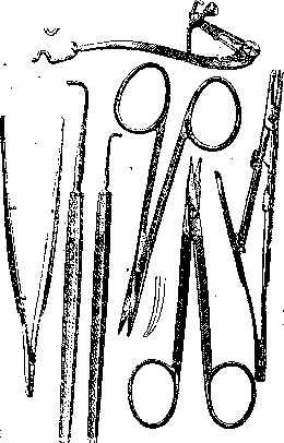
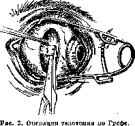
Fig. Instruments for tenotomy operation. which can cause painful sensations spreading deep into the orbit to the place of orbital attachment of the muscles. General anesthesia is used only in exceptional cases in small children. The operation requires very limited instrumentation: eyelid retractor, straight toothed forceps, two hooks for strabismus, small curved and straight blunt scissors, needle holder (Fig. 2) (see Ophthalmological instruments). If, for example, we take T. of the inner rectus muscle, the operation is performed as follows: after anesthesia and appropriate preparation of the operative field, the eyelids are spread with a retractor and the patient's eye is turned outward as much as possible. The operator grasps with forceps the horizontal fold of the conjunctiva near the edge of the cornea, at the level of the inner rectus muscle, and cuts it across with scissors at a distance of 3 mm from the limbus; an opening in the conjunctiva 4-5 mm is obtained, in the area of TEOBR031SH1 48 S of which the sclera is exposed from the underlying layers of connective tissue, the conjunctiva is slightly separated from the sclera upward, downward and along the muscle, up to the lacrimal caruncle, to avoid its subsequent retraction. Then a hook for strabismus is introduced into the wound, end upward, and is pushed upward 1-11/* cm tightly pressed against the sclera, and then sharply turned end down with the calculation that it will penetrate between the sclera and the muscle, catching the latter. The captured muscle is slightly pulled out of the wound and cut off at the very attachment of the tendon to the sclera (Fig. 3). The wound in the conjunctiva is sutured with 1-2 stitches, and a bandage is applied to the eye; the stitches are removed on the 4-5th day, after which the bandage is also removed. A small ecchymosis, which usually occurs at the site of the operation, disappears by the end of the second week.
After T., the eye, freed from the inner muscle, turns a certain number of degrees outward, and the severed muscle, somewhat contracting due to its elasticity, retracts backward, held only by its connections with Tenon's capsule, and grows to the sclera in a new place. As a result, the action of the muscle on the eye is correspondingly weakened. It is believed that cutting the inner rectus muscle corrects strabismus on average by 12-15°, but this is not true for all cases, as fluctuations are possible in both directions and in wide ranges. This depends on the different distance between the limbus and the place of physiological attachment of the muscle, on the strength of the muscle itself, on its connections with Tenon's capsule, etc. In addition, the immediate effect of the operation often differs significantly from later results. The degree of its insufficiency or excessiveness is controlled as follows: if when turning the operated eye toward the nose, it turns out that its adduction has retained its former strength, the result of the operation is considered insufficient. This may first of all depend on the fact that some fibers of the tendon remained uncut. Conversely, if when fixing a finger brought closer to the eyes, the operated eye loses the ability to converge already at a distance of more than 12 cm, this indicates that the effect of the operation is too great, i.e., the strength of the inner rectus muscle is weakened so much that over time one can fear the development of opposite or divergent strabismus. When it is necessary to enhance the effect of the operation, a so-called "auxiliary retraction suture" can be applied for 1-2 days, which on one side is passed through the conjunctiva and episclera, near the outer edge of the cornea, and on the other side is brought out-
to the skin through the outer canthus and tightened enough to put the eye in a state of slight divergence. For the same purpose, Panas proposed preliminary stretching of the muscle expecting its more energetic subsequent contraction, for which the eye is brought into a state of extreme abduction with the help of a hook introduced in the usual way under the tendon, so that the outer edge of the cornea reaches the outer canthus. According to observations by Blagoveshchensky, stretching the muscle can enhance the effect of the operation by another 42°. Slight strabismus remaining after T. can be left uncorrected, especially if there are prospects for the restoration of binocular vision. To reduce the operative effect, the application of a horizontal suture on the conjunctival wound or minimal disruption of the integrity of Tenon's capsule, the application of an "auxillary, adduction suture" in an appropriate manner, etc. is recommended. In addition, to prevent an unexpected excessive effect of the operation, it is advised to take the muscle on a ligature passed through the conjunctiva before the moment of tendon severing, and to tighten or release it as needed. Tenotomy of other rectus muscles is performed in the same way, taking into account their attachments to the sclera. It should be borne in mind that the effect of T. of the outer rectus muscle is generally much weaker than that of T. of the inner rectus. T. of the oblique muscles is a great rarity. As for the indications for T.-see Strabismus.
Related articles
Mentioned in
Cite this page
“Tenotomy.” Soviet Medical Encyclopedia. English translation of Bolshaya Meditsinskaya Entsiklopediya, 1st ed. (Moscow, 1928–1936), ed. N. A. Semashko. https://sovietmedicalencyclopedia.pages.dev/article/tenotomy/