Torticollis
Historical document, translated for reference. It reflects medical knowledge of the 1920s–30s and is not medical advice.
Summary
Torticollis is any displacement of the head in the frontal plane with tilting to the right or left. The clinical details of torticollis are as diverse as the causes that produce them, and they are classified as either congenital or acquired based on when they appear.
Encyclopedia article (1928–1936)
TORTICOLLIS (torticollis, caput obstipum), any displacement of the head in the frontal plane with tilting to the right or left. The clinical details of torticollis are as diverse as the causes that produce them; based on the time of appearance, torticollis is divided into two main groups: 1) Congenital torticollis and 2) Acquired torticollis. I. Congenital torticollis is detected from the moment of birth, and the main cause of its development may lie in changes or deviations from the norm either in the musculature of the neck or in the skeleton. Therefore, a primary muscular form of congenital torticollis (torticollis congenita muscularis) and a primary bony form of congenital torticollis (torticollis congenita ossalis) are distinguished.
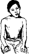
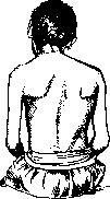
FIG. 1.
Figure 2. The clinical picture, subject to numerous variations, is observed in contracture of both halves of the sternocleidomastoid muscle. With isolated contracture of the anterior or superficial belly, a predominantly rotational turning of the head to the opposite side is observed with lowering of the occiput and raising of the chin. Traction along the course of the posterior or deep belly, however, causes predominantly tilting of the head toward the shoulder on the side of traction. The predominance of traction along the course of one belly or another explains why in some cases torsion of the head predominates, while in others there is tilting of the head to the affected side with weakly expressed torsion. Cases with predominance of traction in the direction of the posterior belly are accompanied by a mild form of total scoliosis of the spine in the opposite direction with small degrees of torsion of the latter. When traction along the course of the anterior belly predominates, a complex scoliosis develops with significant
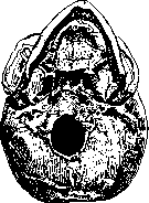
Figure 3
torticollis, as a result of which the cervical part of the spine together with the head is displaced in the frontal plane toward the healthy side (Fig. 2). In all cases of T., asymmetrical development of the skull (Fig. 3) and face is observed due to the deviation of the trajectory of the center of gravity of the head toward the affected side, which lags behind in growth compared to the healthy side. This explains the severe forms of facial asymmetry in adults who were not treated for T. in the early stage of skeletal growth. The etiology of congenital muscular T. cannot at present be considered finally established. However, it can now be said with certainty that the theory of birth trauma to the sternocleidomastoid muscle (Stro-meyer) by no means explains all cases of congenital T., the cause of which Stromeyer saw in the partial rupture of this muscle during extraction of the head (breech position) and during the application of forceps. Hematomas along the course of the m. sterno-cleido-mastoideus in newborns have been known since the time of Dieffenbach, and therefore the possibility of subsequent muscular contracture due to its scarred regeneration cannot be denied. The inflammatory theory of Volkmann, Mikulicz, Kader is also acceptable for explaining a number of cases of T., who saw the main cause of the development of muscular T. in hematogenous infection of the traumatized muscle with subsequent development of fibrous myositis. The possibility of the development of muscular T. on the basis of a similar process after injection of chemical irritants into the muscle has now been proven by the clinic of Girgolav through experiments on rabbits. However, bacteriological studies in children always gave negative results. Petersen considers the cause of the development of muscular T. to be the incorrect position of the head during intrauterine life with its inclination to the right or left, which in his opinion leads to passive contracture and fibrous degeneration of the muscle on the side where the head approaches the shoulder. Volcker adds to this dystrophic changes in the muscle due to compression of the vessels, and Sippel proved the possibility of such a position of the fetal head in the uterus by X-ray examination, after which a child was born indeed suffering from T. Therefore, Petersen and his followers explain cases of postpartum hematoma along the course of the muscle from the point of view of their theory: the contracture position of the head presents an obstacle to the passage of the head, as a result of which tears occur in the degenerated muscle. However, the theory proposed by Petersen does not in essence withstand criticism, since inclination of the head to the right or left during intrauterine life leads to scoliosis of the cervical part of the spine, and therefore the T. observed under such conditions should be classified as the primary bony form, not muscular, which does not exclude the correctness of all other reasoning of Petersen, since on the concave side of the neck the sternocleidomastoid muscle always appears shortened and therefore can easily be traumatized during the act of birth. - Contrary to all the theories presented about the origin of congenital muscular T. are the studies of Couvelaire, who performed histological examination of the sternocleidomastoid muscles in newborns born with signs of muscular T. and who died within the first 24 hours, obviously not having time to undergo any chronic inflammatory process during life. In these newborns, according to Couvelaire's studies, a clearly expressed congenital degenerative process of Zenker's type with abundant development of connective tissue was found in the sternocleidomastoid muscles. Couvelaire's research sheds light on the previously obscure etiology of congenital muscular T. and reconciles the conflicting interpretations of the pathogenesis of T. by the authors of the aforementioned theories. - The course and prognosis of muscular T. without timely treatment in childhood are very discouraging, since due to the violation of the normal conditions of statics of the head and spine, the normal conditions of growth and development of the latter are also violated. The deformation progresses steadily, accompanied by the development of scoliosis and a disfiguring asymmetry of the skull and face. Treatment. As with all deformities, as well as with T., the better the treatment results, the earlier it is started. And since T. with its facial asymmetry is primarily a disfiguring deformation of a person's appearance, early treatment here acquires special value. Treatment is recommended to begin from the first months after birth by means of manipulative correction to hypercorrection with fixation of the results achieved by a plaster bed, which must be repeatedly changed as the treatment progresses. Spitzy recommends in addition to treatment at this age with beds in cases of severe contracture to perform subcutaneous division of the lower ends of the sternocleidomastoid muscle without anesthesia. Spitzy decides on such an early operation due to the intensive growth of the infant in the first year of life with the aim of possibly early prevention of asymmetrical growth of the skull and face. In younger children with milder degrees of T., conservative treatment consisting also of manipulative correction to hypercorrection with fixation of the results achieved by a corset with a cervical and head retainer, which can be fixed at any inclination of the head to the right or left, is sometimes successfully carried out. Good results are obtained from wearing an oblique collar supporting the head from the affected side, as well as wearing a Gourdon cap with elastic traction on the healthy side. - The best results are obtained by surgical treatment in the form of division of the shortened muscle, which is performed either at its lower or upper end. Division of the lower end of the shortened muscle has been practiced since ancient times, and in the pre-antiseptic period of surgery, subcutaneous tenotomy according to Stromeyer was always performed. A curved tenotome was used, which was inserted shallowly near the medial edge of the strongly tensing lower ends of the muscle when the head was tilted to the healthy side, and these strands were divided from the inside outward. Such an operation cannot be recommended, as it cannot be considered either radical or safe. At present, only the method of open division of the lower end of the muscle is recognized. It lacks the negative aspects of subcutaneous tenotomy and is technically so simple that in adolescents and adults the operation is performed under local anesthesia, which greatly facilitates the subsequent correction and application of a plaster cast. With the patient lying on their back, a firm pillow is placed under the shoulders, over the edge of which the head turned to the healthy side hangs; this achieves maximum tension of the shortened muscle. A small skin incision is made parallel to the clavicle and 1 finger above it from the lateral edge of the clavicular portion to the medial edge of the sternal portion. The platysma and the anterior leaf of the muscle sheath are incised, the tendon of the sternal portion is exposed, and under it a wide Kocher probe is passed, on which it is divided layer by layer at the upper edge of the skin incision. Small muscular arterial branches encountered are grasped and ligated. The central end of the divided muscle belly is immediately pulled upward, and the peripheral end is grasped with a forceps and severed at its attachment to the sternum. Similarly, at the lateral angle of the incision, the clavicular portion is exposed and divided on a Kocher probe, which completes the operation on the contracted muscle, if the sclerotic process is limited in nature. However, if the lower ends of the muscle are found to be in scar adhesions with the posterior wall of the muscle sheath, which in turn is scarred to the anterior leaf of the vascular sheath, then by no means can one be limited to the above-mentioned operation. To prevent recurrence of T., it is necessary in such cases to carefully divide the underlying scar adhesions on a probe and excise them down to the exposure of the large vessels of the neck, as well as to widely resect the scar-degenerated ends of the muscle. -- Instead of dividing the lower end of the muscle, Lange recommends dividing its upper end under the mastoid process, as was first proposed by the French surgeon Tillaux. For this purpose, a small incision is made from the specified process downward along the anterior edge of the muscle, through which the upper end of the muscle is exposed in its entire width. A wide Kocher probe is passed under the muscle, on which the muscle is divided immediately below its attachment to the mastoid process. -- At the completion of the operation on the muscle and the surrounding scar tissue, the skin incision is sutured tightly, and it is necessary to proceed to the correction of more or less expressed scoliosis of the cervical spine, which in childhood in the primary muscular form does not appear very pronounced. This correction in the case of operation under general anesthesia should be performed with great caution.
It is much better to achieve the goal through staged corrections with the application of plaster casts, always striving for hypercorrection in the final result to prevent recurrence of T. After correction, which aims to eliminate not only the tilting of the head but also its torsion and displacement in the frontal plane, the achieved result is fixed with a plaster cast encompassing the head, chest, and the shoulder on the affected side. If hypercorrection is not achieved immediately, it is necessary to repeat the corrective manipulations after 10-14 days with the application of a new plaster cast. These manipulations often need to be repeated 2-3 times to achieve the necessary hypercorrection, after which the result must be fixed with a plaster cast for 4 weeks. If hypercorrection is achieved immediately with the head resting on the healthy shoulder, one plaster cast for four weeks is sufficient. After removal of the final plaster cast, it is necessary to begin subsequent treatment to consolidate the result achieved by the operation, which should continue for at least 4 weeks. This treatment consists of the following procedures: a) repeated (at least 3 times), daily, passive positioning of the head in the position of maximum hypercorrection; b) daily persistent exercise in the morning and evening to achieve the same result through active contraction of the stretched and weakened sternocleidomastoid muscle on the healthy side; c) massage and faradization of this muscle to restore its normal function; d) wearing a leather or celluloid collar during the day and night that maintains the hypercorrected position. With such systematic treatment in childhood, excellent results can be achieved, with the disappearance of compensatory counter-curvatures of the thoracic and lumbar sections of the spine and the restoration of symmetrical development of the skull and face. The later treatment is started, the less perfect the results. Fixed compensatory counter-curvatures with torsion can no longer be completely corrected; and asymmetry of the skull and face remains forever in adolescents and adults despite complete correction of the head position. 2. The primary bony form of congenital T. is based on the following causal factors: a) oblique position of the fetal head in the uterus due to pressure on one side of the head by the uterine musculature with a small amount of amniotic fluid, or due to a tumor growing either in the uterine wall or in the immediate vicinity of it; b) unilateral bony adhesions between two or more cervical vertebrae; c) a similar adhesion between the atlas and the occipital bone; d) underdevelopment of half of one of the cervical vertebrae; e) an excessive wedge-shaped hemivertebra inserted between two normal vertebrae; f) asymmetrically located cervical ribs. In this form of congenital T., both sternocleidomastoid muscles are normally developed, and one of them, namely on the side of the neck to which the head is tilted, appears only shortened, without any degenerative changes. Therefore, there is neither contractural fixation of the head nor its torsion here. The head rotates freely in both directions, unless there is an adhesion between the atlas and the second vertebra, and only the tilting of the head toward the convexity of the cervical curvature is limited to a greater or lesser degree. Nevertheless, even with this form of T., asymmetrical development of the skull and face is observed due to the deviation of the trajectory of the center of gravity of the head to the right or left of the midline. Therefore, even with the primary bony form of T., it is necessary to begin treatment as early as possible. Conservative treatment in infants should be conducted, as with the primary muscular form, through staged corrections and plaster beds, and incision of the shortened normal muscle is not required here due to its elasticity. In children who are already walking, quite satisfactory results can also be achieved with conservative treatment, staged corrections, and plaster casts, especially if the curvature of the cervical part is small, and incision of the upper or lower end of the shortened muscle can shorten the treatment period. Good results are also achieved by prolonged wearing of a Gurdon cap with elastic traction. In adolescents and adults, the normal method of treatment should be considered the incision of the upper or lower end of the shortened muscle followed by staged correction and plaster casts until hypercorrection is achieved. Subsequent treatment is carried out in the same way as with primary muscular torticollis. II. Acquired forms of torticollis are divided into the following clinical forms. 1. The muscular form is the result of myositis of the sternocleidomastoid muscle, does not have a persistent character, and is observed either in the form of the well-known suddenly appearing so-called rheumatic (more correctly, rheumatoid) T., or in the form of T. sometimes accompanying general infectious diseases (measles, scarlet fever, diphtheria, typhoid, etc.), or with local conditions, for example, with the direct spread of the inflammatory process of the mastoid process (infection in the middle ear). These cases must be distinguished from other cases of T. in ear diseases; these are cases caused by labyrinthine damage. The patient voluntarily holds the head in a position in which there is no feeling of dizziness. In the rheumatic process, the clinical picture of T. is less typical due to the usual simultaneous involvement of other neck muscles. Local heat treatments and treatment with salicylate preparations quickly lead to recovery here. Isolated infectious myositis of the sternocleidomastoid muscle gives a characteristic clinical picture of muscular torticollis. Local heat treatments are also indicated here; for internal treatment, large doses of urotropine are recommended for 5-7 days. In most cases, such infectious myositis ends favorably, and only in rare cases does the development of a muscle abscess occur. It is also necessary to mention here the gumma of the sternocleidomastoid muscle, which also sometimes gives symptoms of torticollis. Treatment is specific. 2. The bony form of acquired T. is observed as a consequence of various destructive processes affecting the joints on one side of the neck, either between the spine and the head or between individual cervical vertebrae. Such pathological processes are general infectious diseases, osteomyelitis and syphilis of the vertebral bodies, but most often tuberculosis (spondylitis cervicalis). The clinical picture of this form of T. is identical to the clinical picture of the congenital bony form of T., but with more significantly limited mobility of the cervical spine and head. The history and X-ray definitively establish the diagnosis. Treatment should always be carried out very cautiously to avoid exacerbation of the pathological process. In the tuberculous process, manual correction is absolutely contraindicated, and all treatment should be reduced to prolonged elastic traction (Gurdon cap) in childhood, and in adolescents and adults to the use of a corset from which a steel springy rod with a pellot pressing on the lateral surface of the lowered half of the head extends. 3. The neurogenic form of T. Very often T. is purely a nervous disease affecting neurotically or psychopathically predisposed people. In a large number of cases, this is one of the manifestations of the constitution of obsessive states, confirmed by the facts observed during the war in traumatic neurotics. Organic disease of the nervous system is also possible. Regarding this form of the disease, much is unclear in the literature, and many positions have not gone beyond the stage of development and discussion. In this section, there are many terms and an unclear classification, especially in the French, Italian, English, and German neurological and psychiatric literature. The most common terms (out of 40 known in the literature) are torticollis spastica and torticollis spasmodica, with the term torticollis spastica characterizing the form of tonic-type spasms, and torticollis spasmodica the form of clonic-type spasms. But some authors do not make a strict distinction here either (A. Bauer). Skipping similar classifications, two practically important forms should be noted: paralytic and spastic. The first form may arise as a result of flaccid paralysis of one of the sternocleidomastoid muscles and spastic contraction of the other. In flaccid central or peripheral paralysis of one muscle, which is generally very rare, a contractural condition of the other sometimes develops with the head positioned in the typical way for congenital muscular torticollis. In most cases, however, patients learn to replace the function of the paralyzed muscle by combined contraction of other muscles on the same side of the neck. Treatment should be directed in this direction with daily repeated gymnastic exercises and corrective manipulations aimed at stretching the healthy muscle that is in a state of contracture.
Spastic torticollis is observed either in the form of spastic contracture of one of the sternocleidomastoid muscles with the characteristic head position seen in muscular torticollis, or in the form of athetosis (spasmus mobilis) of one of the mentioned muscles, or in a mixed form, in which clonic contractions may give way to tonic tension of the muscles. The pathogenesis of these conditions is explained differently; some authors explain this state as irritation of the cortical rotational center of the head. Erb and Oppenheim explained it as irritation of the conductors in tumors and cysticerci of the brain, cerebellum, and parts of the spinal cord. Disease of the meninges was also indicated. Finally, some forms of torticollis are considered as a result of damage to the corpus striatum. Irritation of the peripheral nervous system is currently given importance by very few authors. The treatment plan for spastic torticollis is developed depending on the diagnosis of the underlying condition, starting with psychotherapy and physiatric procedures and medication treatment and ending with the proposal to remove the cortical rotational center. It should be said that no form of torticollis has occupied surgeons as much as this form. A whole series of operations on the nerves (section of the accessory nerve, posterior roots of the cervical nerves) and on the muscles (Kocher's myotomy, either alone or together with section of the accessory nerve, plastic operation connecting the sternocleidomastoid and trapezius muscles) have been proposed. In view of the modest results of surgical treatment at present, it is generally accepted as a rule to precede surgical intervention with conservative treatment. If it is unsuccessful, surgical intervention can be considered with a strict choice of method. In any case, one must abandon the idea of operating on the central nervous system and limit oneself to intervention on the periphery. 4. Scar torticollis is the result of scar fibrosis either of the muscle itself after a purulent process in it, or of the skin on the corresponding half of the neck after ulcerative processes, phlegmons, extensive lymphadenitis with fistulas, traumatic injuries, and burns. In case of scar fibrosis of the muscle itself, operative lengthening of the muscle according to the method just described is indicated in milder cases. In more severe cases, complete division of the lower end of the muscle is required. In case of scar fibrosis of the skin, excision of the entire affected area of skin and its replacement with a Filatov flap is indicated. 5. Habitual torticollis is caused by a more or less pronounced contracture of the muscles of the right or left half of the neck due to prolonged habitual tilting of the head to one side. Such an asymmetrical head position may depend on both developed professional work habits (carrying bulky loads always on one shoulder with a tilt of the head in the opposite direction) and the constant striving to restore binocular vision in astigmatism or partial clouding of the cornea in one eye, as well as in paresis or paralysis of the muscles of one eye; but it also occurs in labyrinth disease, as has already been indicated above. The success of treatment of these forms is closely related to the elimination of the main causes; but when a clearly expressed muscle contracture already exists, systematic active and passive corrective exercises are necessary to eliminate the muscle contracture.
Related articles
Mentioned in
Cite this page
“Torticollis.” Soviet Medical Encyclopedia. English translation of Bolshaya Meditsinskaya Entsiklopediya, 1st ed. (Moscow, 1928–1936), ed. N. A. Semashko. https://sovietmedicalencyclopedia.pages.dev/article/torticollis/