Cephalhematoma
Historical document, translated for reference. It reflects medical knowledge of the 1920s–30s and is not medical advice.
Summary
This article describes cephalhematoma as a blood tumor in newborns caused by hemorrhage between the skull bone and the periosteum, typically resulting from birth trauma. It details the clinical presentation, diagnosis, and prognosis, noting that while the condition is generally benign and requires no active treatment, it must be distinguished from caput succedaneum.
Encyclopedia article (1928–1936)
CEPHALHEMATOMA (from Greek kephale—head and haima—blood), or "blood tumor" of the head in newborns, is a tumor formed by hemorrhage between the outer surface of the flat bones of the skull and the periosteum covering them (see figure). Most often, cephalhematoma is located on the parietal
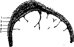
Cross-section of the skull of a frozen cadaver through the blood tumor: 1—periosteum; 2—galea aponeurotica; 3—skin; 4—bone; 5—dura mater.
bones, rarely on the occipital or frontal, and as a rare exception on the temporal and jaw bones. Hemorrhage usually occurs on the parietal bone that is facing forward, toward the symphysis, and therefore most often on the right parietal bone. Older authors confused cephalhematoma with caput succedaneum, and it was first accurately described by Gooch in 1791. The cause of cephalhematoma is birth trauma, but opinions on the mechanism of this factor's influence are conflicting: some believe that during the forward and backward movements that the head makes during contractions, there is first a shifting and detachment of not only the skin but also the periosteum. Numerous small vessels rupture, but while the head is compressed in the birth canal, they do not bleed; upon its exit, they begin to bleed, and the resulting pocket fills with blood. Others see the primary cause in the rupture of vessels resulting from trauma; hence the bleeding, and only then the detachment of the periosteum by blood and the blood tumor. A certain role in the formation of cephalhematoma may be played by the practice of forcibly holding back the head during its exit. In any case, the role of trauma is paramount. As a very rare exception, cephalhematoma is also encountered in older children (due to contusions), but in newborns, it is observed not so rarely—on average in 0.5–1% of all births. It forms more often in children of primiparas, especially older mothers, as well as during rapid births; with the application of forceps, bilateral cephalhematoma is often encountered; cases have also been described in breech presentation. When sawing through a skull with a cephalhematoma, one finds small hemorrhages under the skin, in the aponeurosis, and deeper—a detached periosteum, with an accumulation of blood beneath it, and finally, the exposed and often rough surface of the underlying bone; under the bone—an unchanged dura mater. Shortly after birth, a tumor appears on the child in the aforementioned places, which may gradually increase in size during the first days if the vessel continues to bleed. For the most part, the tumor is unilateral; but sometimes it is bilateral (on both parietal bones); very rarely it occurs in three places (on the parietal and occipital), and 4 cephalhematomas have even been described. At the full development of the tumor, its size ranges from the size of a walnut to a small apple, with a capacity of 5 to 200 g. The shape is usually somewhat elongated, the contours are well-defined, and they never cross the suture lines, because the periosteum is very firmly attached to the bone here. Fluctuation is quickly indicated; the tension of the cephalhematoma varies depending on the amount of blood extravasated. After a few days, a hard, ridge-like thickening appears along the periphery of the cephalhematoma, caused by the fact that the detached periosteum begins to deposit new bone elements. Sometimes the entire detached surface of the periosteum participates in this process, and after its disappearance, the tumor leaves a significant bony thickening that slowly smooths out. The skin over the cephalhematoma is unchanged, the swelling is not painful, and the child's general condition does not suffer. In large cephalhematomas, however, a certain degree of anemia may be observed, as well as pronounced jaundice (local hemolysis). Complications may include suppuration of the cephalhematoma, most often through skin abrasions or crusts on the head: the skin reddens, temperature rises, and the tumor becomes painful; however, sometimes all symptoms except pain are absent, and the question is resolved by a trial puncture. "Internal" cephalhematoma, where the tumor forms between the inner surface of the bone and the dura mater, is considered by some authors to be a kind of complication as well, resulting from a fracture or congenital defect in the bone through which blood seeps and accumulates under the dura mater. Diagnosis of cephalhematoma is not difficult: only in the first hours can cephalhematoma be confused with caput succedaneum, from which it differs by its sharp contours, fluctuation, and prolonged course, as well as by the fact that it does not cross the sutures. Cephaloceles have their own specific localization (fontanelles, bridge of the nose), can be reduced, and pulsate. The prognosis for cephalhematoma is usually favorable, although the course is prolonged, from 3 weeks to 3–6 months or longer. Prevention. One should be careful when applying forceps, gently support the perineum, and especially refrain from forcibly pushing the head back in order to slow its crowning. Cephalhematoma requires no treatment. In the first days—protect it from bumps, use an ice pack if the tumor is clearly growing; later—calm waiting while keeping the head clean. One should not perform punctures to aspirate blood in the first days, as reducing the pressure can renew the bleeding, and furthermore, it can infect the tumor and cause it to suppurate.
1. Chebotarevskaya.
Related articles
Cite this page
“Cephalhematoma.” Soviet Medical Encyclopedia. English translation of Bolshaya Meditsinskaya Entsiklopediya, 1st ed. (Moscow, 1928–1936), ed. N. A. Semashko. https://sovietmedicalencyclopedia.pages.dev/article/cephalhematoma/