Olfactory Nerve
Historical document, translated for reference. It reflects medical knowledge of the 1920s–30s and is not medical advice.
Summary
The olfactory nerve is the first cranial nerve responsible for conducting olfactory sensations. It originates in the nasal cavity and terminates in the olfactory bulb, with detailed anatomical structure and fiber organization described.
Encyclopedia article (1928–1936)
OLFACTORY NERVE, olfactory nerve, the first pair of cranial nerves, conducts olfactory sensations. It originates in the nasal cavity, in the so-called olfactory area, which is located on the inner and lateral surfaces of the nasal cavity; on the lateral surface, this area in the middle sections of the superior nasal concha occupies an area of very irregular shape measuring 1 cm2; on the inner surface (on the nasal septum) it does not extend beyond the lower border of the superior nasal concha. This area is characterized by the presence of special olfactory cells located among the epithelial cells of the mucous membrane; the peripheral processes of these cells are very short, directed toward the free surface of the mucous membrane, where they end in an expansion (olfactory vesicles of van der Stricht). The central processes of the cells go in the deep layers of the mucous membrane, directed upward. On both the lateral and inner surfaces, small branches anastomose with each other, forming true plexuses (Fig. 1), and gather into larger trunks numbering about 20 (fila olfactoria, s. nn. olfactorii mediales
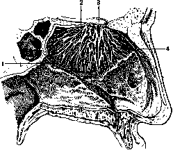
Fig. 1. Internal branches of the n. olfactorii: 1-n. naso-palatinus; 2-ramus internus n. olfactorii; Л-bulbus olfactorius; 4-n. ethmoidalis. et laterales), which penetrate through the openings of the lamina cribrosa into the cranial cavity and end in the olfactory bulb. All fibers of the olfactory nerve are non-myelinated (Rémak's), but differ from sympathetic ones
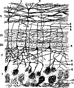
Fig. 2. Structure of the olfactory bulb: /-layer of glomeruli; Л-stratum plexiforme ex-ternum; ///-layer of mitral cells; IV- stratum plexiforme internum; V-layer of myelinated fibers; 1 and 2-granule cells; 3-recurrent collateral; 4-axon of mitral cell; 5-axon of small cell; 6-mitral cell; 7 and 8-small cells with tufts: 9-dendritic tuft of mitral cell; 10-glomerulus; 11-branches of peripheral olfactory fiber.
by their caliber; they are surrounded by an endothelial sheath. Pia mater accompanies the bundles of these fibers from the cranial cavity, forming their neurilemma. Dura mater lines the openings of the lamina cribrosa, and then divides into two layers: one forms the fibrous sheath around the fibers, the other continues into the periosteum of the lamina cribrosa. Arachnoidea does not extend onto the fibers. Bulbus olfactorius (olfactory bulb), where the fibers of the n. olfactorii end, is oval in shape, 8-10 mm in length, 3-4 mm in width, and 2-3 mm in thickness; it is gray in color, consists of cortex and white matter, in its center there is substantia gelatinosa, and in animals a canal lined with ependyma. From periphery to center, the following layers can be distinguished in it (Fig. 2): 1) peripheral layer of fibers of the n. olfactorii; 2) layer of olfactory glomeruli (glomeruli olfactorii), formed by the endings of the fibers of the n. olfactorii and the branches of the dendrites of the proper cells of the bulbus olfactorii; the transmission of olfactory sensations from the first neuron to the second occurs here; in addition, there are small cells with horizontal axons ending in neighboring glomeruli-association cells; 3) stratum moleculare, s. plexiforme externum, formed by the dendrites of mitral cells and special cells-cellules empanachees Cajal's, sending dendrites into the glomeruli, and an axon into the tractus olfactorius; 4) layer of mitral cells, whose dendrites branch into the glomeruli, and axons go into the tractus olfactorius, forming the second olfactory neuron; 5) stratum plexiforme internum or layer of collaterals belonging to cellules empanachees; 6) layer of myelinated fibers, formed by the axons of mitral cells and cellules empanachees, as well as centripetal fibers from the brain commissure, ending in the 4th and 5th layers. Between the fibers are granule cells with numerous dendrites; their axons end in the 3rd layer. Physiology and pathology-see Olfaction, by E. Kononova. OH, international unit of electrical resistance. O.- resistance of a column of mercury with a cross-section of 1 mm and a length of 106.3 cm at 0°. For large resistances, the derived unit megohm = 1,000,000 ohms is used. Abbreviated designation for O.-Ω.
Related articles
Mentioned in
Cite this page
“Olfactory Nerve.” Soviet Medical Encyclopedia. English translation of Bolshaya Meditsinskaya Entsiklopediya, 1st ed. (Moscow, 1928–1936), ed. N. A. Semashko. https://sovietmedicalencyclopedia.pages.dev/article/olfactory-nerve/