Paranasal Sinuses (air-containing sinuses, adjacent to the proper)
Historical document, translated for reference. It reflects medical knowledge of the 1920s–30s and is not medical advice.
Summary
The article describes the anatomy of the paranasal sinuses, including their location, structure, and relationship to surrounding tissues. It details the pathology of sinus diseases, their causes, symptoms, diagnosis methods, and complications.
Encyclopedia article (1928–1936)
Paranasal sinuses, air-containing sinuses, adjacent to the proper nasal cavity on each side: maxillary cavity (see), or upper jaw, frontal, anterior and posterior ethmoid cells, and sphenoid. The frontal sinus (sinus frontalis) is located in the thickness of the frontal bone (see). In 3-5% of cases, the frontal cavity is absent, more often on one side, rarely on both. In men, the sinuses are larger than in women. The frontal sinus is divided in the middle into two halves, often of unequal size. Sometimes additional septa are found. The anterior wall, corresponding to the supraorbital arch, is often very thick and strong. The posterior wall--the cerebral--is covered with elevations, the lower--orbital--is the thinnest of all. The frontal sinus is intimately connected by its drainage passage with the anterior cells of the ethmoid bone and ends in a funnel, which then passes into the drainage passage communicating with the nasal cavity (ductus naso-frontalis). It ends in the infundibulum or in front of it, which depends on the size of the bulla ethmoidalis. In the first case, probing is possible, in the second it is very difficult.-Ethmoid cells (cellulae ethmoidales) are located in the ethmoid labyrinth and throughout their course border the inner wall of the orbit, from which they are separated by a very thin plate (lamina papyracea). The number of cells ranges from 3 to 12. Their size varies. Their belonging to the anterior or posterior is determined by the exit openings: if the opening is located under the middle turbinate in the middle nasal passage, these are anterior cells; if in the posterior-superior space of the superior nasal passage, these are posterior cells.-The sphenoid, or sphenoidal sinus (sinus sphenoidalis) lies in the body of the sphenoid bone (see). In it, the upper, lateral, lower, posterior and anterior walls are distinguished. The upper is turned toward the brain, its frontal lobe with the olfactory bulb, chiasma opticorum and the brain's appendage with the sella turcica. This wall is very thin, and the optic nerves can enter the sinus. The internal carotid artery passes through the thickness of the lateral wall. The lower forms the roof of the nose and nasopharynx above the choanae. The posterior is slightly concave. The anterior wall is the thinnest, stands frontally. On it is located the exit opening (ostium sphenoidale) with a diameter of 1 to 4 mm. This opening opens into the recessus spheno-ethmoidalis and is located a few millimeters below the upper wall. In front of it, it is usually not visible, as the recessus spheno-ethmoidalis bends to the side and is covered by the posterior end of the middle turbinate. The opening is easier to see 1) if the middle turbinate is bent outward, 2) if the posterior end of the nasal septum is concave inward. When probing the main cavity, a probe slightly bent at the end is passed along the line connecting the spina nasalis inferior with the middle of the lower surface of the middle turbinate. The distance from sp. nasalis infer. to the sinus ranges from 6-7 to 9-9.5 cm. The structure of the mucous membrane and physiological significance are similar to the maxillary cavity (see). Pathology. Diseases of the paranasal sinuses consist of inflammatory processes of the mucous membrane in acute or chronic form, bone lesions and tumors. Etiology. Most often, the cause of acute inflammations of the paranasal sinuses is influenza. Bruck found in influenza 70% involvement of the maxillary, 64% of the sphenoid, 50% of the ethmoid and 25% of the frontal sinus. Less frequently, the cause of sinusitis is the spread of inflammation from adjacent bone walls to the mucous membrane in injuries (especially gunshot wounds), in syphilis and tuberculosis and tumors of both the paranasal sinuses and the nasal cavity itself. Numerous recent studies, especially by Russian authors, have shown that all kinds of harmful professional influences (dust, smoke, gases, vapors, etc.), causing processes in the nose, contribute to an increase in the number of diseases of the paranasal sinuses in persons exposed to these professional hazards. Also unfavorable in terms of the occurrence of sinusitis are general diseases such as tuberculosis, syphilis and others. Sinusitis mainly affects adults. In children, sinusitis is rare (usually after infectious diseases, e.g., scarlet fever). With repeated infections, sinusitis tends to recur. This especially concerns the frontal sinus.-Pathological anatomy. Acute and chronic, catarrhal, purulent and atrophic forms of inflammation of the sinus mucosa are distinguished (see Maxillary sinusitis). Inflammatory changes may not be limited only to the mucous membrane itself but may extend to the bone, causing inflammatory and hyperplastic changes in it (osteomyelitis). Inflammatory processes can also spread to neighboring organs (brain, orbit) or be complicated by septic disease of the entire body. The spread of the inflammatory process occurs through the veins or by the spread of the process along the veins or by thrombosis of the veins and subsequent breakdown of the infected thrombus. In rare cases, the cause of the spread of the process is pre-existing abnormal openings (dehiscences). The presence of pus in the cavity is not always proof that it formed there itself: it can flow into the cavity from some other cavity, forming the so-called pyo-sinus. This is most often observed in the maxillary cavity when pus flows into it from the overlying frontal and ethmoid cavities. Symptoms. Patients' complaints usually consist of unilateral runny nose with abundant liquid, bloody or purulent discharge. Sometimes the discharge is insignificant or may be completely absent. Complaints of headaches without definite localization depending on the affected cavity are common. These pains can have several causes. In acute sinusitis, pains are a) of a neuralgic nature. Attacks usually begin in the morning from 9-10 o'clock and continue until 3-4 o'clock in the afternoon. Such pains are especially often observed in inflammation of the frontal sinus, b) Pains of the sinus walls of a constant nature, but not very strong. When pressure is applied to the sinus walls, they intensify. In chronic sinusitis, pains are insignificant, dull or completely absent. This absence of pain is more often observed in diseases of the maxillary and ethmoid sinuses. On the contrary, pains are almost always present in inflammations of the frontal and sphenoid sinuses. Complaints of loss of smell due to closure of the olfactory fissure and sometimes of the sensation of an unpleasant odor are common. As secondary signs, complaints of nasal obstruction, subsequent catarrhal diseases of the nasopharynx, pharynx, larynx, bronchi, etc. are observed. Elevated temperature usually depends on the underlying infectious disease (influenza). General excitement, redness of the face, irritability, increased pulse can also sometimes be caused by sinusitis. Diagnosis is based on a careful analysis of anamnestic data and examination of the nose. Unilateral runny nose, especially purulent, is always highly suspicious of disease of the paranasal sinuses. The main task is to determine from which cavity the pus is being discharged. After thoroughly coating the middle nasal passage with cocaine-adrenaline, an attempt is made to notice at what position of the head pus reappears: the appearance of pus in the horizontal position is characteristic of the frontal sinus, when tilting the head in the opposite direction and downward--for the maxillary cavity. Subsequently, an attempt is made to probe the frontal sinus through the natural opening with a probe bent at a 90° angle, and if this is successful, a thin tube is inserted and the frontal sinus is irrigated. The presence of pus in the irrigation fluid serves as clear evidence of involvement of the frontal sinus. Unfortunately, anatomical conditions allow such irrigation only in 50% of cases. The presence of pus in the superior nasal passage (above the middle turbinate) is characteristic of disease of the posterior ethmoid cells and the sphenoid sinus, which usually occur together. Probing of the sphenoid sinus (see above) confirms the assumption.-Additional research methods include diaphanoscopy (transillumination), used in diseases of the frontal and maxillary sinuses, and X-ray photographs. The value of additional methods is relative, and their data can have significance only in the presence of other symptoms of the disease. (Special research methods for the maxillary cavity--see Maxillary sinusitis.) Inflammation of the frontal sinus (frontitis). The etiology is the same as for maxillary sinusitis (see). Path-anatomically, the mucous membrane is not changed throughout, but in places. The fronto-nasal passage is often temporarily impassable, which depends on the swelling of the mucous membrane in the middle nasal passage. The formation of cysts and polyps is rare. More often than in diseases of other cavities, bone changes are observed, both of a primary nature, e.g., as a result of trauma, due to the protruding position of the frontal sinus, and of a secondary-inflammatory origin. The latter depend on the pressure of pus on the mucous membrane and the spread of the inflammatory process from it to the bone. Diseases of the bone may consist of periostitis, osteitis, osteomyelitis with subsequent necrosis and formation of fistulas. The latter more often originate from the lower wall of the frontal sinus, located on the skin at the inner angle of the eye or on the upper eyelid. More rarely, perforation occurs on the posterior wall with the process spreading to the meninges and brain. Due to the closure of the fronto-nasal canal, a collection of discharge in the frontal sinus can occur, with subsequent expansion of its walls.
(The accumulation of pus is called empyema of the frontal sinus, accumulation of mucus is called mucocele; the presence of air or its accumulation in the frontal sinus is called pneumosinus or pneumatocele of the frontal sinus.) The transition of the inflammatory process to the frontal bone and other flat bones of the skull gives a severe picture of osteomyelitis of the flat bones of the skull, fortunately observed very rarely. Subjectively, in acute cases, headaches are always observed, often very severe. When pressing in the upper-inner corner of the eye, the pain intensifies. There is also noted a slight discharge of pus from the nose, nasal obstruction, anosmia, less often eczema of the nasal entrance and nasal bleeding. Among the objective signs is the presence of pus at the anterior end of the middle nasal turbinate, flowing out when the head is in a straight position. Most often, purulent discharge can be observed in the morning. Also noted is atypical hypertrophy of the anterior end of the middle turbinate and swelling of the mucous membrane on the lateral wall of the middle turbinate. Often noted is swelling of the anterior wall of the frontal sinus and the upper eyelid, less often deviation of the eyeball downward and outward. Diagnosis in clear cases is not difficult. Most often it is necessary to differentiate from neuralgia of the second branch of the trigeminal nerve. In neuralgia, the ramus supraorbitalis, passing at the border of the middle and inner thirds of the upper wall of the orbit, is painful on pressure, while the bone is not painful; in inflammatory processes, the opposite is true. The presence of eyelid swelling also speaks for the inflammatory nature of the disease. Trial probing and washing of the frontal sinus in the presence of pus confirm the diagnosis. If probing is not possible, one can try to push aside the middle turbinate with a Killian mirror (nasoscopy) or remove the anterior end of the middle turbinate, which facilitates access to the naso-frontal duct. The latter two procedures are at the same time therapeutic measures, as they facilitate the outflow of pus from the frontal sinus. Transillumination and X-ray films in the anteroposterior direction can help in making the diagnosis. The latter presents particular difficulties in closed empyemas. Treatment in acute forms without sharply expressed general and cerebral phenomena is conservative. It consists in smearing the middle nasal passage and the naso-frontal duct with cocaine-adrenaline to expand it and facilitate the outflow of secretions. For the same purpose, instillation of drops of the same composition is prescribed. From the outside, dry heat is applied to the forehead, blue light, and internally aspirin, phenacetin, antipyrin, etc. In prolonged acute cases and in chronic cases, intranasal surgery is recommended
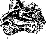
Fig
Formation of a mucoperiosteal flap according to Halle.
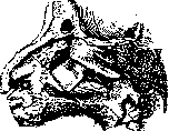
Figure 2. Displacement of the mucoperiosteal flap downward and backward according to Halle.
Opening of the frontal sinus according to Halle. The operation consists in forming a flap of mucous membrane from the area in front of the middle turbinate and removing bone in the anterior part of the nose (agger nasi) with special burrs and chisels, followed by expansion of the passage with a sharp curette (fig. 1-7). The operation is technically very difficult and requires thorough knowledge of anatomotopographic relationships. With the slightest error, it is easy to reach the lamina cribrosa and the cranial cavity. Trial opening of the frontal sinus is performed from the outside. If pathological changes are found in it, one can immediately proceed to the radical operation. The skin incision is made along the inner part of the eyebrow with a bend onto the root of the nose. After the periosteum is separated, an opening is made in the anterior wall (Hajek) or in the inferior wall (Boenninghaus). Indications for radical operation are cases not amenable to intranasal treatment, as well as cases with existing external fistulas or bone changes. Inflammations of the frontal sinus of traumatic origin (gunshot wounds) or complicated by diseases of the meninges, orbit, or in sepsis also require external operations. Up to 60 methods of external opening of the frontal sinus have been proposed. Practically, they can be divided into methods with opening through the anterior wall, inferior wall, through both, methods with a bridge and osteoplastic methods. Of all these methods, the most commonly used for small and medium cavities is opening through the inferior wall with mandatory removal of the anterior ethmoid cells (fronto-ethmoidal trepanation by A. F. Ivanov). For very large cavities, the method of Killian is used. The operation is performed under local anesthesia or general anesthesia. The incision, starting from the middle of the eyebrow inward, curves around the orbit and ends on the frontal process of the maxilla. After the periosteum is separated from the anterior and inferior (orbital) surface of the frontal sinus, an opening is drilled in the upper-inner corner of the orbit, where the frontal sinus can almost certainly be found.
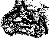
Fig. 3. Introduction of a Ritter's bougie.
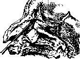
Figure 4. Introduction of a burr.
Figure 5. The inferior wall of the frontal sinus has been removed.
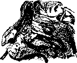
Figure 6.
Introduction of a curved curette. Upon entering the sinus, the opening is expanded by removal of the inferior (orbital) wall. The pathologically altered mucous membrane is examined and removed. The entire mucous membrane should not be removed. Then the bone of the nasal process of the maxilla, the anterior ethmoid cells are removed, and wide communication with the nose is created, into which a drain is inserted, which is removed after 5-6 days. The wound on the forehead is sutured tightly. This method gives a good cosmetic and therapeutic effect. The operation according to Killian. The skin incision is as described above. On the periosteum in the area of the eyebrow arch - two parallel incisions outlining the future bridge supporting the skin of the forehead from collapse (fig. 8 and 9). Subsequently, the bone of the anterior and inferior wall of the frontal sinus, the nasal process of the maxilla, the anterior ethmoid cells, and all the diseased mucous membrane are removed subperiosteally. Unfortunately, it is not always possible to preserve the formed bridge well: it necrotizes, sequestra form, fistulas appear, and healing is delayed. The goal of preventing the anterior wall from collapsing is also not achieved. Therefore, in cases of recurrence, and especially in cases complicated by diseases of the orbit or brain, one has to, disregarding cosmetics, remove the anterior and inferior walls of the frontal sinus (Riedel's method). Further treatment in all methods - washing the frontal sinus and smearing the formed passage with solutions of argyrol (2-5%). As a preliminary stage of all operations, resection of the anterior end of the middle turbinate is recommended. As a complication after radical operation, and sometimes independently, osteomyelitis of the frontal bone develops, accompanied by high temperature, severe general condition, diffuse swelling of the entire forehead with the formation of subsequent abscesses. Fortunately, this severe complication is rare. Treatment consists in as early as possible radical intervention according to the general principles of surgery. If the patient survives, the operation has to be repeated due to the formation of new foci. Mucocele of the frontal sinus - obstruction of the outlet opening and accumulation of mucous content in the frontal sinus, stretching the walls of the sinus and thinning them.
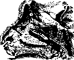
Fig 7. The mucoperiosteal flap is in place.
PARANASAL
sinuses (pneumosinus) - accumulation of air in the frontal sinus with obstruction of the opening and stretching of its walls. The causes are unknown. It is assumed that the basis is ordinary catarrhal phenomena with obstruction of the outlet opening (Brieger). Inflammation of the ethmoid cells (ethmoiditis) is usually combined with inflammation of other paranasal sinuses: the anterior ethmoid cells with the frontal sinus, the posterior ones with the main sinus. In etiology, often influenza, in children - scarlet fever. Pathological changes in acute inflammation of the ethmoid cells are expressed by hyperemia, thickening of the mucous membrane, and seropurulent exudate. Since the mucous membrane of the ethmoid cells, except for the convex surface and the free edge of the middle turbinate, is thinner and looser than the mucous membrane of other sinuses, inflammatory changes quickly pass to the deep layers of the mucous membrane, and edemas easily occur. In acute inflammation, there is a gelatinous swelling of the mucous membrane, resembling polyps, but softer and more diffuse than them and easily passing. Often the inflammation can spread to the bone, caus
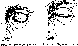
according to Killian.
incisions.
its necrosis with the formation of external fistulas at the inner corner of the eye or causing inflammation of the orbital contents. - Chronic inflammations of the ethmoid cells are catarrhal and purulent. The product of such inflammation is usually nasal polyps - thickened mucosal elevations on a broad base or on a stalk. Thickening of the ethmoid bones is also observed if the inflammation spreads to the bone. In case of disease of the posterior ethmoid cells, a serious complication may arise from the nearby optic nerve (neuritis retrobulbaris). Progressive blindness can also develop in cases of concealed inflammation of the posterior ethmoid cells (Wolfovich). Atrophic forms with the formation of crusts on the mucosa, depending on the resorption of bone on the basis of ostitis rarefaciens, are less common. - Symptoms of acute inflammation consist in tenderness on pressure over the root of the nose, a dull sensation in the same place, and especially increased lacrimation. In chronic forms, there is often pus in the middle nasal passage, complaints of nasal obstruction, polyps, and secondary lesions of the pharynx and larynx. Disturbances of smell are common in diseases of the posterior cells. The diagnosis is made on the basis of examination of the nose. The finding of pus and polyps in the middle nasal passage, excluding disease of the neighboring sinuses (maxillary and frontal), indicates
Related articles
Cite this page
“Paranasal Sinuses (air-containing sinuses, adjacent to the proper).” Soviet Medical Encyclopedia. English translation of Bolshaya Meditsinskaya Entsiklopediya, 1st ed. (Moscow, 1928–1936), ed. N. A. Semashko. https://sovietmedicalencyclopedia.pages.dev/article/paranasal-sinuses-2/