Ethmoid Bone
Historical document, translated for reference. It reflects medical knowledge of the 1920s–30s and is not medical advice.
Summary
This historical article from the 1928-1936 Great Medical Encyclopedia describes the anatomy, development, and structure of the ethmoid bone (os ethmoidale), detailing its vertical, horizontal, and labyrinthine parts.
Encyclopedia article (1928–1936)
ETHMOID BONE (os ethmoidale), an unpaired, median, symmetrical bone located anteriorly to the sphenoid bone in the corresponding notch of the frontal bone. It thus takes part with only a small portion of itself in the formation of the base of the cerebral cranium, and with its main mass in the formation of the facial cranium—specifically the nasal cavity and partly the orbit. Its shape is very complex. Three main parts are distinguished in it: 1) the vertical plate (median), 2) the horizontal plate, which intersects the former almost at its upper edge, and 3) two roughly four-sided prisms fused with the lower surface of the horizontal ethmoid plate and its lateral edges, elongated in the anteroposterior direction and suspended in the nasal cavity (labyrinthi ethmoidales). The ethmoid bone develops through the ossification of the posterior parts of the nasal capsules, the anterior part of which remains cartilaginous throughout life in the form of the nasal cartilages. The cartilaginous primordium of the ethmoid bone has the same shape as the adult bone. The process of ossification of the cartilaginous primordium begins in the fifth month of embryonic life. It proceeds from four centers: two for the lateral parts of the bone and two for the rest. First, lateral ossification points appear in the lateral parts of the labyrinths in the form of vertical or oblique trabeculae, which subsequently, by connecting, form the cellulae ethmoidales (ethmoidal cells) and their conchae (Fig. 1). Both labyrinths completely ossify by the time of birth; by this time they are connected to each other by a fibrous plate (Fig. 2); the remaining ossification centers appear only by the end of the first year after birth. During this period, 5 point-like islets can be observed on each side, about 1/4 mm in diameter each. From them, the crista galli, horizontal, and perpendicular plates subsequently develop. In the perpendicular plate, the process of ossification thus proceeds from top to bottom. There are often two additional
Figure 1. Frontal section through the cartilage capsule: 1-lamina perpendicularis; 2-ossification of the labyrinth; 3-crista galli; 4-cartilaginous framework. (After Vorobyov.)
ossification points: one for the apex of the crest, the other at the edge of the ethmoidal cleft. Ossification of the perpendicular plate ends in the fifth year of life. In the sixth year, the parts of the bone ossifying from separate centers fuse together. By the age of 25, the perpendicular plate is still not fused with the corresponding _________________ cran- T, p _ ru m cni
constantly in old age.—In the majority of carnivorous mammals and some primates, the ethmoid bone does not take part in the formation of the orbit. It is located entirely between the frontal and palatine bones, which are joined into a single whole, and thus they lack the lamina papyracea. In its structure, the ethmoid bone differs from other bones in that it consists exclusively of thin plates of compact substance, between which small air cavities are located. Therefore, the ethmoid bone is extremely light. Traces of spongy tissue are found with difficulty in the crista galli and in the upper and lower parts of the perpendicular plate. The bone tissue of the ethmoid bone, especially in its lateral masses, forms plates and scales that are very thin, extremely fragile and brittle, connecting with each other in the most bizarre manner and bounding a system of more or less tortuous cavities known as the ethmoid cells (cellulae ethmoidales), which form the labyrinth of the ethmoid bone.—The vertical plate (lamina perpendicularis) represents in shape an irregular pentagon divided by the horizontal plate into two parts, one of which lies above the horizontal plate and the other below it. The upper part of the vertical plate has a triangular shape with rounded edges and somewhat resembles a rooster's comb (without notches) (crista galli) (Figs. 3 and 4). The base of the crista galli merges with the horizontal plate of the ethmoid bone; a sickle-shaped process of the dura mater is attached to its upper edge. The anterior edge, thick and almost vertical, ends in 2 processes (processus alares); it forms the foramen caecum with the frontal bone (Fig. 3), if the latter is not entirely formed by the frontal bone. The crest as a whole forms a solid support for the posterior wall of the frontal sinuses. The lower part of the perpendicular plate connects below with the vomer, posteriorly
Figure 3. Lamina cribrosa from above: 1-foramen caecum; 2-ethmoidal cleft; 3-frontal bone (orbital surface); 4-sphenoid bone; 5-lamina cribrosa; 6-projection of canalis ethmoidalis ant.; 7-crista galli; 8-foramen ethmoidale ant.; 9-impression of a. meningeae ant. (After Testut.)
with the vertical crest of the sphenoid bone, and anteriorly with the nasal process of the frontal bone, the nasal bones, and the cartilages of the nasal septum. It divides the nasal cavity into two parts and contains on both of its surfaces depressions, often barely noticeable, for the vessels and nerves of the mucous membrane of the olfactory part of the nose. The horizontal plate is quadrangular in shape, more elongated from front to back than in the transverse direction, and connects the two labyrinths of the ethmoid bone. It has numerous openings through which branches of the olfactory nerve pass from the cranial cavity. It is called the cribriform plate (lamina cribrosa) (hence the name of the ethmoid bone). There are 25-30 openings in it on each _________________ side. Some of these left view: 1-anterior surface of massae latera- T TJ „ T les; 2-processus uncina- i 1 . . „ r „ 0 tus; 3-lamina papyracea; openings lead only 4-cellulae ethmoidales nn„ r„„ post.; 5-sulcus ethmoida- to depressions, NOT hs PsL' 6~crista galli into through-canals (Fig. 4 - 5). The lateral parts suspended from the outer parts of the cribriform plate are located outside the midline between the nasal cavity and the orbit. The labyrinth of the ethmoid bone has 6 surfaces: outer, inner, upper, lower, anterior, and posterior. The outer surface is flat, smooth, and forms part of the medial wall of the
Figure 4. Ethmoid bone,
The ethmoid bone.
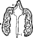
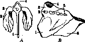
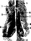
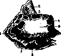
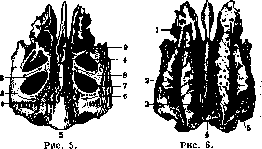
Figure 5. Ethmoid bone from above: 1 - lamina perpendicularis; 2 - crista galli; 3 - lamina papyracea; 4 - lamina cribrosa; 5 - posterior margin of the perpendicular plate; 6 - sulcus ethmoidalis post.; 7 - ethmoidal cell; 8 - sulcus ethmoidalis ant.; 9 - infundibulum. Figure 6. Ethmoid bone, view from below: 1 - middle concha; 2 - processus uncinatus; 3 - lamina cribrosa; 4 - lamina perpendicularis; 5 - middle nasal meatus. orbit and is called the paper plate of the ethmoid bone (lamina papyracea or os planum). It connects above with the frontal bone, below with the maxilla, anteriorly with the lacrimal bone, and posteriorly with the sphenoid and palatine bones (Fig. 4). Its internal surface forms the greater part of the outer wall of the nasal cavity. Two thin plates extend downwards and inwards from this surface: the superior and middle conchae of the ethmoid bone—concha superior, s. Morgagni, et concha media (concha suprema is underdeveloped and usually absent). The superior concha is small and occupies only the posterior part of the internal surface. The space between the superior concha and the internal surface of the ethmoid bone forms the superior nasal meatus (meatus nasi superior), and between the middle concha and the ethmoid bone lies the middle nasal meatus (meatus nasi medius) (Fig. 6). The upper surface of the ethmoid bone presents on the intact skull semi-cells of very irregular shape, which articulate with the semi-cells of the frontal bone. Here there are two transversely arranged cavities that connect with the notch of the frontal bone and form the anterior and 24 £2 AND Figure 7. Ethmoid, frontal, and sphenoid bones from below: 1 - facies orbitalis; 2 - concha nasalis media; 3 - ala parva ossis sphenoidalis; 4 - fissura orbitalis sup.; 5 - foramen rotundum; 6 - facies spheno-maxillaris; 7 - canalis pterygo-palatinus; 8 - foramen ovale; 9 - foramen spinosum; 10 and 11 - laminae lateralis et medialis processus pterygoidei; 12 - corpus ossis sphenoidalis; 13 - concha sphenoidalis; 14 - processus vaginalis; 15 - hamulus pterygoideus; 16 - spina angularis ossis sphenoidalis; 17 - facies infratemporalis ossis sphenoidalis; 18 - concha nasalis sup.; 19 - facies temporalis alae magnae; 20 - facies orbitalis; 21 - processus zygomaticus ossis frontalis; 22 - lamina perpendicularis ossis ethmoidalis; 23 - glabella; 24 - processus alaris ossis ethmoidalis. (According to Vorobyov.) posterior foramina of the ethmoid bone (foramina ethmoidalia anterius et posterius). The very anterior cell, constant in its shape and position near the crista galli, from which it is nevertheless separated by a part of the perpendicular plate, leads by its wide, gradually tapering origin (infundibulum) into the frontal sinus (Fig. 5). On the inferior surface of the ethmoid bone, the lower margin of the middle concha, the middle nasal meatus, and the rough surfaces by which it connects with the maxilla are visible, as well as a very thin plate attached to the anterior surface of the middle nasal concha, extending from here from front to back and as if lengthening the concha—this is the uncinate process (processus uncinatus). Its lower margin reaches almost to the inferior nasal concha. The uncinate process diagonally crosses the entrance to the maxillary sinus (sinus maxillaris) and strongly narrows it (Figs. 6 and 7). The anterior surface represents depressions or semi-cells connecting anteriorly with the lacrimal bone. Here, at the point of intersection of this surface with the lower one, the uncinate process begins (Fig. 8). The posterior surface is quadrilateral, rough at the site of connection with the body of the sphenoid
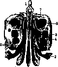
Figure 8. Ethmoid bone, anterior view: 1 - crista galli; 2 - ethmoidal labyrinth; 3 - processus uncinatus; 4, 5, and 6 - ethmoidal cells; 7 - upper surface of the ethmoid labyrinth.
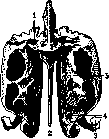
Figure 9. Ethmoid bone, posterior view: 1 - articular surface for articulation with the sphenoid bone; 2 - lamina perpendicularis; 3 - processus uncinatus; 4 - middle concha; 5 - superior concha.
bone and the orbital process of the palatine bone. Here one or two more semi-cells are encountered, which on the intact skull form the corresponding cells of the sphenoid bone (Fig. 9). The number of cells in the ethmoid bone is variable: on average from 7 to 9; a decrease in their number is associated with an increase in their volume. According to their position, they are divided into anterior, middle, and posterior. The largest middle cell, adjacent to the uncinate process, is called the bulla ethmoidalis. A cleft is formed between it and the uncinate process. The narrow, lower, more horizontal part of the cleft is called the semilunar hiatus (hiatus semilunaris). It connects the cavity of the middle nasal meatus with the maxillary sinus. The wider anterosuperior part of the cleft, called the infundibulum, communicates with the frontal sinus and the anterior ethmoidal cells. The posterior and middle cells open into the superior nasal meatus, the anterior ones into the middle meatus. The posterior ones communicate with the sphenoidal sinus, the anterior ones with the frontal sinus. Some anatomists distinguish cellulae frontales, lacrimales, sphenoidales, palatinae et maxillares. The ethmoid bone receives its blood supply through the anterior and posterior ethmoidal arteries, two on each side, sometimes from the supraorbital artery. The anterior, more powerful one supplies the anterior and middle cells. Venous outflow proceeds via veins of the same name emptying into the ophthalmic vein. The lymphatic vessels of this region have two pathways of outflow: upper and lower. The upper goes together with the lymphatic vessels of the eyelids to the lymph glands of the parotid region. The lower goes partly forward with the lymph flows of the external parts of the nose into the buccal, submandibular, and parotid lymph nodes, and chiefly backward into the deep cervical lymph nodes (angular lymph node) and partly into the lateral retropharyngeal lymph nodes. It is innervated by the posterior ethmoidal nerve (from the nasociliary nerve) and orbital branches (sphenopalatine ganglion). The syntopy of the ethmoid bone is clear from the above: the ethmoid bone borders anteriorly with the frontal bone, lacrimal, nasal, and maxilla, posteriorly with the sphenoid bone, below with the vomer and palatine bone; special attention should be paid to the fact that the ethmoid bone is closely connected with the orbital cavity and the anterior cranial fossa—its upper wall is at the same time the cranial wall. Its lateral wall is the wall of the orbit. This proximity determines the nature of complications in diseases of the ethmoid bone. The pathology of the ethmoid bone boils down most often to diseases of the mucous membrane lining all its cells, from which the process also passes to the bone walls (acute or chronic ostitis). The ethmoid labyrinth is of enormous importance for the pathology of the nasal cavity, as it represents a bizarre combination of cells separated one from the other by a paper-thin plate, sometimes communicating with each other and with the nasal cavity, and sometimes completely isolated. Being caught by some acute process, the ethmoid cells rid themselves of it with difficulty, which in turn often serves as the cause of a wide variety of diseases of the nasal cavity, neighboring accessory sinuses, nasopharynx, larynx, bronchi, lungs, and even the heart. In diseases of the ethmoid bone, its cells are frequently destroyed. As experience shows, this is not associated with danger to the patient, with the exception of those extremely rare cases when a collapsed labyrinth or a part of it falls into the trachea (Malyutin). In ethmoiditis (see Accessory nasal sinuses) (acute, chronic, catarrhal, and purulent), a strong enlargement and protrusion of the ethmoid bulla can occur due to the accumulation of mucus and blockage of the exit from the cell. Bone cavities of the same nature, filled with liquid contents, can also form in other cells. They are known as bone cysts—rhinocele. The pathological process in the ethmoid bone leads to two opposite states of the ethmoids: firstly, the bone can become excessively softened; secondly, sometimes, on the contrary, it becomes sclerosed. In both cases, operations on the ethmoid bone can lead to complications; thus, during intranasal interventions, the lamina papyracea may be damaged, which can break even without direct action on it, when biting off the transverse cell walls attached to it with forceps. Even during injections into the cavities of the ethmoid bone, if they are performed forcibly, a similar complication may occur. Damage to the optic nerve is possible in cases where its canal is in direct relation to the posterior ethmoidal cells, which occurs in the majority of cases. In rare cases, the canal can pass entirely through the posterior cells. Among other diseases of the ethmoid bone, the frequent formation of mucous and fibrous polyps in ethmoiditis should be noted. Luetic periostitis and tuberculous ulcers are also encountered (secondary; primarily they usually appear on the nasal septum). Among neoplasms, osteomas of the ethmoid bone are found at a young age. Cancer and sarcoma of the ethmoid bone are also frequent diseases. Isolated fractures of the ethmoid bone are extremely rare. Usually, fractures of the ethmoid bone accompany so-called oblique fractures of the base of the skull. Fractures of the vertical plate are frequently encountered with bruises of the nose and especially with fractures of the nasal septum. Operations on the ethmoid bone: 1) destruction of bone septa and cell walls for a wider outflow of pus and 2) removal of the conchae. Access to them: 1) intranasal (endonasal) or 2) extra-nasal—through the medial wall of the orbit (extranasal).
Related articles
Mentioned in
Cite this page
“Ethmoid Bone.” Soviet Medical Encyclopedia. English translation of Bolshaya Meditsinskaya Entsiklopediya, 1st ed. (Moscow, 1928–1936), ed. N. A. Semashko. https://sovietmedicalencyclopedia.pages.dev/article/ethmoid-bone/