Palatine Bone
Historical document, translated for reference. It reflects medical knowledge of the 1920s–30s and is not medical advice.
Summary
The palatine bone is composed of horizontal and perpendicular plates forming part of the hard palate and lateral nasal wall. It develops from connective tissue and participates in various pathological processes including congenital clefts and tertiary syphilis.
Encyclopedia article (1928–1936)
PALATINE BONE, os palatinum, consists of two plate-like parts: horizontal (pars horizontalis) and perpendicular (pars perpendicularis), connected to each other at a right angle. Schematically, the P. b. is depicted in the form of the letter L, where the horizontal line corresponds to the horizontal part of the bone, and the vertical line to its perpendicular part. Two such letters, facing each other with their horizontal plates, give an idea of the relationship between the two palatine bones. Figure 1 shows such a simple scheme, but


Figure 1.
Figure 2.
Figure 1. Scheme of the relationship of parts, processes, and ridges of the palatine bone: /-horizontal part; 7/-vertical part, or perpendicular part; .?-pyramidal process; 2- orbital process; 3-main process; a-nasal ridge; b-inferior concha ridge, c-ethmoidal ridge (middle concha). Figure 2. Posterior-outer surface of the perpendicular part of the palatine bone: 1- pyramidal process; 2-orbital process; 3-main process; a-sphenopalatine incisure; b-pterygopalatine groove. only somewhat complicated by the addition of conventional designations of various processes and ridges, by means of which the P. b. comes into connection with other bone formations. The P. b. lies behind the maxilla and supplements it in the formation of the hard palate (horizontal part) and the lateral wall of the nose (perpendicular part). In the diagram, the horizontal part is designated by the Roman numeral I. This part constitutes the posterior section of the hard palate. Its upper, or nasal surface, facies nasalis, is smooth and slightly concave; the lower, oral, or palatine surface, facies palatina, is rough and resembles the roughness of the palatine process of the maxilla, with which the P. b. is connected by the transverse palatine suture (sutura palatina transversa); the connection between the horizontal parts of both P. b. occurs in the median palatine suture, which from the side of the nasal cavity gives a high nasal crest (crista nasalis) for connection with the nasal septum. The posterior free projection of this crest is called the posterior nasal spine, spina nasalis posterior.-The vertical, or perpendicular part (II) is part of the outer wall of the nose. On its inner, nasal surface, facies nasalis, there are two ridges (b and c), of which the first (crista conchalis) serves for attachment of the inferior concha, and the second (crista ethmoidalis) for the middle concha. The outer surface of the perpendicular part is called the maxillary, facies maxillaris, because it adjoins the nasal surface of the body of the maxilla. At this place, the P. b. with its vertical plate covers the posterior semicircle of the entrance to the maxillary sinus. The vertical plate at the top ends in two processes, one of which enters into the composition of the posterior part of the orbit and is therefore called the orbital (processus orbitalis). The second is covered by the main bone and is therefore called the main (processus sphenoidalis). Between the two mentioned processes there is a deep sphenopalatine incisure (incisura sphenopalatina), which turns into an opening of the same name due to the covering of the incisure by the main bone. This opening connects the nasal cavity with the pterygopalatine fossa (fossa pterygopalatina). From the place where the two parts of the P. b. join, the pyramidal process (processus pyramidalis) originates, which together with the pterygoid process of the main bone forms part of the pterygoid fossa (fossa pterygoidea). On the palatine surface of the pyramidal process there are openings called the greater palatine (foramen palatinum majus) and the lesser palatine (foramina palatina minora). Through these openings, the pterygopalatine canal (canalis pterygo-palatinus) opens here, which is formed from the pterygopalatine groove (sulcus pterygo-palatinus), located (fig. 2) on the outer surface of the perpendicular part of the palatine bone, after the attachment here of the maxilla and the pterygoid process. The pterygopalatine canal at the top opens into the pterygopalatine fossa. The P. b., like the maxilla, belongs to the investing bones of the facial skeleton, developing from a connective tissue base. The center of ossification is usually one- at the junction of the horizontal and perpendicular plates.-Age differences of the P. b., besides its general growth, consist in the ratio of the length of the horizontal and perpendicular parts: in a newborn, the length of both parts is almost equal; later, there is a tendency for faster growth in the perpendicular plate, and in an adult, the latter is almost twice as long as the horizontal. The P. b., inserted between the maxilla and the pterygoid process of the main bone, according to Braus, exerts a regulative formative influence on the development of the middle part of the facial skeleton, opposing its growth to the growth of neighboring bones in their common bone sutures.-The P. b. participates in a number of pathological processes of practical importance. Thus, in congenital clefts of the hard palate, the P. b. mostly also lack a median suture. In the rarer partial non-fusions of the hard palate, the palatine bones may either have a normal connection between them or the defect may be limited only to their area. If there is also a defect in the corresponding area of mucosa, plastic surgery may be required (see. Cleft palate).--Traumatic injuries to the P. b. are most often gunshot wounds and are mainly encountered in war, accompanying injuries to the deep parts of the facial skeleton. Finally, mention should be made of necrotizing processes in the P. b. in tertiary syphilis of the nasal cavity and palate. With sequestration of pieces of P. b., especially its horizontal part, perforation of the posterior part of the hard palate may occur. In persistent defects after appropriate courses of specific treatment and after testing the blood for absence of infection (Wassermann reaction), appropriate plastic operations are performed. I. Sechulsky. PALATE, palatum (see fig. 1), is a plate separating the oral cavity from the nasal cavity and nasopharynx. This plate is divided into two parts: the anterior, which has a bony basis and is called the hard P. (palatum durum) (fig. 3, 4, 5), and the posterior, which has as its basis connective (palatal aponeurosis) and muscular tissue and is called the soft P. (palatum molle) (fig. 1, 2, 4).-The hard P. separates the oral cavity from the nasal cavity, serving as the ceiling for the former and the floor for the latter. Figure 3 gives a clear representation of the constituent parts of the hard P. Upon examination, it is seen that the main mass of the bony plate of the hard P. is the palatine process of the maxilla (processus palatinus). To this process in front adjoins the intermaxillary, or incisive bone (os incisivum), corresponding to the anterior part of the alveolar arch with four incisors. Behind, the hard palate ends
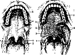
Figure 1.
Figure 2.
Figure 1. General view of the palate with the oral cavity maximally open (cheeks are cut); 1-uvula; 2- anterior palatine arch; <?-palatine tonsil; 1-palatine suture; 5-transverse folds of the palate. Figure 2. Mucous and submucous layer of the palate: 1, 2 and 3-palatine mucous glands; 4-palatine tonsil between two arches; 5-muscles of the soft palate; 6-palatine suture; 7-transverse folds of the palate. with the horizontal parts of the palatine bone (see), adjoining the palatine processes of the maxilla. The posterior edge of the hard palate has the appearance of two arches, open backward and connected at their inner ends in the form of a projection called the posterior nasal spine (spina nasalis posterior). Such a composition of the hard P. is revealed by a series of sutures, along which the connection of its parts occurs. Along the middle line of the hard palate
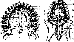
Figure 3.
Figure 4.
Figure 3. Bony composition of the hard palate from the oral cavity side: 1 - incisive foramen; 2 - median palatine suture; 3 - transverse palatine suture; 4 - greater palatine foramen; 5 - pyramidal process; 6 - hamulus of the pterygoid bone; 7 - posterior nasal spine; 8 - lesser palatine foramina; 9 - horizontal part of the palatine bone; 10 - palatine process of the maxilla; 11 - incisive palatine suture; 12 - incisive bone. Figure 4. Vessels and nerves of the hard palate: 1 - palatine median suture; 2 - palatine nerve; 3 - connective tissue base of the soft palate; 4 - uvula; 5 - mucous membrane of the soft palate; 6 - hamulus of the pterygoid bone; 7 - greater palatine foramen with vessels and nerves passing through it; 8 - palatine artery; 9 - incisive foramen with nasopalatine arteries and nerves. The median palatine suture (sutura palatina mediana) runs along the middle. It divides the hard palate into two halves - right and left, passing between the palatine processes of the maxilla, between the horizontal parts of the palatine bone, and between the right and left halves of the incisive bone. Along the course of the median palatine suture, sometimes throughout its entire length, sometimes only in individual segments, there is excessive development of bone tissue in the form of a ridge (torus palatinus). In the transverse direction, two sutures run. The anterior suture separates the incisive bone from the palatine processes of the maxilla and is called the incisive suture (sutura incisiva). The ends of the serrated line of this suture disappear as they approach the canine sockets or the spaces between the lateral incisors and canines. This suture may completely disappear with age due to the fusion of adjacent bones. Between the palatine processes of the maxilla and the horizontal parts of the palatine bone passes the transverse palatine suture (sutura palatina transversa). The hard P. has a series of canals and small canals opening on its oral surface. Through them vessels and nerves emerge. Through the greater palatine foramina (foramen palatinum majus) (Figure 4) (see Palatine bone) pass vessels and nerves of the same name (a., v. and nn. palatini) (Fig. 3,4) and supply the hard P. These vessels and nerves run close to the alveolar margin and may be in danger of damage during operations for cleft lip (see). Through the lesser palatine foramina (foramina palat. minora) (Fig. 3,8) pass arteries and nerves of the same name, supplying the soft P. The palatine arteries are branches of a. palat. descendens, which passes through the pterygopalatine canal and originates from a. maxillaris intermedia. In addition, the soft P. also receives branches from the palatine and pharyngeal ascending arteries a. (palatina ascendens and a. pharyngea ascendens). Both originate from the external carotid artery. The veins of the palate partly empty into the veins of the nasal mucosa, partly into the veins of the tongue, partly into the venous pterygoid plexus, and partly into the veins of the tonsils. The lymphatic vessels of P. pass laterally from the tonsils and fauces and empty into the superior groups of deep cervical lymph nodes. The origin of the palatine nerves appears more complex. If regarding the innervation of the hard P. authors are in agreement that it occurs through nerve branches passing through the pterygopalatine ganglion from the second branch of the trigeminal nerve, then regarding the supply of the soft P., especially its motor branches, authors are not in agreement. Some of them (Batuev, Broman, Keibel, Corning and others) believe that the innervation of the soft P., at least partially, occurs through the facial nerve (nervus petrosus superficialis major and through gangl. sphenopalatinum), from which the palatine nerves originate. Other authors (Braus, Tonkov and others), mainly based on experimental studies by Yagita on dogs, as well as on clinical observation that paralysis of the facial nerve is not accompanied by paralysis of the soft P., believe that the motor innervation of the soft P. occurs through the vagus nerve. However, Yagita himself once obtained contraction of the soft P. upon irritation of the root of the facial nerve, which ceased after transection of the nervus petrosi superficialis major. Apparently, the innervation of some muscles of the soft P. may also occur from the facial nerve. In the anterior part of the hard P., where the palatine processes of the maxilla and the halves of the incisive bone meet in the median palatine suture, is located the incisive foramen (foramen incisivum) (Fig. 3 and 4). It leads into the unpaired incisive canal (canalis incisivus, s. nasopalatinus), which, going upward, divides into two and opens into the nasal cavity through two openings, one on each side of the nasal septum (Figure 5). Through the incisive C.N.E.L. from the nasal cavity onto the oral surface of the hard palate emerge thin nasopalatine arteries and nerves (aa. and nn. nasopalatini), spreading in the anterior parts of the palate. In addition to vessels and nerves, the contents of the incisive canals are two separate, often obliterated Stenson's ducts, belonging to the rudimentary Jacobson's organ. The oral surface of the hard P. is uneven, with a series of elevations, depressions, and small grooves for vessels and nerves, and elevations between the grooves. In connection with this, the mucous membrane of the oral surface of the hard P. holds extremely tightly to its underlying layer. The hard P. from the nasal side is shown in Fig. 5. We see a horizontal section of the maxilla made below the attachment of the inferior nasal concha. The boundary between the hard P. and the alveolar process is a slightly convex outward line. Laterally from it lies the floor of the maxillary sinus with its dental crypts. Medially is located the nasal surface of the hard palate. It is smooth, and the mucous membrane covers it with an even layer. Corresponding to the median palatine suture rises the nasal crest (crista nasalis). Along this
Figure 5. Bony composition of the hard palate from the nasal side: 1 - anterior nasal spine; 2 - medial wall of the maxillary sinus; 3 - section of the pyramidal process; 4 - outer lamina of the pterygoid process; 5 - hamulus of the pterygoid bone; 6 - transverse palatine suture; 7 - posterior nasal spine; 8 - horizontal part of the palatine bone; 9 - pterygopalatine canal; 10 - floor of the maxillary sinus with dental crypts; 11 - palatine process of the maxilla; 12 - nasal crest; 13 - nasal openings of the incisive canal.
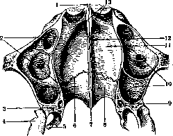
The nasal septum is established on the crest. In front, the crest ends at the anterior nasal spine (spina nasalis anterior). The projection of the crest at the back is called the posterior nasal spine (spina nasalis posterior). The transverse palatine suture is also clearly visible. Similarly, the following formations are prominently displayed: the pterygo-palatine canal (canalis pterygo-palatinus), the pterygoid hamulus (hamulus pterygoideus), which is an important point of attachment for the muscles of the soft palate; the cut of the pterygoid process; the lateral plate of the pterygoid process (lamina lateralis processus pterygoidei); the main constituent parts of the hard palate—the palatine process of the maxilla and the horizontal part of the palatine bone—are also clearly visible. The soft palate anatomically and physiologically belongs to the pharynx (see). In front, the soft palate detaches from the posterior edge of the hard palate, on the sides it is connected to the lateral walls of the pharynx, and behind it ends with a free edge that repeats the shape of the posterior edge of the hard palate. The median projection on the posterior edge, varying in width and length and called the uvula (uvula) (figures 1, 2 and 4), resembles the configuration of the posterior nasal spine. The soft palate changes its position and shape depending on the function it performs at one moment or another. In a state of rest, when breathing occurs through the nose, the soft palate arches downward onto the posterior part of the tongue, thus isolating the oral cavity from the pharyngeal cavity. The value and reliability of this isolation is especially enhanced when food is being chewed in the oral cavity, while breathing continues through the nose and pharynx. During the act of oral breathing, as well as at certain moments of the act of swallowing, the soft palate straightens to form a more horizontally positioned plate, tightly pressed against the posterior wall of the pharynx, separating the nasopharynx from the oral part of the pharynx and the oral cavity. At another moment of the act of swallowing, the soft palate separates the oral cavity from the pharynx. The mechanism of the latter phenomenon is due to the fact that the muscles of the soft palate, which are part of the palatoglossal arches, connect with the transverse muscle of the tongue, forming a contractile ring that can, upon contraction, eliminate its own lumen. The mucous membrane of the palate along the median line has a narrow whitish strip, the palatine suture (raphe palati), which is better expressed in front and represents a fold, while behind it sometimes has the appearance of a groove. In the anterior part of the hard palate behind the incisors, the mucous membrane forms transverse folds of the palate (plicae palatinae transversae), or palatal rugae (rugae palatinae). In the submucosal layer of both the hard and soft palate, and on both surfaces of the uvula, there is a layer of mucous glands (glandulae palatinae). At the border of the hard and soft palate on both sides of the median line, there are two small depressions into which the ducts of the palatal glands open. The mucous membrane of the palate from the side of the oral cavity is covered with stratified squamous epithelium, while from the side of the nasal cavity and nasopharynx it is covered with stratified cylindrical epithelium.

Profile of the palate. If one considers the palate in a sagittal section, one can notice the arched direction of the anteroposterior line. The descent forward goes to the interincisal point on the alveolar process, the descent backward goes along the soft palate in a state of rest. The character of the anterior descent depends mainly on the position of the incisor bone. The posterior descent V:
Figure 6.
Figure 7.

Figure f>. Normal profile of the hard palate in a frontal section: 1 to 4 maxillary sinus; 2- nasal cavity; 3- nasal septum. Figure 7. High and narrow palate: 1- nasal cavity; 2- nasal septum; 3- maxillary sinus. varies depending on the mobility of the soft palate. According to Osokin, the hard palate reaches its greatest height in newborns at the border of the anterior and middle thirds, in children 10 years old - in the middle of the second third, in adults - at the border of the middle and posterior thirds. In a frontal section of the hard palate at its highest point, an arch is obtained, concave toward the oral cavity (figures 6, 7, 8 and 9). The frontal profile of the palate, as can be seen from the mentioned figures, appears extremely diverse in individuals. The main curvature of the palate in the frontal plane is schematically shown in figure 6. It has the form of a uniformly flattened arch. Such a form of the palate is considered normal. Figure 8 shows changes in the frontal section of a normal palate depending on age. According to Franke (Franke), line 1 corresponds to the palate of a fetus
Figure 8. Age-related profile of the hard palate in frontal section (according to Franke): 1-fetus \}0 cm in length; 2-child two years old; 3-individual 18 years old; 4-elderly person with atrophied alveolar process. 30 cm; line 2-palate of a two-year-old child, in whom the development of the alveolar process has already clearly defined itself; line 5-palate of an 18-year-old individual; line 4-palate of an elderly person. The last line appears very flattened due to atrophy of the alveolar process after tooth loss. Thus, a certain dependence of the age-related height of the palate on the power of development of the alveolar process is revealed. The height of the palate is usually measured at the level of premolars in younger age and at the level of the first molar in mature age. Siebenmann's palatometer facilitates the measurement of the palate, giving simultaneously its height and width. Having the figure, one can calculate the index tt
, width In contrast to the flattened arch, a narrow and elongated arch is considered characteristic of a pathologically developing palate. The simplest scheme of such a palate in frontal section is given in figure 7. This form of the palate is accompanied by other pathological changes in the shape of adjacent formations: curvature of the nasal septum (figure 7,2), low position of the floor of the maxillary sinus (figure 7, 3), elongated shape of the facial skeleton, leptoprosopia and others. Among the most common theories explaining the origin of a high and narrow palate (hypsistaphylia), two should be mentioned, which mutually exclude each other. The theory of Bloch-Korner (Bloch, Korner) sees the cause in the absence of normal nasal breathing and in the presence of oral breathing, as for example occurs in persons with adenoid vegetations in the nasopharynx. The constantly open mouth causes constant pressure on the alveolar process, causing more intensive growth downward and narrowing its arch. Another theory is that of Siebenmann, who considers a high and narrow palate a phenomenon accompanying the long shape of the face, which is predominantly a racial feature and can be inherited. In particular, Siebenmann and his followers do not attribute formative value to the pressure on the upper alveolar process by the cheeks when the mouth is constantly open, considering such pressure negligible. In their opinion, the main influence on the formation of alveolar processes is exerted by the pull of the masticatory muscles, the action of which changes little during oral breathing. Of great interest are the works of Franke, who to some extent aligns with Siebenmann. On the basis of his numerous studies of living persons with a high palate, as well as museum skulls, Franke comes to the conclusion that a high palate occurs almost equally frequently with both oral (figure 9, 1,2, 3) and nasal breathing (figure 9, 5). Franke wanted to find an explanation for the development of a narrow and high palate in the study of the laws of normal and abnormal development of the palate, maxilla, and nasal septum. According to him, a narrow and high palate occurs because in some cases, with defective growth of the palate, the alveolar processes grow normally and outpace the growth of the palate. At present, the theory of Bloch-Korner is the most widespread. A high and narrow palate is often limited in front by the alveolar process,
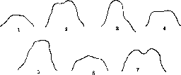
Figure D. Various profiles of the frontal section of the hard palate depending on the growth processes of the latter (according to Franke): 1, 2 and 3- profile with oral breathing; 4 and 5- profile with nasal breathing; 6 and 7- profile from macerated skulls.
the halves of which converge at an angle at the central incisors, giving the hard palate in this place a pointed V-shaped form (see Teeth, fig. 23). Another deformation of the hard palate is found in rachitic compression of the alveolar arch and flattening of its anterior segment. In such cases, the hard palate takes the form of a trapezoid (see Teeth, figure 24). However, the greatest number of all kinds of palate deformities results from a disturbance in proper development in the early stages of embryonic life. These primarily include median clefts of the palate (see Cleft Palate). Cleft palate is the extreme form of deformity affecting the entire palate and often involving the alveolar process and lip. On the other hand, sometimes only a hint of a cleft of the palate is encountered, expressed only by a slight bifurcation of the uvula. Between these two forms there is a whole series of intermediate forms of partial closure of the palate. Sometimes, with the apparent integrity of the mucous membrane, there may be a congenital bone defect in the area of the hard palate and a muscular one in the area of the soft palate. Practically important are primarily congenital defects of all layers of the palate, as they strongly disturb nutrition and speech and require for their elimination surgical intervention or a prosthesis (see Cleft Palate). Congenital deformations also include certain phenomena in the area of the palatine arches. Thus, fistulae are sometimes encountered with an opening below the tonsil, behind it, or in the tonsil itself. The fistula may also have an external opening on the neck, at the medial edge of the sternocleidomastoid muscle. According to the research of Hammar and Venglovsky, this kind of fistula has the closest relation to the pharyngeal pouches and the development of the thyroid gland. Congenital defects in the area of the palatine arches may also manifest as absence of the anterior or posterior arch, partial or complete, formation of pits or even through clefts. In most cases, the mentioned deformations do not disturb the function of the organ. Fistulae in the tonsil area, opening on the neckt may be the subject of surgical intervention. Traumatic injuries to the palate are not frequent. Their types are as follows: a) Injuries from sharp objects during eating; they are not great, mostly heal by themselves. b) Injuries from chemical substances in attempts at poisoning; these injuries are less significant than in the lower parts of the digestive tract, where these substances remain for a longer time. In acute cases of injury, it is recommended to neutralize the poisonous substance, in later cases to prevent improper scarring. c) Gunshot injuries to the palate are most often encountered in war, less often in attempts at suicide by shooting in the mouth. In the latter cases, colossal injuries are inflicted, and death occurs from bleeding or damage to other important organs of the head. Milder cases, leaving behind deformations, may require plastic operations or prosthetics. d) Accidental injuries to the palate in children from falling on sharp objects (stakes, harrow tooth, etc.) have no great practical significance, as they usually heal by themselves. Foreign bodies in the area of the palate are found mainly in the tonsils (see). In rare cases, parasites in the area of the palate have also been encountered and described. The fight against parasites is carried out in cases of suppuration - by incision; in cases of a recognized non-suppurating echinococcal cyst, a formalin solution is introduced to kill the parasite and then the cyst is removed, if technically possible.
Inflammatory processes on the palate are very frequent and diverse. Pathological processes of the teeth, tonsils, small injuries during food intake - all these moments can cause acute or maintain chronic inflammations of the palate. Besides local causes, inflammatory conditions of the palate are also caused by general ones: various infections (measles, scarlet fever, rubella, smallpox, etc.), diseases of the stomach, generally all factors contributing to the development of stomatitis. The most frequent causative agents of inflammatory processes on the mucous membrane of the palate are staphylococcus, streptococcus, pneumococcus. In addition, there is a whole series of other causative agents that at times cause a specific form of inflammatory processes on the palate, for example thrush, aphthae, pemphigus and diphtheria, Vincent's angina (see. 857", "HEI Angina), gonorrhea, etc. Furthermore, we encounter on the palate most infectious granulomas. - For lesions of the soft palate with tuberculosis and syphilis, see Pharynx. The mucous covering of the hard palate is also affected by tbc, mostly secondarily, spreading here from deeper parts of the respiratory and digestive tracts. Diagnosis is not difficult when examining the whole body, when carefully studying the periphery of the affected area, where tubercles can be discovered. - Treatment is general strengthening, and in corresponding cases local in the form of cauterizations, curettage, etc. The hard palate is also affected by syphilis. Especially unpleasant is its tertiary stage, often leading to necrosis of all tissues and perforations, mainly in the posterior part of the hard palate and in the anterior part of the soft palate, which leads to great disturbances in phonation and food intake. After appropriate courses of specific treatment and with proven absence in the body of an active virus, if necessary, recourse is had to plastic surgery or prosthetics. - Scleroma affects all parts of the palate, but mainly the nasopharyngeal surface of the soft palate, leading to deformation of the palatal curtain, and sometimes to ulcers, which can be recognized only histologically. - Leprosy of the palate is recognized by characteristic nodules and bacteriologically. Of the necrotic processes of an inflammatory nature affecting the bony vault, besides gumma, one can also point to osteomyelites, originating from necrotic lesions of the alveolar process. - Actinomycosis of the palate in humans is a rare phenomenon, usually of secondary origin when spreading from the neck and other parts of the oral cavity. Hypertrophic processes of the hard palate are expressed in excessive development of bone tissue in the area of the median palatine suture, as a result of which a more or less prominent ridge forms in place of the suture. - Hypertrophies of the soft palate are concentrated mainly in the tonsils, which can sometimes reach considerable sizes. - Atrophic processes on the mucous membrane of the palate are expressed by its thinning, decreased secretion of mucus, dryness, and can depend on various causes (long-term irritation by a prosthesis, in some persons due to smoking, with prolonged mouth breathing). Atrophy of the muscular layer of the arches usually occurs as a phenomenon accompanying paralysis of the soft palate. - Atrophy of the bony palate is expressed in its thinning and is a manifestation of old age. - New growths of the soft palate - see Pharynx. Of the new growths of the hard palate, the following deserve attention: a) Benign: 1) Mikulicz's disease (see) in the form of two symmetrical swellings, located on both sides of the median suture; 2) so-called palatine pearls, located in the thickness of the bony part of the suture of the middle of the palate and resulting from a late, occurring in embryonic life, union between the jaw processes with the entrapment inward of their epithelial covering. The considerable size of these tumors sometimes gives rise to their incorrect interpretation as a malignant new growth. b) Malignant tumors of the hard palate usually develop secondarily, spreading from neighboring parts (alveolar process, soft palate, tongue, nasal cavity). Sometimes the palatine glands can be the starting point for the development of cancer. At first, the tumor is covered with normal, extremely tense mucous membrane. Then, the tumor, having destroyed the vault, can break into the nasal cavity, simulating a gumma and complicating the diagnosis. With a precisely established diagnosis and technical possibility, recourse is had for treatment to appropriate operations.
i. Sendulsky.
Related articles
Mentioned in
Cite this page
“Palatine Bone.” Soviet Medical Encyclopedia. English translation of Bolshaya Meditsinskaya Entsiklopediya, 1st ed. (Moscow, 1928–1936), ed. N. A. Semashko. https://sovietmedicalencyclopedia.pages.dev/article/palatine-bone/