Organ of Corti
Historical document, translated for reference. It reflects medical knowledge of the 1920s–30s and is not medical advice.
Summary
The organ of Corti is the terminal apparatus of the cochlear branch of the auditory nerve, serving as the primary organ of hearing. Located within the cochlear duct, it contains specialized hair cells and supporting structures that transduce sound vibrations into nerve impulses.
Encyclopedia article (1928–1936)
ORGAN OF CORTI (Organon Cortii), named after the Italian histologist Corti, who first described it in detail [synonym papilla acustica basilaris (G. Retzius)], represents the terminal apparatus of the cochlear branch of the auditory nerve (ramus cochlearis nervi acustici) and is the organ of hearing. It is located on the basilar membrane of the cochlear duct, ductus cochlearis (see Inner Ear), along its entire length from the base
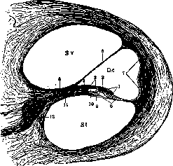
Figure 1. Cross-section of a cat's cochlea with the organ of Corti: Sv-scala vestibuli; St-scala tympani; Dc-ductus cochlearis; 1-organ of Corti; 2-membrana tectoria; 3-labium vestibulare; 4-lamina spiralis int.; 5-lamina spiralis ossea; 6-membrana Reissneri; 7-ligamentum spirale; 8-membrana basilaris; 9-labium tympanicum; 10-sulcus spiralis int.; 11-nervus cochlearis; 12-ganglion spirale. (After Lavdovsky.)
to the apex, making 2 1/2 spiral turns along with it (hence the old name organon spirale, papilla spiralis Huschke). In a cross-section through the cochlear coil (Fig. 1), the cochlear duct has the shape of a triangle or, more precisely, a sector of a circle, the apex of which attaches to the end of the spiral bony lamina (lamina spiralis ossea); the opposite arc adheres to the bony wall of the cochlea; the upper side is formed by Reissner's membrane (membrana vestibularis Reissneri), bordering the scala vestibuli, and the lower side by the edge of the bony lamina spiralis and the basilar membrane (membrana basilaris, s. lamina spiralis membranacea), facing the scala tympani. On the upper surface of the basilar membrane, closer to the lamina spiralis, there is a cushion-like thickening—the organ of Corti. The shape of the cochlear duct changes from the base to the apex: the angle attached to the lamina spiralis becomes smaller and, in the upper turn of the cochlear duct, appears flattened; its height in the basal turn is 0.5 mm, in the upper turn 0.35 mm; the length of the tympanic wall, conversely, increases from 0.45 to 0.8 mm (Retzius). The thickened edge of the lamina spiralis ossea (limbus) has a notch or groove (sulcus spiralis) at its free end, bounded by two lips: the upper (labium vestibulare) and the lower (labium tympanicum); on the surface of the vestibular lip, narrow and long elevations run in a radial direction, separated by furrows [auditory teeth (Gehörzähne Huschke)]; the tectorial membrane (membrana tectoria), draped over the organ of Corti, also originates here; the lower lip consists of two lamellae, between which the branches of the cochlear nerve pass; they emerge onto the surface through openings in the upper lamella not far from the edge (habenula perforata). The lower lip transitions directly into the basilar membrane, which heads toward the wall of the cochlea and ends in a conical elevation, the spiral ligament (ligamentum spirale). The width of the basilar membrane increases from the base of the cochlea (210 μ) to the apex (360 μ); one distinguishes in it (Fig. 2) an inner zone, covered by the organ of Corti (habenula tecta), and an outer one (zona pectinata); its base consists of radially running fibrils, which are thin and intertwined in the covered part, and thicker (1-2 μ) and arranged extremely regularly in the outer part, forming the so-called auditory strings. The length of the strings in humans is 135 μ in the basal turn, 220 μ in the middle, and 234 μ in the upper (Retzius); their number: according to some—13,400 (Hensen), according to others—24,000 (Retzius). The strings are soft and easily bendable during life, but become brittle after fixation; they dissolve in acetic acid, and their nature is not fully clarified; above, the strings are covered by a thin boundary membrane in which nuclei are visible; below, by a similar but thicker one, which transitions into the tympanic covering (tympanale Belegschicht, Retzius), consisting of connective tissue with cells and containing blood vessels in the inner zone. The entire cochlear duct is lined inside with a single-layered epithelium, a modification of which is the organ of Corti (Fig. 3). The organ of Corti includes: Corti's arches, Deiters' supporting cells, the reticular membrane formed by both, sensory or hair cells with nerve endings, and the tectorial membrane; Hensen's and Claudius' cells are located external to the organ of Corti (Hensen, Claudius). Corti's arches are formed by two pillar cells (Pfeilerzellen), the lower ends of which are pushed apart, while the upper ones converge; a triangular space—the tunnel—remains between them. Each pillar consists of a triangular base, in which the nucleus is located, a narrow body, and an expanded head, on which there is a cuticular cap; the main mass of the pillar is formed by skeletal fibrils that run along its length from the base to the cuticular cap (Fig. 4); a joint forms between the heads, resembling an elbow
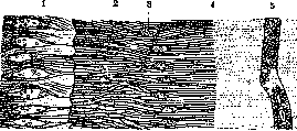
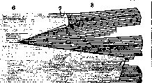
Figure 2. Basilar membrane after removal of the organ of Corti (surface view): 1-auditory teeth on the vestibular lip of the limbus spiralis; 2-inner part of the sulcus spiralis with translucent medullated nerve fibers; 3-foramina nervosa with severed nerves; 4-edge of the labium tympanicum; 5-vas spiralis; 2-4-habenula perforata; 3, 4 and 5-habenula tecta (region of Corti's arches); 6-5-zona pectinata with torn strings; 7-homogeneous membrane covering the strings; 8-strings of the basilar membrane. (After Ebner.)
Figure 4. Organ of Corti of a guinea pig: skeletal fibrils of Corti's arches (1 and 2) and Deiters' cells (3, 4 and 5). (After Kolmer.) (Waldeyer). The bases of the inner pillars lie on the lower lip of the spiral lamina, and the outer ones on the basilar membrane, covering it up to the beginning of the strings. The length of the inner pillars in the basal, middle, and upper turns is 48-68-70 μ; the outer ones—62-100-103 μ; the distances between the legs—48-81-90 μ; the height of the tunnel—42-45-49 μ. The number of inner pillars is greater than the outer ones—5,600:3,850 (Retzius), 6,000:4,500 (Waldeyer). Deiters' supporting cells lie external to Corti's arches in three rows; their basal part, containing the nucleus, is wide and cylindrical in shape; but toward the middle, the cell body narrows abruptly and forms a thin process, which heads toward the surface, making a spiral bend, which is why the cell cannot be captured in its entirety in a section; the process transitions into a biscuit-shaped cuticular plate, the so-called phalanx. Deiters' cells also contain a fibrous skeleton, which in the process gathers into a thick supporting thread of Retzius, and at the upper end again disperses into individual fibrils and supports the phalanx. Between the outer pillars and the first row of Deiters' cells, there is also a free gap—Nuel's space (Nuel). The cuticular caps of Corti's arches and Deiters' cells, fusing together, form the reticular membrane (membrana reticularis), covering the organ of Corti (Fig. 5). Within its composition, one can distinguish phalanges of the 1st (processes of the outer pillars), 2nd, 3rd, and 4th orders (of Deiters' cells); they are arranged in a checkerboard pattern such that round openings remain between their lateral notches, into which the upper ends of the sensory cells are inserted; only the phalanges of the 4th order fuse into a continuous closing plate. As a whole, the reticular membrane forms a frame for the sensory cells. Hair cells (Haarzellen), or sensory cells, like the cells of the maculae and cristae acusticae, have the shape of a thimble, sometimes with a thin process extending from the base, and are located between the thin processes of Deiters' cells, resting their bases on their bodies; they do not reach the basilar membrane, as was previously believed. In their body, closer to the base, is the nucleus, and in the upper section—a rounded dark inclusion (Hensen's body), which has not been cytologically studied; on the free surface, there is a bundle of hard, non-ciliated hairs (about 20), arranged in the shape of a horseshoe. External to Corti's arches, between Deiters' cells, are 3 rows of outer sensory cells; their total number
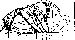
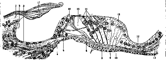
Figure 3. Cross-section of the human organ of Corti: 1-labium vestibulare limbi spiralis; 2-sulcus spiralis; 3-epithelium covering it; 4-passage of nerves through the labium tympanicum (habenula perforata); 5-inner and 6-outer pillar of Corti's arch; 7-Deiters' supporting cells; 8-fibrous layer of the basilar membrane; 9-cellular layer on the tympanic side; 10-upper boundary layer; 11-ligamentum spirale; 12-Claudius' cells; 13-Hensen's cells; 14-outer auditory cells; 15-Nuel's space with a passing nerve bundle; 16-inner auditory (hair) cells; 17-tectorial membrane. (After Ebner.)
is 13,000 (Retzius) or 18,000 (Waldeyer); internal to the arches—1 row of inner ones—3,500 (Retzius), 3,300 (Waldeyer). The frame for the inner cells is formed by the cuticular processes of the inner pillars of Corti's arches and the single row of inner supporting cells. Nerve endings of the usual type have been discovered on the sensory cells (Lenhossék, Retzius); a nerve fiber, branching out, encompasses the cell and terminates in button-like thickenings. The nerves originate from bipolar cells of the spiral ganglion at the base
E. Kononova
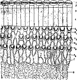
Figure 5. Part of the human Organ of Corti (surface view): 1 - internal auditory (hair) cells; 2 - internal pillars of the Corti arches; 3 - external pillars; 4 - flat processes (phalanges) of the internal pillars; 51, 52, 53 - first, second, and third rows of external auditory cells; 61, 62, 63 - phalanges of the first, second, and third rows of supporting cells; 7 - closing plates; 8 - Hensen's cells. (According to Retzius.) Nerve fibers from the spiral crest (crista spiralis) emerge through the nervous foramina (foramina nervosa) of the lower lip and are directed toward the sensory cells; they can be seen in the tunnel and the Nuel space, where they travel accompanied by connective tissue cells. An essential part of the Organ of Corti is the tectorial membrane (membrana tectoria) of epithelial origin, consisting of thin fibrils soldered into one common mass, soft but extremely elastic; it is attached to the epithelium of the upper surface of the vestibular lip over a considerable extent; its free part, initially thicker (20-25 μ), tapering toward the end, has a length from 120 μ (basal turn) to 240 μ (apical turn) and covers the surface of the Organ of Corti. It is assumed that during life, the hairs of the sensory cells are immersed in the tectorial membrane and are tightly connected to it (Hensen, Wittmaack); thus, the vibrations of the endolymph are transmitted through its mediation to the hair cells. Outward from the last row of Deiters' cells is located a high cylindrical epithelium, forming the outer part of the cushion-like thickening of the Organ of Corti, the so-called Hensen's cells; they pass further outward into a layer of low cuboidal Claudius's cells, which wrap around the outer wall of the cochlear duct. This wall consists of connective tissue, passing into the periosteum of the bony cochlea, and in its middle, an elevation is noticeable (prominentia spiralis); it is covered with a single-layered epithelium, which becomes multi-rowed above the elevation. In this place, the wall contains a large number of blood vessels (stria vascularis), with capillaries branching even within the epithelium (Retzius). Reissner's membrane in humans is very thin (3 μ) and consists of connective tissue covered with low epithelium.
Physiology. The Organ of Corti is the organ of hearing; sound waves are transmitted by the sound-conducting apparatus (tympanic membrane, chain of auditory ossicles, oval window) to the perilymph and endolymph of the cochlea and are perceived by the auditory receptors—the hair cells of the Organ of Corti; perception is carried out through the mediation of the hairs, specifically by their bending. The mechanism of transmission of endolymph vibrations to the hairs has not been precisely clarified. According to the widespread theory of Helmholtz, the basilar membrane, consisting of stretched strings of different lengths, is an analyzer that decomposes complex sound vibrations into simple tones; each tone, depending on its pitch, causes a resonating vibration of a specific section of the basilar membrane, which entails an oscillatory movement of the hair cells and deformation of the hairs upon which the tectorial membrane rests. The role of the Corti arches can be represented in the following way (ter Kuile): the internal pillars, resting on the bony lip (labium tympanicum), do not take part in the movement of the membrane, while the external ones, during its vibration, perform rotational movements around an axis passing through the stem of the internal pillar; this movement, through the mediation of the reticular membrane, is transmitted to the hair cells inserted into it, the hairs of which, upon displacement of the cells, bend and rub against the tectorial membrane. According to Helmholtz's theory, low sounds set in motion the long strings located in the apical turn of the cochlea, and high sounds—the short strings of the basal turn; this point was disputed by many authors (Meyer, ter Kuile, Ewald); Ewald, based on the observation of vibrations of an artificial membrane corresponding in dimensions to the basilar one, assumed that the basilar membrane vibrates as a whole, nodes and antinodes form on it, and each tone corresponds to a specific sound pattern (Schallbild). Experimental verification of Helmholtz's theory has been performed many times. A number of authors removed the apical or basal turns of one cochlea with complete removal of the other and tested the auditory perceptions of the animal; in some cases (Munk, Baginsky), the results confirmed Helmholtz's theory, in others—they contradicted it (Stepanov). Also, contradictory results were obtained in experiments on the destruction of sections of the Organ of Corti by prolonged exposure to sounds of a certain pitch: some authors (Wittmaack, Yoshii) found destructions that confirmed Helmholtz's theory, others (Popov)—the opposite. The very possibility of resonance in the strings of the basilar membrane was questioned in view of its weighting by the thick boundary membrane, which is also of different thickness at the base and the apex (Ebner). Therefore, recently, authors have been leaning toward the idea that it is mainly the tectorial membrane that resonates (Shambaugh, Plate), which is closely connected during life with the auditory hairs.
[Diagram of] the brain stem and the pons Varolii: 1 - frontopontine tract; 2 - pyramidal tract; 3 - temporo-occipital tract of the pons.
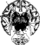
Long crossed tract, connecting the cerebral cortex with the opposite half of the cerebellum. Corticopontine tracts include frontal, temporal, and occipito-pontine fiber systems. The frontopontine system (Arnold's bundle) originates in the frontal lobe; authors' opinions differ regarding the exact place of its origin: according to some data, it begins on the outer surface in the anterior sections of the frontal gyri, according to others—in the posterior sections, and finally, according to a third opinion, the fibers originate on the lower and inner surfaces of the frontal lobe. In the white matter of the frontal lobe, the frontopontine system occupies the outer sagittal layer; in the internal capsule (capsula interna), it passes through the lower sections of the anterior limb (see figure), in the brain stem (pedunculus cerebri), it is located in the inner fifth part of its base, and then terminates in the nuclei of the base of the pons Varolii. The temporopontine system (Turck's bundle) originates in the middle sections of the second and third temporal gyri, passes through the lower sections of the corona radiata and the
sublenticular part of the internal capsule (pars sublenticularis capsulae internae) into the brain stem, where it occupies the outer fifth part of the base; it terminates in the upper-posterior nuclei of the base of the pons. Some authors believe that the occipitopontine system also goes in the composition of this system, i.e., fibers from the occipital lobe (from which gyri, it is not exactly known). These systems are especially developed in humans in connection with the great development of the cerebral hemispheres. The corticopontine tracts connect the cerebral cortex through the pons Varolii with the opposite hemisphere of the cerebellum, which also indicates their functional significance—the possible participation of various sections of the cerebral cortex (visual, auditory, etc.) in the work of the cerebellum; this is most clear regarding the frontal lobes, where the higher cortical centers of equilibrium are located, which send their stimuli to the cerebellum, and in the case of its destruction, can take its function upon themselves.
Related articles
Mentioned in
Cite this page
“Organ of Corti.” Soviet Medical Encyclopedia. English translation of Bolshaya Meditsinskaya Entsiklopediya, 1st ed. (Moscow, 1928–1936), ed. N. A. Semashko. https://sovietmedicalencyclopedia.pages.dev/article/organ-of-corti/