Achondroplasia
Historical document, translated for reference. It reflects medical knowledge of the 1920s–30s and is not medical advice.
Summary
This article from the 1928–1936 Soviet medical encyclopedia describes achondroplasia, a congenital condition characterized by dwarfism and disproportionate limb shortening. It details the distinctive physical features, including a large head, short limbs, and a trident-like hand, and discusses historical theories regarding its etiology.
Encyclopedia article (1928–1936)
ACHONDROPLASIA, achondroplasia (from Greek a- negative particle, chondros- cartilage and plasis- formation); synonyms: nanism chondrodystrophicus, mikromelia, rachitis foetalis, chondrodystrophia foetalis, Parrot-Marie disease—a disease characterized by dwarfed growth with a sharp and highly original disturbance of the normal proportions of the body. The credit for isolating this interesting growth anomaly into a separate pathological form belongs to Parrot (1876). Pierre Marie's classic work, appearing in 1900, was no less significant for the further development of the doctrine concerning this disease. The characteristic feature of this disease is the discrepancy between the length of the limbs and the size of the head and trunk in the absence of other defects of build and intellect. The head and trunk of the patient are of normal size for the given age, while the upper and lower limbs are sharply shortened. Such disproportionate dwarfs with short limbs were already known in antiquity (the Egyptian god Ptah represents a typical image of mikromelia). The hands have a peculiar appearance in this disease: the index and middle fingers are little separated from each other, as a result of which the hand resembles a trident (main en trident). The fingers differ from each other much less in length than in the norm, which gives the hand a square-like appearance (main carree). The feet are short and broad. On the limbs, most often on the lower ones, curvatures are noted in many cases. Often the central parts of the limbs are most shortened, i.e., the humeral and femoral bones (micromelia rhizomelique, P. Marie). Almost all authors note the high position of the head of the fibula, which is explained by the relatively lesser shortening of the fibula than of the tibia. The trunk in A. is usually not affected and remains approximately of normal size, with a flat back, reduced shoulder blades, and sharply protruding buttocks. The chest and ribs are developed normally in most cases, but cases of A. with chest deformity in the form of a protruding and curved sternum have also been described, since bone dystrophy is sometimes expressed not only in the long bones but also affects the bones of the chest. The spine in typical cases is not affected; often it is straighter than in the norm, with less sharply expressed normal curves; in some cases lordosis is noted.
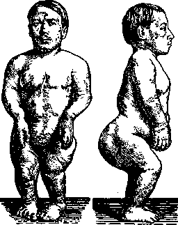
Achondroplasia. Patient 22 years old; height 91.9 cm (case of D. M. Rossiyskiy from the Clinic of Internal Diseases, 1st MSU). The pelvis is reduced in all its dimensions, and due to the anteverse position of the pelvis, the sacrum is raised upward by its posterior surface. From the side of the skull, macrocephaly and brachycephaly with prominent frontal and parietal tuberosities are noted. The face is usually broad but small in size and seems small in comparison with the skull. The nose in mikromelia is often wide with a low, depressed nasal bridge; the hard palate is high, domed, and epicanthus is often noted. Ears and teeth show no deviations from the norm. The development and eruption of teeth occur in a timely manner. From the side of the skin, internal organs, nervous system, sensory organs, and sexual apparatus, deviations are usually also not noted. The state of the psyche is normal in most cases, and only in relatively few cases is a lowering of the psyche with features of mental deficiency noted. Cases of A. are also observed that concern, for example, only the lower limbs or some single limb. The basis of A. lies in a disturbance of the correct course of the endochondral development process of the bones, consisting mainly in the fact that the multiplication of cartilage cells at the boundary of the cartilage and the forming bone proceeds very slowly, insufficiently, and stops early. The bone lesions in A. are usually quite symmetrical. The epiphyses of the bones are voluminous, extremely massive; the diaphyses are strong and solid. All normal bone irregularities, grooves, and roughnesses at the places of muscle attachment are sharply expressed. On the skulls of adults, underdevelopment of the base of the skull in the sagittal direction is noted (hence the depression of the nose), narrowing of the foramen magnum, and an excessively vertical position of the shortened basilar bone. Histological examination of the limb bones in A. shows a peculiar picture in the zones of endochondral ossification in the form of a very narrow zone of proliferation of cartilage cells, with the multiplication of the latter almost absent; individual cartilage cells and groups of them are separated by wide layers of the matrix, often bearing signs of fibrous transformation. In addition, ingrowth of connective tissue of the periosteum between the epiphysis and the diaphysis often occurs, which completely stops endochondral ossification and the longitudinal growth of the limb bones. A. is considered by all authors to be a congenital disease. In most cases, A. appears in the family by chance, but there are also indications of familial and hereditary A., and in some cases it has even recurred in several generations. The question of the pathogenesis and etiology of A. is not yet elucidated. Until the 70s of the last century, A. was considered a manifestation of congenital syphilis or intrauterine rickets. Some authors (Parrot, Bouch, Mayet, etc.) saw in A. a purely local disease as a result of the congenital dystrophy of the embryonic cartilage, which is a manifestation of the degeneration of the organism. Other authors (Lannois, Apert) considered A. to be an original physiological variety of man, just as in the animal world there are dachshunds and bulldogs. A theory of atavistic transmission of properties from distant ancestors of man—pygmies (Leriche, Poncet) even arose, according to which achondroplasics represent the descendants of an extinct tribe of ancient pygmies. Finally, there are theories that see the cause of achondroplasia in infection or intoxication, presumably transmitted to achondroplasics by the maternal organism. Some theories assume a direct action of some infectious or toxic agent on the osteogenic cartilage; according to other theories, a disturbance of the correct vital activity of the epiphyseal cartilage is a secondary phenomenon, closely dependent on the dystrophy of the general cause. According to the opinion of Pierre Marie, achondroplasia is the result of the abnormal activity of certain internal secretion glands.
Related articles
Mentioned in
Cite this page
“Achondroplasia.” Soviet Medical Encyclopedia. English translation of Bolshaya Meditsinskaya Entsiklopediya, 1st ed. (Moscow, 1928–1936), ed. N. A. Semashko. https://sovietmedicalencyclopedia.pages.dev/article/achondroplasia/