Glanders
Historical document, translated for reference. It reflects medical knowledge of the 1920s–30s and is not medical advice.
Summary
Glanders is an infectious disease primarily affecting horses and other equids, caused by the bacterium Burkholderia mallei. The disease can be transmitted to humans, making it a zoonotic concern with significant historical importance in veterinary medicine.
Encyclopedia article (1928–1936)
Glanders (Latin malleus, adapted from Greek malis - malignant disease), is mentioned as early as the 4th century as an infectious disease of odd-toed ungulates. The classification of G. as a zoonosis, i.e., its ability to infect humans, was established only in the early 19th century. The obsolete name 'Wurm' (German Wurm, French farcin) was previously used to denote the chronic form of G. in horses with predominant skin lesions. The causative agent of G., discovered in 1882 by Löffler, is B. mallei (see illustration, plate to Syphilis article, fig. 7) and exhibits considerable polymorphism; in the diseased organism it usually appears as a rod of medium length, 2-5 μ and medium thickness, 0.5-1.0 μ, with rounded or slightly pointed ends. In very young cultures, smaller round or oval forms are encountered, and in old ones - often threadlike formations, sometimes branching and even with club-shaped thickenings at the ends, for which reason B. mallei is by many classified as streptothrichs. B. mallei does not possess active motility, does not form spores, is Gram-negative, and weakly takes aniline dyes in simple aqueous solutions, better - in solutions with the addition of mordants, such as alkaline Löffler's methylene blue, carbolic Ziehl's fuchsin, etc. With moderate staining, the protoplasm stains unevenly, appearing granular; when stained by Neisser's method (methylene blue - vesuvine), grains similar to diphtheritic ones are found; in old cultures, the smallest grains at the limit of visibility are found, capable of growing into ordinary vegetative forms. The biological significance of all these granularities has not yet been sufficiently clarified. B. mallei is aerobic and only facultatively anaerobic to a weak degree; at temperatures below 20° and above 45° it does not develop, optimum 30-40°; for growth on artificial nutrient media it prefers a weakly acidic reaction (according to Vyshelessky, optimum pH 6.0-7.4); during growth it secretes a mucous substance; it is capable of developing on most common laboratory media, especially with the addition of glycerin. Glanders cultures on potato media, with rare exceptions, form a characteristic yellowish pigment, which, gradually darkening, takes on various brownish or reddish shades, as a result of which their translucent colonies acquire a honey or amber appearance. On coagulated serum, initially yellowish colonies are also obtained, but which whiten over time. Serum agar, as well as gelatin, are not liquefied. On meat-peptone-glycerin-agar, in the incubator, already on the second day after sowing, small grayish transparent mucous colonies appear, which with further growth may take on a slightly yellowish tint, but usually, merging, they give a grayish continuous mucous coating. In condensation water, as in all liquid media, a delicate turbidity, mucous sediment, and a thin film on the surface are formed. Even in old cultures on meat-peptone-glycerin-broth, used for the preparation of mallein (see), the turbidity never becomes lumpy, and the sediment retains its mucous character, while the liquid itself darkens and takes on a yellow-brown tint. B. mallei coagulates milk at 37° in 10-12 days with neutral reaction, but when grown on litmus milk serum according to Petrushka it causes its reddening. The resistance to physical and chemical agents of B. mallei as a non-spore-forming microbe is very insignificant. Direct sunlight kills it within 24 hours and later. It dies in pure dilutions after 10 minutes when heated to 55° (protein coagulation temperature), in secretions of the diseased organism - after longer periods or at higher temperatures (up to 100°). When drying, it loses viability within 2 weeks-3 months, depending on the composition, humidity, temperature of the environment and light conditions. All chemical substances used in disinfection practice kill it in pure dilutions in ordinary concentrations, in secretions and other pathological materials - only those that do not coagulate proteins, similar to sublimate, formalin, etc. Chemically indifferent substances, 80% aqueous solution of glycerin and 10% aqueous solution of urea kill B. mallei with prolonged shaking at 37° due to their high osmotic pressure, without destroying its antigenicity. Epidemiology G. is in complete dependence on its epizootology, since except for laboratory infections, people become infected exclusively through contact with glandered animals (horses) or objects contaminated by them. In areas free from glandered horses, human diseases with this disease do not occur, but in most countries G. occurs as an enzooty. Their extent and intensity depend primarily on the size and rationality of the veterinary-sanitary measures applied. Secondly, the epizootology of G. is connected with the social and agricultural structure of the country; in capitalist states with individual, predominantly small livestock ownership and predominant private horse breeding, control over G. is significantly hampered, and such broad preventive measures as mass malleinization and the establishment of special 'mallein farms' (see below) are generally not feasible. The main reservoir of the glanders virus in nature is the body of glandered horses, in which with natural infection glanders in most cases takes a chronic and even latent form, which creates many carriers and disseminators of infection. From other odd-toed animals, G. takes in donkeys, with rare exceptions, an acute course, and in mules more often than in horses, acute and subacute. As for the susceptibility of animals to G in general, large cattle, domestic rats and birds are considered naturally immune. With artificial infection, it is possible to cause only transient local lesions in large cattle and pigeons. Among small ruminants, sheep are also not naturally susceptible to the disease, while goats can become infected from glandered horses; experimental infection of both species only occasionally leads to generalization of the infection. Camels can suffer from G. in acute or subacute form. Pigs are very little susceptible even to artificial infection. In dogs, vaccination with glanders material in most cases causes only self-healing local abscesses; general infection was observed only in young, old or exhausted individuals in a few spontaneous cases and after eating glanders meat. Cats belong to the most susceptible to G. animals; the disease in them usually takes an acute and subacute form, regardless of the method of natural or experimental infection (see below - diagnosis of G.). The same susceptibility is observed in large wild representatives of this family (lions, tigers, leopards, etc.), which often died in zoos from feed G. Among rodents, spontaneous cases of G. have not been observed, but most of them (except domestic rats) are to one degree or another susceptible to G. with laboratory infection. Domestic white mice, mistakenly considered naturally immune, actually suffer from glanders in a prolonged form. In field, forest mice and in ground squirrels, infection is achieved very easily and proceeds in acute form. The susceptibility of rabbits is subject to large individual fluctuations: in some, vaccination with glanders material is ineffective or gives only local external abscesses, in others generalization of infection with subacute or acute course is observed, and in exceptional cases even septicemia without the formation of internal foci. Guinea pigs are considered very susceptible; the ineffectiveness of vaccination with definitely glanders material is in them an exceptional rarity; generalization of infection in them after subcutaneous vaccination leads to death in 3-6 weeks, and after intraperitoneal - in 1-2 weeks. No stable or at least long-lasting acquired immunity against G. has been observed. After clinical recovery (the question of the possibility of bacteriological recovery, as with tuberculosis, remains still open), animals sometimes show increased resistance to reinfection in the sense of a milder course of the disease. Temporarily increased resistance to G. can also be experimentally achieved with the help of more or less weakened or killed glanders microbes (for example 'parasol' of Marker, i.e., killed with glycerin or urea culture), as well as with the help of glanders 'toxins' obtained by various methods (including 'mallease' of Konev, i.e., anti-formalin solution of glanders bacilli), but it has not yet been possible to build practically applicable preventive vaccinations on this possibility. The same was given by experiments with serum of hyperimmunized against glanders animals. - Under natural conditions, infection with G. usually occurs through the digestive tract. In this case, glanders bacilli penetrate the body either through the lymphatic apparatus of the nasopharynx or, most often, through the intact walls of the intestine, after which, entering the bloodstream, they cause primary foci in the lungs.
Direct infection through the respiratory apparatus is theoretically possible, but is denied by most researchers, since the dried and atomized glanders virus is devoid of virulence. The external skin coverings and open mucous membranes often serve as portals of entry with the slightest violation of their integrity, but even in an undamaged state, as experiments have shown, they can sometimes be permeable to the glanders virus. Glanders is not inherited, but intrauterine infection of the fetus is apparently observed due to hemorrhages in the placenta during the mother's glanders bacteremia.
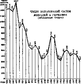
A. Vladimirov. Statistics and Geographical Distribution. The following figures give an idea of the extent of G. in the second half of the 19th century: in the French army in 1874 out of every 1,000 horses died from glanders-11.2, in 1875-9.2. With the introduction of systematic measures to combat it, it began to decrease sharply. In Germany, for example, the number of glanders diseases among horses decreased from 1220 in 1886 to 310 in 1909 (see figure). «91 During the World War, G. reached extremely large proportions, and in early 1915 on the eastern front of Germany, it was necessary to kill approx. 2,000 horses with glanders each month. With the organization of the central veterinary service and the implementation of appropriate measures, G. in the German army quickly decreased: in 1921 the number of diseases among horses decreased to 0.28 per 1,000, in 1922-to 0.07, in the following years-to 0.02. Diseases among people were observed in individual cases almost exclusively among persons having contact with sick horses. Thus, in Prussia, according to official data, cases of disease among horses and people were registered (Table 1; according to E. Lührs): Table 1. Horses Years People Years 1902 .... 1909 .... 1903 .... 1910 .... - 1904 .... 1911 .... 1905 .... - 1912 .... - 1906 .... 1913 .... 1907 .... 1919 .... 1908 .... 1920-22 . . In pre-revolutionary Russia, glanders was registered very highly. According to official data in Russia, horses with G were killed: 1908-17,618; 1909-16,948; 1910-16,345; 1911-21,774; 1912-28,075. Among people in pre-revolutionary Russia, approx. 200 cases of G were registered annually (Table 2). Table 2. Number of registered cases of G. in pre-revolutionary Russia for the period 1903 to 1912 (absolute numbers). i Number Number 1 Years of Years of disease disease 1903 ...... | 1904 ...... ; 1905 ...... ! 1906 ....... More than half of all cases were registered in the former Novorossiysk district (former Kherson, Taurida and Yekaterinoslav provinces and the Don Voisko region). In the USSR, according to official data, 106 cases of G were registered in 1926, 41 cases in 1927. By individual republics and on transport, the diseases are distributed as follows (Table 3): Table 3. Republics 1926 1927 Georgia ......... 43 34 The mortality rate in hospitals in pre-revolutionary Russia for the period from 1906 to 1912 averaged 69% (out of 666 treated in hospitals, 460 died). By individual years it ranged from 60% to 86%. Stefan (Odessa) establishes mortality in the acute form of G. at 100% (more than a hundred observations), in the chronic form-40-50%.
I.
Dobreitser. Pathological Anatomy. Glanders bacilli, having entered the animal organism by one means or another, cause primarily two kinds of reactive phenomena: a very rapid and abundant influx of phagocytes to the site of their penetration and the formation of a more or less significant edema around this site. Depending on the predominance of one or the other of the mentioned phenomena, as well as on the affected tissue, those path.-anat. elements arise which are most frequently encountered in G: pustules, nodules, diffuse infiltrates, lymphangitis and lymphadenitis, ulcers, abscesses, scars. Pustules are found both on the skin and on mucous membranes; on a limited area of the papillary layer of the skin, an exudate forms around the penetrating glanders bacilli, which bulges the epidermis in the form of a flat dome; the pustule is grayish-yellow in color, usually surrounded by a red hyperemic band and contains a viscous fluid rich in leukocytes and glanders bacilli [see separate table (p. 695-696), fig. 1-3]. Unlike smallpox pustules, on which they resemble very much, they do not have a 'navel'. Pustules on mucous membranes present a completely analogous picture.-Ulcers, the most characteristic path.-anat. changes in G, are formed due to the extraordinarily rapid and abundant influx of phagocytes, mainly epithelioid cells, to the site of penetration of glanders bacilli, which is facilitated by the strong edema caused by the metabolic products of Bac. mallei. Macrophages partly enter into direct active contact with the bacilli, but in a larger part form a passive barrier, which depending on the duration of the process turns into connective tissue, then into fibrous scar tissue and finally into calcified capsule. The youngest nodules present in appearance as if an ecchymosis with a small gray translucent elastic center. Fully developed nodules contain liquid or caseous pus; they are no longer surrounded by edema and their capsule is firmly fused with the surrounding tissue; often they merge into more or less extensive groups. The picture of diffuse infiltrates of interstitial tissue depends on their location (skin, lungs, etc.); they begin with stasis in dilated lymph vessels and impregnation of the surrounding tissues with fluid containing glanders bacilli and phagocytes. At the edges of the infiltrated area, the formation of connective tissue is observed very early, which may end in complete sclerosis of the entire area if the abundance and virulence of glanders bacilli do not give the upper hand to suppurative phenomena.-Nearby acute glanders processes, as a rule, reveal lesions of lymph vessels, first in the form of perilymphangitis, and then in the form of inflammation of the vessel walls and occlusion of the lumen by the clotted contents of the vessel. Subsequently, in chronic course of the disease, the affected lymph vessels together with the surrounding connective tissue may undergo such intense sclerosis that they turn into dense fibrous cords, which stand out particularly prominently in horses with cutaneous G.-Lymphadenitis in the area of affected organs is characterized by the fact that they begin with enlargement of the entire gland with signs of hyperemia and edema, but yet without the formation of definite foci. Such foci are outlined only later in the follicles. At the same time, the sclerotic process begins, not only encapsulating the foci in the follicles but also spreading to the interfollicular and surrounding connective tissue, as a result of which the glands take on the characteristic appearance of dense nodular packets, fused with adjacent tissues (fasciae, etc.).-Abscesses may form on the basis of pustules, single or merging nodules, diffuse infiltrates and lymph cords. In such a case, an oily pus, resembling synovial fluid, accumulates inside the phagocytic barrier, and upon its rupture outward, characteristic glanders ulcers with undermined edges and dense (greasy) granulating bottom are obtained. Sometimes from more deeply located abscesses, pus is discharged through tortuous fistulas, and in the lungs, after forming cavities, through the bronchi. Depending on the localization of the glanders process, a more or less characteristic path.-anat. picture is obtained. On the skin in humans, as primary phenomena at the site of infection, papules or nodules are found, which quickly undergo ulceration, which in acute cases has a tendency to progress and is often accompanied by the formation of diffuse infiltrations of erysipelas or phlegmonous character, and the participation of extraneous microbes is not excluded. Multiple eruption of glanders pustules of the above-mentioned type is observed in humans only as a secondary phenomenon, conditioned by the generalization of the infection. In horses suffering from chronic glanders, the sclerotic thickening of affected areas of the skin, especially on the limbs and on the head, can reach extraordinary deforming dimensions (elephantiasis malleosa).-Of the mucous membranes, those lining the upper respiratory tract are most frequently affected, especially the nasal cavity, both in humans and in animals. In the overwhelming majority of cases, not as primary affections, but in the order of generalization of the infection, pustules and nodules appear, undergoing disintegration and transformation into ulcers, which are less inclined to heal than on the skin, and, gradually spreading, destroy not only considerable areas of the mucous covering but also the adjacent cartilage and bone. Sometimes, however, more or less extensive star-shaped scars are encountered. The conjunctiva of the eye is not primarily affected, but sometimes serves as the portal of entry for G. The mucous membrane of the digestive tract is rarely affected.-In the lungs, Bac. mallei causes either primarily (carried from the intestine) or secondarily (entering the bloodstream from other foci) first of all the formation in the interstitial tissue of isolated or merging nodules. Along with young nodules, old encapsulated and even calcified ones may be present. When nodules form in the immediate vicinity of the respiratory tract, glanders bacilli can penetrate through their swollen mucous membrane and mix with the mucus secreted by them. Sometimes in the lungs, wedge-shaped lobular infarcts are formed due to occlusion of small or medium vessels. Sometimes the process involves an entire lobe, which turns into a hepatized mass with embedded purulent or caseous foci (pneumonia malleosa). In prolonged cases, the foci may turn into dense shrunken scar tissue. On the other hand, the formation of openly communicating with the bronchi cavities is possible, with the participation of extraneous microbes. Fat emboli have been observed in cases of death from acute glanders. The spleen is often enlarged, but at the same time it does not always contain nodules (in 42% in horses according to Bollinger). In laboratory animals, they represent a constant phenomenon.-In the liver, macroscopically visible nodules are encountered less frequently than in the spleen (14% in horses). They are located in the center of the lobules, which indicates their hematogenous origin.-The kidneys are affected even more rarely (10% according to Bollinger). In the sexual organs, primary glanders foci in females may be encountered in the vagina and perimetrium. Secondary affection of the testicle and epididymis is observed both in horses and in humans. When male guinea pigs are infected in the abdominal cavity, as a rule, bilateral specific inflammation of the tunica vaginalis already sets in after 2-3 days, extending to the epididymis and testicle (see below - Strauss' reaction).-The central nervous system is apparently very rarely involved in the glanders process.-Glanders changes in the muscular apparatus are observed more frequently in humans than in horses. They develop mainly in the intermuscular spaces of the lower extremities. Particularly characteristic here are more or less extensive encapsulated abscesses.-Glanders changes in bones have been described repeatedly in humans and in horses. In the latter, the ribs seem to be the favorite site, on the inner, concave side of which thickenings are formed, caused by ossifying periostitis around the softened glanders focus. But glanders nodules have also been found under the periosteum and in the bone marrow in the area of other bones; in humans, several cases of osteomyelitis malleosa have been described. In acute G. in tubular bones, the marrow takes on the appearance of embryonic tissue (possible cause of fat embolism in the lungs).-G. of the joints-almost a constant phenomenon in artificially infected small laboratory animals; it is also observed in humans. The course of G. has been studied in most detail in horses, in which it is most pronounced. Acute G. of horses begins after an incubation period of 3-5 days with a sudden sharp rise in temperature-sometimes up to 42°, often accompanied by chills. The animal immediately falls into prostration: weak pulse, accelerated superficial respiration, loss of appetite. Visible mucous membranes become strongly injected. After 1-3 days, specific local symptoms appear. On the nasal mucosa [see separate table (p. 695-696), fig. 4 and 5], ecchymoses erupt, on the site of which pustules quickly form, bursting after a few hours and turning into ulcers.
From this moment on, there is abundant discharge from the nostrils, first liquid, serous-purulent with an admixture of blood, but soon, as the ulcerative process spreads, becoming increasingly bloody-ichorous; to it are added clots of fibrin and pieces of necrotized mucous membrane; blocking the nostrils, it makes breathing difficult, which becomes snuffling, wheezing. With the spread of the process to the larynx, suffocation may occur. Simultaneously with the symptoms from the nose, glanders changes may also develop on the skin. In various parts of the skin, painful edematous tumors appear, which however soon subside, giving way to abscesses, chancroidal ulcers, and indeterminate purulent surfaces. The nearest lymph vessels and lymph glands are involved in the glanders process with similar phenomena: first edematous swelling, then suppuration, often with breakthrough to the outside and formation of typical glanders ulcers. Animals quickly lose weight, losing up to 40 kg per day. Usually at the end, lobar pneumonia develops in them and they die on the 8-30th day of the disease. Cases of recovery from acute G. or its transition to chronic form are not observed. The chronic form represents in horses approximately 90% of all cases of G. disease. In it, it is difficult to determine the duration of the incubation period, as it b. ch. for a long time it proceeds completely hidden without any noticeable signs. The duration of the manifest period of chronic G. in horses is apparently measured in months and years, but it is difficult to judge it, as in all civilized countries, glanders-infected animals are immediately destroyed. Due to the slow course, the development of pathological processes can be so isolated in their localization that separate types are obtained, distinguished by practitioners under the names of cutaneous, nasal, or pulmonary G. In reality, complete pathological-anatomical isolation of localization is rare. The division into types is justified from a clinical point of view. In humans, G. proceeds in the overwhelming majority of cases in acute form, sometimes even more violently than in horses, with 2-3 days of incubation and fatal outcome in 6-8 days; usually the disease lasts 2-4 weeks. General painful phenomena occur after the specific changes at the site of infection have reached a certain degree of development (ulcers, lymphangitis, etc.) or in the absence of visible local processes, after indefinite precursors. The following are observed: moderate elevation of temperature, rarely accompanied by chills, headache, pains in muscles and joints, sometimes swelling of joints, to which may be added cough with admixture of blood to sputum and other symptoms, which often lead to error and are taken for signs of beginning typhoid fever, rheumatism, influenza, lobar pneumonia, sepsis, etc., until the typical changes for G. on the skin, on the mucous membranes or in muscles suggest the correct diagnosis. On the skin in different places, especially often on the face, the above-mentioned pustules appear (which at first glance have some resemblance to smallpox), turning into chancroidal ulcers. In the connective tissue and in the muscles, abscesses form, sometimes exposing even tendons and bones. On the mucous membranes, pustules and ulcers also appear, often but by no means constantly in the nasal cavity, from where the process can spread, on the one hand, to the further respiratory passages and the oral cavity, and on the other hand, to the conjunctiva of the eye and the meninges. The lungs are also often affected, and finally with generalization and the onset of glanders bacteremia, all parenchymatous organs, serous membranes, etc. are affected. The outcome of acute G. in humans is always fatal. Chronic G. in humans in many respects is analogous to chronic G. of horses. All pathological processes proceed slowly and are interspersed with regressive phenomena: firm encapsulation of nodes, cleansing and healing of ulcers, etc. When infection occurs through the skin, the course of the disease often resembles that of horses, for a long time maintaining a local character: ulcers, lymphangitis, lymphadenitis, sometimes complicated by erysipelas. But in the further course of the disease, the G. bacteria mostly penetrate into the bloodstream, which causes elevation of temperature and causes the appearance of new foci: intermuscular abscesses, inflammation of joints, foci in the lungs, nodules and small ulcers on mucous membranes, with the lesion of the nasal mucosa, contrary to the indications of many textbooks, not belonging to the constant manifestations of chronic G. in humans, but being absent in at least half of all cases. Periods of abatement and exacerbation are very uneven, as a result of which the fever curve has a completely irregular shape with elevations of varying height and duration and with intermissions. Dragging on for many months and even several years, chronic G. in humans does not so rarely, as was previously thought, end in clinical recovery. In most cases, it takes a fatal outcome due to complications with other infections or due to transition to acute form. The clinical diagnosis of G. is often very difficult in the absence of any characteristic external manifestations of it. This applies also to the 'hidden' G. of horses and to the initial form of G. in humans. In the latter case, it was repeatedly confused with syphilis and with tbc. It is extremely important to establish in the anamnesis whether the patient had contact with horses. But even in the presence of visible or accessible to research changes, it is often impossible to do without bacteriological confirmation of the diagnosis. Simple bacterioscopy is not sufficient even for a preliminary conclusion, as B. mallei has no characteristic morphological signs. Cultures are easily obtained when sowing material from fresh closed foci. To isolate B. mallei from blood is possible only during bacteriemic feverish attacks. The organism of susceptible animals often represents a more sensitive reactivity to B. mallei than artificial nutrient media. Therefore, control inoculations are used not only for more precise determination of the isolated cultures, but also as an independent diagnostic method. The most suitable laboratory animals for this purpose are guinea pigs. They differ by almost absolute susceptibility to glanders, which in them proceeds as a rule in acute form. The diagnosis can be still more accelerated if for inoculations males are taken, as in them (as shown by Strauss) after intraperitoneal infection already in 2-3 days begins the typical swelling of the descended into the scrotum epididymis and testicles with accumulation in them of a purulent, rich in glanders bacilli, curd-like secretion, accessible to further bacteriological research (Strauss' reaction) (see separate table, Figure 1. Three large glanders pustules - on the cheek, at the angle of the lower jaw and under the ear lobe. Figure 2. Glanders pustules and ulcers of the face. Figure 3. The same patient - on the skin are scattered pustules, near the elbow - hemorrhage, marked swelling of the left knee. Figure 4. Nasal septum of a horse with single and confluent glanders ulcers. Figure 5. Nasal septum of a horse with radiating and tree-like branching scars. Figure 6. Strauss' reaction 5 days after inoculation into the abdominal cavity. (According to Savvaitov) fig. 6). To cause this reaction in guinea pigs is possible also by subcutaneous infection and even by rubbing the test material into the shaved skin. The latter two methods of inoculation, usually used when working with contaminated materials, prolong the incubation period and are not needed when the experiment is set up at once on several guinea pigs, as they in general are little sensitive to other infections. The use of cats for diagnostic inoculations of G. was the basis of the so-called 'Russian accelerated method', developed by Russian veterinarians and formerly often practiced by them. It consists in that several cats, if possible young, are infected with the test material into an artificial skin pocket on the nape. Already in two days one of the cats is killed and with its muscle tissue from liver and spleen, sowings are made onto potato medium. Thus, it was often possible to make a diagnosis already on the 4th day. In case of failure of the first attempt, they continued to kill the cats of this series, as they did not die themselves from the infection. This method is used at present only in exceptional cases when guinea pigs are not available, as manipulations with cats are inconvenient, and with glanders cats also unsafe. The suitability for diagnostic inoculations in G. of other small animals, as dogs, white domestic mice, field mice, ground squirrels, has been proved in practice by different authors, but the methods recommended by them have not found wide laboratory application. Of immunoreactions in the diagnosis of G., the first was introduced in 1890 the allergic test by means of mallein (see), a preparation which by the method of preparation and by the methods of application is analogous to tuberculin. In veterinary practice earlier subcutaneous malleinization was widely applied, at present intradermal, conjunctival (eye) and interpalpebral are preferred.
In humans, all these methods are inconvenient to apply, but mallein has been repeatedly used with success for the skin reaction (according to Pirquet). Serological diagnosis of Glanders began in 1897, when Vladimirov found that the agglutination reaction obtained with sera from both diseased and healthy horses acquires diagnostic significance only after diluting it at least 1,000 times. This method was applied in practice in horses and humans until 1909, when it was replaced by the more reliable complement fixation reaction, which was thoroughly developed methodologically by Schutz and Miessner. The application of other serological reactions—precipitation (Dedyulin, Vladimirov; 1900), determination of the opsonic index (Fursenko; 1908), conglutination (Pfeiler and Weber; 1912)—showed their unsuitability for widespread practice. Treatment. As for the therapy of Glanders, innumerable attempts to find more or less reliable therapeutic means to this day have not been successful. In states conducting preventive measures against Glanders, the treatment of glandered horses is prohibited by law. In humans, symptomatic treatment is applied in all cases: excision or cauterization of external nodes, opening of abscesses, cauterization of ulcers (with zinc chloride, etc.), disinfection of the nasal and oral cavities (with hydrogen peroxide, etc.). All surgical interventions are accompanied by necessary precautions so that glandered bacilli from the foci being destroyed do not penetrate further into the body through wound surfaces (abundant moistening of the latter with tincture of iodine). Among medicinal substances, mercury rubs, iodine, arsenic, collargol, chemotherapeutic preparations (salvarsan, Bayer "205"), and others were used in chronic Glanders in humans. For the most part, they did not have a therapeutic effect, and those cases in which a general improvement in the patient's condition was observed are not very convincing due to possible remissions in chronic Glanders without medical intervention. Specific biological preparations also promise little. An antitoxic or antibacterial serum against glanders has not been developed, and the use of active preparations (mallein, faraous, and vaccines killed by various other methods) appears unsafe due to the allergic condition of the chronically ill body and the possible exacerbation of the process during the reaction. Apparently, in all cases of human recovery, the regimen (nutrition, rest) played the main role, promoting the uninterrupted action of the body's cellular and biochemical protective means. Prevention. Personal preventive measures against glanders infection for doctors and laboratory personnel come down to the general precautions that are mandatory when working with any acute infections. Persons having contact with horses in areas not free from Glanders must be promptly and repeatedly properly instructed regarding Glanders, possible routes of its transmission, and measures of personal precaution when handling horses suspected of being infected with Glanders. State prevention aims to eradicate Glanders in horses. Until this goal is fully achieved, precisely regulated veterinary-sanitary measures are carried out and early clinical diagnosis, and above all, systematic detection of hidden glanders by means of mallein and serum reactions with the slaughter of diseased animals and destruction of their carcasses. The successes in the fight against Glanders in the USSR are based on the fact that in areas unfavorable for Glanders, by order of central government bodies, mass malleinization of the entire horse population is carried out (a measure not implemented in capitalist countries), and reacting horses, as well as those with only suspicious signs, are subject to immediate destruction. As for reacting horses without clinical signs, the so-called "mallein reactors", a measure is applied to them, which again can only be implemented in a socialist state and has great economic significance. Such horses are grouped and isolated in special "mallein farms", where they, under constant veterinary supervision, are used for normal agricultural work, with the understanding that at the slightest manifestation of Glanders they will also be subject to destruction. A. Vladimirov.
Related articles
Mentioned in
Cite this page
“Glanders.” Soviet Medical Encyclopedia. English translation of Bolshaya Meditsinskaya Entsiklopediya, 1st ed. (Moscow, 1928–1936), ed. N. A. Semashko. https://sovietmedicalencyclopedia.pages.dev/article/glanders/