Dohle Inclusions
Historical document, translated for reference. It reflects medical knowledge of the 1920s–30s and is not medical advice.
Summary
Dohle inclusions are distinctive structures found in neutrophilic leukocytes, described by Dohle in 1912. They appear as grayish-blue spots and are associated with various infections, though not specific to any particular disease.
Encyclopedia article (1928–1936)
DOHLE INCLUSIONS (Dohle), distinctive islands described by Dohle (1912) in the body of neutrophilic leukocytes and appearing when stained by Giemsa as round, pear-shaped, angular, etc., spots of grayish-blue color. When stained with methylene blue (Löffler, Manson, etc.), they are colored blue, and with methyl-green-pyronin according to Unna in a reddish color. They are often described as condensed areas of protoplasm; they are characterized by the absence of a reaction to oxidase. The appearance of these formations is apparently due to pathological changes in the protoplasm of leukocytes, mainly associated with various infections. The initial assumption that Dohle inclusions should be interpreted as parasitic formations or products of their degeneration (some, for example, saw degenerated spirochetes in them), as well as the opinion that they are specific to scarlet fever, were not confirmed. They are found not only in other infectious diseases (mainly septic), but occasionally also in healthy people. Similar formations in the form of irregular strands are found in most neutrophils of normal frogs.
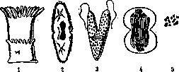
Figure 1. Transverse division of an anemone. Figure 2. Transverse division of a slipper infusorian. Figure 3. Longitudinal division of the flagellate Euglena. Figure 4. Diagram of cell division and karyokinesis. Figure 5. Resting and dividing mitochondria of infusorians.
into two parts, or daughter individuals, but in Protozoa cases of multiple division (schizogony) or simultaneous disintegration of an individual into many daughter individuals are not uncommon. Division usually consists of transverse constriction of the body (see figures 1 and 2), but there are entire groups of animals where it proceeds longitudinally (some anemones, all flagellates; see figure 3).-Division of organs in the bodies of animals, strictly speaking, is observed only in the embryonic state, in the rudiments of organs, and moreover mostly has a pathological character. The best example may be the embryonic splitting of finger rudiments, which leads to polydactyly, or multiplicity of fingers. Apparently, by this path of embryonic splitting, the multiplication of the number of nephridia in the segments of some Oligochaeta occurs, but here this phenomenon becomes normal for the animal. Division of cells is a necessary condition for the development of multicellular organisms, the entire body of which arises as the final result of the division of a single original egg cell. Division is accomplished by gradual constriction of the cell body (see figure 4), and usually there is a pause between two successive divisions, during which the daughter cells grow to the size of the maternal one. Only in the early stages of development of Metazoa eggs, during the period of cleavage, cell divisions follow one another rapidly without intermediate growth pauses. Most of the cell organelles also undergo division during the last division. This is especially clearly evident in the nucleus, chromosomes, and centrosome (see figure 4), the division of which usually somewhat precedes the complete separation of the cell body. At present, the ability to reproduce by division has also been proven for many other cell organelles. Thus, many researchers describe the division of chondriosomes or mitochondria (see figure 5). These very constant cellular inclusions elongate and become constricted either during cell division itself or in the intervals between divisions. The same should be said regarding the various plastids of plant cells. As early as Schimper (1881) and A. Mayer (1883), came to the conclusion that plastids never arise anew in cells, but develop by growth and division of pre-existing plastids. This has been proven for such differentiated forms of plastids as chloroplasts, especially in lower plants. Other forms of plastids apparently arise through the growth of small colorless leucoplasts, and those in turn multiply by division. In recent times, reproduction by division has even been discovered for such complex cell organelles as stinging capsules (Chatton in the flagellate Polykrikos; 1914). Finally, there are indications that new contractile vacuoles of infusorians, during division of the animal, may not be formed anew but by budding off a portion of a pre-existing vacuole. Thus, the ability to divide is proven for almost all cell organelles.
Related articles
Cite this page
“Dohle Inclusions.” Soviet Medical Encyclopedia. English translation of Bolshaya Meditsinskaya Entsiklopediya, 1st ed. (Moscow, 1928–1936), ed. N. A. Semashko. https://sovietmedicalencyclopedia.pages.dev/article/dohle-inclusions/