Diphtheritis
Historical document, translated for reference. It reflects medical knowledge of the 1920s–30s and is not medical advice.
Summary
Diphtheritis is a general pathological process that can occur in various inflammatory conditions of mucous membranes, not exclusively in diphtheria. The term should not be used as a synonym for diphtheria itself, as it lacks specific etiological and nosological definition.
Encyclopedia article (1928–1936)
DIPHTHERITIS (from Greek diphthera-membrane), a term often incorrectly used to denote the infectious disease diphtheria; in reality, it represents a shortened designation for the general pathological process, namely - diphtheritic inflammation, which can develop both in diphtheria and in other etiologically different inflammatory lesions of mucous membranes (e.g. in the intestine-in uremia, dysentery, in the uterus-in septic endometritis). One can also speak of D. of a wound, if it is covered with a diphtheritic | | coating, which in this case may not contain diphtheria bacilli (in case of a wound being infected with diphtheria bacilli, one already speaks of diphtheria of the wound). Finally, diphtheria in some cases is accompanied not by D., but by ordinary catarrhal inflammation. Thus the term D. in etiological and nosological respects does not represent anything definite, and therefore should not be used as a synonym for diphtheria. DIPHTHERIA. Contents: Etiology....................
400 Statistics and geographical distribution
445 Diphtheritis (from Greek diphthera - skin membrane, film), an acute infectious, epidemic disease caused by diphtheria bacilli of Löffler (Loftier), which are most commonly localized primarily on the mucous membrane of the pharynx, nose, or nasopharynx, less frequently on the mucous membrane of the larynx and trachea, and very rarely on the conjunctiva of the eye and the mucous membrane of the vagina. The inflammatory process is accompanied by the formation of false membranes (plaques), which are tightly fused with the underlying necrotized tissue of the mucous membranes. The same process can primarily affect wound surfaces and even the surface of a hajji. But wherever the process is localized, the disease, in addition to primary local disorders, is characterized by more or less severe toxemia due to the entry into the body of toxin secreted by diphtheria bacilli. Etiology. Diphtheria bacilli (Bacillus diphtheriae Klebs-Loffieri, Corynebacterium diphtheriae) were first described in 1883 by Klebs on sections from false membranes. In 1884, Löffler isolated them in pure cultures. They are the causative agents of the disease. Diphtheria bacilli belong to the group of bacilli united under the general name Corynebacterium, as proposed by Lehmann and Neumann, and are characterized by the presence of granularity in the protoplasm and the formation of club-shaped swellings on one or both ends of the rod. The same group includes diphtheroid, tuberculous, and fusiform bacilli. According to the American classification, the name Corynebacterium is given to diphtheria and similar pseudodiphtheria bacilli. According to the degree of polymorphism and the expressiveness of Babes-Ernst's polar grains and their relation to glucose, galactose, and sucrose, the following six species of the genus Corynebacterium are distinguished. 1. C. diphtheriae - polymorphic, have typical grains at the ends of the rods, decompose glucose and galactose, are pathogenic for humans and virulent for guinea pigs. 2. C. pseudodiphtheriae - completely similar to the first species, but not virulent. 3. C. Hofmann-Wellenhof's - shorter, non-polymorphic rods with very weak grain formation, non-virulent, do not decompose the three carbohydrates listed above. 4. C. segminis - have typical Babes-Ernst grains, are little polymorphic, do not decompose carbohydrates, are non-virulent. 5. C. xerosis - also have typical grains and are little polymorphic, but decompose glucose and sucrose, are non-virulent. 6. C. acne - have some similarity to diphtheria bacilli, but are not always typical, decompose all three carbohydrates and differ from the five species listed above in that they are anaerobic. Diphtheria bacilli have very different lengths, from 1-2 μ to 6 μ. French authors note short, medium, and long races of diphtheria bacilli. The thickness of the diphtheria rod, on average about 0.8-1.0 μ, increases unevenly toward one or both ends of the rod; sometimes the thickening is negligible, but sometimes swellings are formed, which gives the rods, especially in older cultures and on less favorable nutrient media, the shape of a club or a gymnasical dumbbell. In photographs of colonies on agar or coagulated blood serum, the rods are arranged in the form of intersecting spread fingers. When stained, the rods often look like two or more segments with weakly staining interstices. Club-shaped swellings, segmentation, as well as the rarely branched forms of the rods are observed mainly in older cultures. Such forms are called involutionary. Diphtheria bacilli are immobile; they do not have flagella, capsules, or spores; they stain by Gram's method, but with prolonged treatment with alcohol they are easily decolorized. With special staining by Neisser's method (acetic acid blue with additional staining with bismarck brown), on a light brown background, the dark blue Babes-Ernst grains stand out sharply at the ends of the rods, which are important for recognizing true diphtheria rods from false ones [see separate table (pp. 423-424), Fig. 5 a, b, and c]. Diphtheria bacilli develop much better with free access of oxygen than under anaerobic conditions. The optimum growth is at t° 35-37° and at pH of the medium = 7.8. When growing on broth, a rather coarse, brittle film is formed, which easily falls to the bottom of the vessel; by the end of the day, the reaction of the broth approaches acidic (due to the decomposition of carbohydrates present in the nutrient medium) so that by the end of 2-3 days it returns to the original alkalinity, and then exceeds it. On agar, small, round, translucent colonies are formed, giving on the slanted surface of agar a delicate, bluish plaque, which in older cultures can acquire a more or less pronounced yellow-brown tint. On coagulated blood serum, the colonies are somewhat larger and coarser, of pearly-gray color. They do not liquefy coagulated serum and gelatin; they grow well on milk without changing its appearance. On coagulated serum, diphtheria bacilli retain their viability and toxin-forming capacity for a long time (6 or more months). They perish on a liquid medium at 60° for 10 minutes, at 70° for 5 minutes; in a dried state they withstand the action of heat at 95-100° for several minutes, and in semi-dried films for an hour. Diphtheria bacilli dried in a thin layer perish in scattered light in a few days; direct sun rays kill them in a few hours; but in a dark place and covered with a layer of dried mucus or film they remain viable for several months. At temperatures below 0° they withstand well, remaining in a frozen state for a long time. Solutions of mercuric chloride 1:1,000 and phenol 5:100 kill diphtheria bacilli in 20 seconds, 3% lysol in 10 minutes, alcohol in 1 minute. Diphtheria bacilli are very virulent for guinea pigs, rabbits, birds, and cats, and much less so for horses, cattle, goats, dogs, mice, and rats. There is a known parallelism between the virulence of diphtheria bacilli for guinea pigs and their pathogenicity for humans, consequently for determining the latter, inoculations of cultures isolated from humans are used in guinea pigs (methods of Igleton and Baxter with pure diphtheria cultures, of Gevens and Powell without isolation of pure cultures: injection of 0.2 cm3 of culture from coagulated serum into the skin of a guinea pig). It is very characteristic of diphtheria bacilli their secretion of specific diphtheria toxins, which are obtained by growing cultures on broth of proper composition and alkalinity for 7-10 days. These toxins, freed from the bodies of bacilli by filtration (through Chamberland or Berkefeld candles), are used for immunizing horses with the aim of preparing therapeutic serum, as well as for vaccinating people to give them immunity to diphtheria (see below). To characterize diphtheria toxin according to P. Ehrlich, the following three of its doses must be established by experiments on guinea pigs: 1) the minimum lethal dose (dosis letalis minima, Dlm) for guinea pigs weighing 250 g, killing them on the 3-5th day; 2) the lethal limit, or Lt dose (Limes Tod) - the amount of toxin which, mixed with one unit of antitoxin (AE) (one antitoxic unit, according to Behring, neutralizes one hundred minimum lethal doses of toxin) kills a guinea pig weighing 250 g in the same time as Dlm, i.e., in 3-5 days; 3) the zero limit, or L0 dose (Limes Null) - that amount of toxin which, mixed with one AE, injected under the skin of a guinea pig weighing 250 g, does not cause at the site of injection the slightest infiltration. Diphtheria toxin is thermolabile: it is destroyed at t° above 60°; at 90° it is already destroyed in 10 minutes, at 100° even faster. But its weakening already occurs slowly at room temperature (20-22°), even at the temperature of a refrigerator (+6°). The addition of 0.5% phenol or toluol (so that the toxin is covered with a layer of 2-3 fingers) contributes to the long preservation of toxicity, especially on ice and in a dark place; formalin in the amount of 4 pro mille and more, on the contrary, contributes to the weakening of the toxicity of the toxin. Ehrlich first proved that the weakening of the toxicity of diphtheria toxin does not always go parallel with the loss of its antigenic properties (see Antigens), i.e., despite the decrease in toxicity [which is expressed in the need to increase the dose of toxin to kill a guinea pig (increase of Lt)], it continues to neutralize the same amount of antitoxin (L0 dose does not change) and in the same degree causes the formation of antitoxins when immunizing animals (transformation of toxins into toxoids, see Anatoxin). Bacillus carriage. As early as Löffler in the beginning of his research on diphtheria discovered diphtheria bacilli in the mucus from the pharynx of healthy people. At the same time, as further research showed, diphtheria bacilli are found in the pharynx and nose of healthy persons, not only those who had direct contact with the sick, but also those who had no contact with sources of infection. For those around the patient, bacillus carriage ranges from 8% (Kober) to 50% (Welch), and for those who had no contact with the sick - from 2.5% (Kober) to 24% (Muller), but the number of carriers of virulent bacilli is much smaller.
Thus, according to data from the British Medical Research Council, among 610 people who had close contact with patients with D., 15% were found to be carriers, all of whom were virulent; among 10,883 people who had more distant contact-8% carriers, of which only 1% were virulent, and finally among 7,974 people who had no contact with patients-3.4% non-virulent carriers and only 0.6% virulent. Thus, virulent bacillus carriers are much fewer than avirulent ones. This gives rise to the desire to limit practical measures in the fight against D. only to measures aimed at virulent bacillus carriers. Theoretically, this desire seems quite correct, since it is obvious that only bacilli of the species Corynebacterium diphtheriae (see above), pathogenic for humans and virulent for guinea pigs, can play a certain role in pathology. However, detailed studies by Bashenin and Birger with their colleagues showed that with repeated determinations of virulence of diphtheria cultures from the same individual, conducted over a long period of time, heterogeneous results are obtained, sometimes positive, sometimes negative, without any discernible reasons explaining such irregularity. Moreover, the aforementioned researchers noted that in some closed children's institutions, no cases of the disease D. are observed for a long time, despite the constant presence of virulent bacillus carriers, while in others, on the contrary, cases occur in the presence of only avirulent bacillus carriers. Of course, this contradiction is explained by the imperfection of the method for determining the virulence of diphtheria bacilli. However, one has to put up with the imperfect method, which gives a certain percentage of errors, and limit sanitary measures aimed at virulent carriers, since the isolation of all diphtheria bacillus carriers is practically impossible even in closed children's institutions. Patients with D. continue to retain diphtheria bacilli in the throat and nasal mucus for quite a long time after their recovery. Indeed, in almost half of the recovered patients, bacilli are found in the throat mucus within 2 weeks, in a quarter within 3 weeks, and in 10% within 5 weeks from the onset of the disease. Later, the number of carriers decreases sharply, but in individual cases, bacillus carriage was observed for 4-9 months. Bacillus carriage has been described even for several years (Pochon, Neisser). Fortunately, such prolonged bacillus carriage is a great rarity. Unfortunately, there are no reliable means to free carriers of diphtheria bacilli. The best results are obtained by the extirpation of enlarged tonsils (before the operation, it is necessary to inject diphtheria antitoxin to prevent the wound from being infected with diphtheria). It should be noted that patients and convalescents mainly secrete virulent diphtheria bacilli, and only later, starting from the 5-6th week from the onset of the disease, avirulent diphtheria bacilli begin to predominate in their mucus. This determines the main role in the spread of diphtheria of patients and convalescents and the comparatively minor role of healthy bacillus carriers. Discharge from the hospital of convalescents from D. is usually based on two or three bacteriological examinations that gave negative results. When it is impossible to rely on bacteriological research, discharge is allowed after 2 weeks from the disappearance of all clinical manifestations of the disease. (See also Bacillus carriage.) Epidemiology. D. has a contagious nature, spreading either directly from person to person or through the medium of objects contaminated with diphtherial secretions (mucus from the throat and nose, shedding membranes). Rare cases of diphtheria epidemics arising through food products, in particular through milk (Neisser, Sobernheim and Nagel), have been described. Spiegelberg and Litterer (Spiegelberg, Litterer) found virulent diphtheria bacilli in chickens, and the latter author observed the successful transfer of bacilli to chickens both from chickens themselves and from a sick person. The presence of diphtheria bacilli in horses has also been proven (Kliewe, Westhues). However, only contact transmission of the infection is of practical importance, as a result of which diphtheria epidemics never give such sudden and widespread outbreaks as water epidemics of typhoid fever or cholera. The spread of diphtheria bacilli through the air in droplets splashed by patients with D. and bacillus carriers during screaming and coughing has been proven by experiments by Jellinek (Jellinek; 1924), who found diphtheria bacilli on cough plates at a distance of 35 cm from a bacillus carrier. Diphtheritis was isolated as a separate nosological unit only in 1826-28 by French researchers Bretonneau and Trousseau. However, descriptions of 'malignant angina' affecting mainly children and giving epidemic outbreaks at times are found even in the Talmud and in the earliest writers of the Christian era (Aretaeus, Aetius and others). In the XVI and XVII centuries, D. was undoubtedly widespread in Europe, especially in 1618-42, as well as in the XVIII century. The first reports of D. in America date back to the XVIII century. At the beginning of the XIX century, D. was extremely poorly developed in Europe. It was so little known that even 15 years after the publication of Bretonneau's works, the famous Viennese pathologist-anatomist Rokitansky (Rokitansky) considered D. a 'French invention'. Only in 1850 was there no doubt about the existence of D. in Europe. From then until 1890, epidemics of D. rapidly increased in both Europe and America, after which the epidemic curve began to decline. According to the research of Kisskalt, the spread of D. in Europe in the XIX century proceeded from the east. In Russia, another epidemic wave of D. was observed, reaching its maximum by 1910. Then there is a continuous decline in the curve, reaching its lowest point in 1919. In the last 2-3 years, an increase in the diphtheria epidemic has again been noted in Europe, America and other countries, expressed in an increase in the number of cases (especially in an increase in their severity). But the rise of the epidemic curve is still very slow. Thus, the epidemic curve of D. periodically rises and falls under the influence of some still unexplained conditions. There are attempts to explain this interesting phenomenon, observed not only in diphtheria but also in other infectious diseases. The influence of seasonal factors is confirmed by the seasonal fluctuation of this curve, which rises in the autumn-winter season. Gottstein seeks an explanation in the change of generations of more resistant to D. with less resistant ones. Susceptibility to D. depends on age. The decrease in susceptibility to D. with age is explained by the contact of the mass of the population with the causative agent of the disease through bacillus carriers, as a result of which constant unnoticed immunization occurs. Polish researchers (Hirszfeld, Brokman) found that a child's susceptibility to D. also depends on his belonging to one or another blood group (see Hemagglutination). Thus, if a child belongs to the same blood group as one of his parents who has a positive Schick reaction, then he also has a positive reaction and is therefore susceptible to D. and remains so for life, despite active immunization or even a past case of D., which gives him only temporary immunity. On the contrary, if a child belongs to the blood group to which the parent with a negative Schick reaction belongs, then, even if he has a positive Schick reaction, he easily acquires immunity to D., both through immunization and independently upon reaching maturity. Thus, the role of constitution emerges.
S.
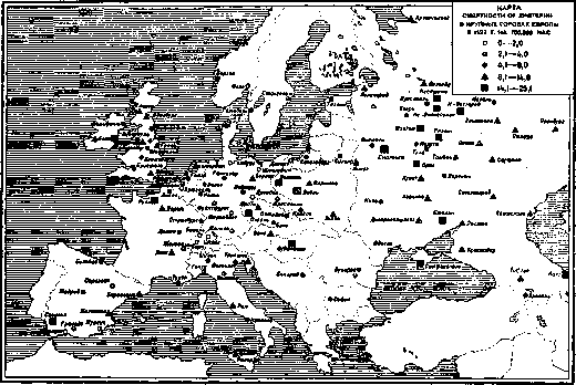
Korshun. Statistics and Geographical Distribution.- 1. Mortality. Statistics from different countries show a sharp decrease in mortality from D. after the introduction of serum treatment (1894) and a gradual decrease in subsequent years. Table 1 shows mortality rates from D. for various years in different countries (on average per year per 100,000 population). Table 1. ~----____^^ Years Countries ----____ 1861-65 1871-75 1881-85 1891-95 1896- 1900 1901-05 1906-10 1911-13 1914-18 1919-23 1924-27 England....... 63.6 32.0 32.2 30.6 22.2 16.3 12.7 15.2 11.7 7.2 Belgium . . 119.1 98.2 78.4 49.1 27.1 21.5 17.5 16.8 - 10.1 3.6 * Germany . - - 114.0 46.1 33.0 23.7 21.0 29.2 10.7 5.0 Holland - 40.6 31.0 15.6 13.6 7.8 7.1 8.3 6.6 3.1 Denmark . . 80.5 32.9 59.5 131.3 31.0 13.0 9.9 8.1 8.7 12.7 4.8 Spain . . - - 45.6 25.1 20.1 23.5 20.4 12.4 7.1 Italy . . - - 50.5 24.2 13.9 15.6 10.3 12.4 7.8 7.1* Norway . - 72.6 69.1 14.5 13.2 21.2 18.6 12.8 20.6 2.9 * Switzerland - 64.8 46.2 29.2 21.0 14.5 11.0 11.6 4.1 Sweden . . 71.9 37.0 84.6 60.8 43.9 33.8 15.3 13.8 16.9 15.7 3.1 Scotland 81.6 60.7 44.2 36.7 21.2 17.0 17.9 17.9 16.4 12.6 9.8 Japan . . - - 15.2 9.6 9.8 9.8 8.2 6.5 6.5 - - .- 29.7 22.6 15.6 15.4 8.1 Australia . - - - 16.1 12.5 12.0 5.9 * In Italy i ж op вегии for 1924-2 5 ГГ. И I Belgium for 19 24-25 and not only with D., but also with some other epidemic diseases. Bruckner connects the rise and fall of the epidemic curve with 35-year periodic climate changes, with the increase in the diphtheria wave apparently coinciding with humid-cold climate waves. The dependence of the spread of D. on the atmosphere- For all these countries, a decrease in mortality from D. after 1894 is noted. In some of the countries (England, Germany, Holland, Denmark, etc.), an increase in mortality from D. can be noted during the period of the world war or in the years immediately following it. The last period (1924-27) for all countries is characterized by an unprecedentedly low mortality from D. Figures 1 and 2 show the curves of mortality from D. by year in England and Switzerland (from 1876), Germany (from 1892), and the U.S.A. (from 1899). In England for the period from 1876 to 1902, regular periodic increases and decreases in eoj IIIIIIIIIIIIIIIMII 1 Mortality from D. in England and Switzerland (per 100,000 population) / в!,7 „ ^ j 1 K _ _П____________________1+1. 1 Цвййцация д 34 \ 1 1/4 1л if-- \ .....___ л»г..н« .qV,J г \ \/ y '\#ъ_____ BC Ч^ . v^tf^S Г \K\j * ------------------ Switzerland H ". - _ „.....___:|::£:::^:1... \Г f 5il 3 5T9tJ.j6ls.tl S 5 T $ t 1 5 i 7 UfI 5 I ^'Шб 18S0 1890 1900 1910 1920 Figure 1. mortality from D. are noted, with the latter during this period never falling below 25 per 100,000 population. The maximum increase was observed in 1893 (38.9). Starting from 1903, the curve changes and gives a steady step-like decrease (in the process of decrease there are temporary increases). A slight increase is noted for the period from 1914 to 1920, after which there is a further decrease in the curve -п И 1 1 1 N ! 1 1 1 1 1 1 1 1 -1 1 1 1 1 1 1 1 1 1 II M 1 1 1 - 13fr 120- ~^1 етгг Mortality from D.in Germany and U.S.A.>1 □- Уп [ 1 \ __[ ]_i ___________ Germany \ л t ^ *> ерики 3S ^ \ < *\ 7, <н "K Го ь t я. |7 !> i J, г, в лы j 492 1900 1910 19^0 Figure 2. to 7 per 100,000 population. Mortality curves in other countries are of roughly the same character. In Switzerland, mortality rates from D. for the period from 1876 to 1901 were significantly higher than in England (fluctuations from 27 to 81 per 100,000 pop.); a steady decrease in the curve begins from 1902, and from 1922 mortality from D. in Switzerland is lower than in England. In Germany for the period from 1892 to 1919, mortality rates from D. are higher than in England and Switzerland; a sharp and subsequently steady decrease in the mortality curve begins from 1895. A pronounced increase in mortality is observed in 1915-17, after which there is a rapid decrease in the curve, and mortality rates equalize with those in England and Switzerland. The mortality curve from D. in the U.S.A. almost repeats that of Germany both in the height of the indicators and in its course. It lacks the increase during the years of the world war.-The order of arrangement of individual countries by the level of mortality from D. is different before and after the world war. In Tables 2 and 3, individual states are arranged in descending order by the level of mortality from D. for the periods from 1901 to 1913 and from 1922 to 1926 (see also Figs. 3 and 4). On the basis of an analysis of pre-war materials on the geographical distribution of D. (for 1890-94 and 1906-10), S. A. Novoselsky comes to the conclusion that "the spread of diphtheria in Europe increases in the direction from west to east." Materials for 1901-13 (Table 2) generally confirm this conclusion (with the exception of Romania, where mortality from D. is registered relatively low). A different geographical distribution of D. is observed in recent years (Table 3); the highest mortality rates are observed in western European countries: in Spain, England, Scotland and Ireland, Italy. Central and eastern countries: Austria, Czechoslovakia, Yugoslavia, Bulgaria, Poland and Romania give significantly lower mortality rates. Among non-European countries, the U.S.A. both before the world war and in recent years occupy one of the highest places in terms of their mortality rate from diphtheria; Japan, New Zealand give average mortality rates. Table 4 shows mortality rates from D. in some major European cities for the period from 1880 to 1927 (per 100,000 population). Table 5. Mortality from D. This table gives an idea of the decrease in mortality from D. after the introduction of anti-diphtheria serum (in Leningrad and Moscow less pronounced than in Western European cities) and of the low figures of this mortality in recent years (see Fig. 5). Table 5 provides a comparison of mortality from D. for 1925-27 in a number of major cities in different parts of the world. Among European cities, the lowest mortality rates are observed in cities in Germany (with the exception of Berlin), Switzerland and Sweden (see Fig. 6 - mortality from D. in European cities in 1927). Mortality from D. in non-European cities in general remains at the same level as in European cities. The highest rates are observed in some cities in Africa, low rates are given by cities in Australia. 1927 for many cities of all parts of the world is characterized by an increase in mortality from diphtheria. Table 6 (see art. 407) gives an idea of the movement of mortality from diphtheria in major cities of different parts of the world in 1925-27 (per 100,000 population). Europe Lodz..... Budapest . . . Dublin .... Moscow . . . . Leningrad . . Rome...... Berlin . . . . Prague ..... Glasgow .... Vienna...... Paris..... London .... Copenhagen . Warsaw . . . Krakow .... Sofia ..... Strasbourg . . . Belgrade .... Madrid .... Geneva .... Königsberg . .
Durich..... Brussels . . . Dresden .... Hamburg . . . Basel..... Munich .... Oslo...... Nuremberg . . 1925. 1926. 1927. 19.8 18.2 9.9 13.0 23.3 10.7 15.5 17.3 15.6 10.5 15.6 8.0 10.0 14.0 10.1 9.4 13.6 3.2 5.7 13.0 9.0 10.4 12.1 11.3 12.5 10.9 3.5 5.1 10.6 6.4 8.5 9.6 10.4 11.7 8.4 4.9 6.9 8.1 9.0 8.2 8.0 6.9 13.2 7.9 10.3 8.0 7.5 5.7 6.3 5.7 - 3.6 5.4 3.8 3.9 0.0 0.0 3.2 5.3 3.6 2.1 0.5 1.7 1.6 1.2 1.9 1.1 1.9 3.2 1.7 1.9 1.4 0.0 0.7 3.5 2.6 0.6 2.0 0.8 0.4 1.8 0.8 0.0 Asia Nagasaki....... Tokyo ......... Calcutta....... Singapore....... Bombay........ Africa Suez ......... Cairo......... Alexandria ..... Johannesburg..... North America Montreal....... Toronto ....... Panama........ New York....... South America Bogota......... Buenos Aires..... Lima......... Australia Sydney ........ Auckland........ 1925. 10.5 3.3 4.0 0.5 11.8 36.3 11.4 2.3 16.5 12.1 10.0 11.3 9.2 2.6 5.0 1.6 1926. 1927. 11.1 7.7 5.0 2.1 0.6 11.8 30.2 12.6 4.1 20.3 17.2 6.0 8.1 13.8 13.0 4.7 11.6 9.4 8.1 1.8 0.5 69.7 44.1 38.9 5.8 31.3 24.5 13.5 12.0 15.1 11.7 3.0 7.8 5.2 in Moscow, Leningrad and Odessa (on average per year per 100,000 population). table. e. Years Moscow Leningrad Odessa 1881-1885 . 1886-1890 . 1891-1895 . 1896-1900 . 1901-1905 . 1906-1910 . 1911-1913 . 1914-1918 . 1919-1923 . 1924-1927 . 63.0 77.5 73.0 63.8 45.8 49.6 45.3 29.2 20.2 15.3 110.0 58.6 53.2 109.0 62.6 56.2 22.7 30.6 27.2 10.2 40.2 62.3 21.2 29.5 13.5 5.5 In general, across all these cities, a gradual decrease in mortality from diphtheritis can be noted, with particularly low rates in recent years. The mortality curves from D. in individual years (fig. 7) give an idea of the periodic rises and falls in its incidence (cyclical nature of diphtheritis epidemics). - Upon reviewing all the tables presented above (1-6), particularly low mortality from diphtheritis in the recent period can be noted. This can be explained by two facts: 1) a major epidemic of D. before the war, and for many states and cities also during the world war, followed by a natural decrease in the incidence of D. due to the cyclical nature of diphtheritis epidemics; 2) changes in the population composition (sharp decrease in the child population after the world war) and the resulting decrease in the incidence of D. The most recent years (1927-28) for individual states and cities are characterized by the beginning of a new wave of diphtheritis. Mortality from diphtheritis by sex and age is illustrated in the following table (table 7). Table 7. Mortality by sex and age in Moscow in 1926 (per 100,000 of each group). Among individual age groups, the highest mortality is found in children under 5 years. Mortality of children from D. under 5 years of age by individual age groups can be illustrated by the following figures (table 8). Table 8. Mortality by sex and age of children under 5 years (per 100,000 pop.). And Prussia Age 1905-09. 1906-10. m. f. both sexes m. both sexes 0-1 year . 1-2 years . 3-3 » 3-4 » 4-5 » 35.0 95.0 99.0 114.0 108.0 30.0 86.0 98.0 120.0 111.0 34.0 84.0 99.0 119.0 109.0 136.6 201.9 152.3 } 115.2 109.6 169.3 137.1 107.3 122.8 185.6 144.7 111.3 Age Male Female Both sexes 0-12 months..... 96.7 78.8 87.9 1- 4 years .... 82.7 80.6 5- 9 » .... 10.1 23.5 16.8 10-14 » . . . . 1.4 0.7 15-19 » . . . . 0.9 0.5 20-59 » . . . . 60 years and older . 3.7 1D Boys under 5 years of age show somewhat higher mortality from D. than girls. In older ages, mortality among women is higher than among men. From Figures 3 and 4. In England during the indicated period, the highest mortality was observed in the age group from 3 to 5 years; it was somewhat lower in children from 1 to 3 years; lowest in the under 5 age group—in children under 1 year (figure 8). In Prussia, the highest mortality was noted in the age group from 1 to 2 years, followed by age groups from 2 to 3 years and under 1 year. 2. Case fatality (mortality). By individual countries, the number of deaths per 100 registered cases in recent years can be seen from table 9. Case fatality from D. shows large variations by individual countries: from 3-4 per 100 cases (New Zealand, Denmark), to 15-18 (Czechoslovakia, Bulgaria) and '24 (Japan). Case fatality in eastern countries is generally higher than in western ones. However, the completeness of case registration significantly influences the level of the case fatality indicator. Hospital mortality from D.
Table 9. Case fatality from D. in individual countries (per 100 cases). Countries 1921. 1922. ! 1923. 1924. 1925. 1926. 1927. New Zealand...... 4D 3.7 9.4 5.7 7.2 13.2 10.2 8.9 10.7 14.3 24.1 3.9 4.4 11.8 6.7 7.8 9.8 14.5 11.0 10.1 13.6 23.3 3.5 3.4 13.8 5.6 6.8 11.6 11.0 10.5 7.3 14.4 24.4 3.0 3.9 9.7 5.3 6.0 10.2 14.4 10.3 18.2 15.1 24.2 3.1 3.0 7.7 5.4 5.8 9.6 16.1 10.1 16.3 11.8 24.3 2.7 2.9 4.5 5.9 9.1 8.6 14.1 15.1 24.5 3.6 1.3 5.1 6.9 5.3 9.6 11.0 14.0 14.6 14.9 24.2 Leningrad for the period from 1886 to 1926 is expressed by the following indicators (table 10). Table 10. Case fatality in hospitals in Leningrad (per 100 cases). Years Case fatality Years Case fatality 1886-1890 . . . 1891-1895 . . . 1896-1900 . . . 1901-1905 . . . 9.6 18.0 25.6 22.1 1906-1909 . . . 1910-1914 . . . 1915-1921 . . . 1922-1926 . . . 19.5 14.6 16.9 15.8 Variations in case fatality by individual periods range from 9.6 to 25.6 per 100 cases. Case fatality decreases with increasing age. Table 11 shows case fatality by age groups in Leningrad hospitals for the period from 1886 to 1909. The greatest danger from D. is represented by the age group under 5 years. In the group of children under 5 years, the highest case fatality is observed in the age group under 1-2 years. If one traces the change in case fatality from D. over a long period among younger age groups, a gradual decrease can be seen (cf. fig. 9). For cases in Leningrad, the corresponding figures are given in table 12. The decrease in case fatality after 1895 coincides with the introduction of anti-diphtheritic serum. Case fatality in D. varies depending on the presence or absence of an epidemic. Thus, in the Virchow Hospital (in Berlin) in recent years, the case fatality (per 100 cases) was:
Figure 6.
in 1924 - 5.0, in 1925 - 8.7, in 1926 - 17.4, in 1927 - 12.8, in 1928 (up to November 15) - 16.4 (see also figs. 10 and 11). Deicher explains the increase in mortality from diphtheritis by the presence of a large number of severe septic forms of diphtheritis that are difficult to treat with diphtheria serum, which is observed at the beginning of the diphtheritis epidemic. An increase in mortality during epidemics was also noted by other authors. 3. Morbidity. For the period from 1919 to 1927, the following number of cases of D. was recorded in various countries in absolute figures (table 13). In a number of countries (USSR, Bulgaria, Hungary, Italy, Poland), a gradual increase in the number of cases of D. has been observed in recent years. From table 14, it can be seen in which order various countries are arranged according to the height of morbidity per 10,000 population for the last 3 years. In recent years, diphtheritis is most frequently registered in Europe - in Denmark, England and Scotland, and among non-European countries - in Australia, New Zealand, Canada and S.A. S. A number of countries - Romania, Yugoslavia, Belgium, etc. - give very low morbidity rates. The USSR gives an average morbidity rate. Table 13. Countries 1919 1920 1921 1922 1923 1924 1925 1926 1927 England . . . 66,508 Belgium . . Bulgaria . . - - Germany . . - - 37,949 Holland . Denmark .... ! 9,904 Italy . . . ; 11,103 - Lithuania .... - - Poland . . . 4,130 Romania . . 2,238 Finland . France . . | 10,009 14,598 Switzerland . Sweden . . . 26,448 Japan . . . ! 14,290 14,522 13,968 12,776 13,858 15,210 : - 204 235 139 472 119,831 106,192 Australia . . 9,915 New Zealand . . ^ t 1 1 Deaths 1 1 from 1 M in Leningrad and Odessa [per 100,000 population] 90- 80- 70-60- n o№~ *o- 80-20- ao- \ \ \ и in m ей 1КГ pj\ \ Moscow. Ml \ л3 ___________ Leningrad /] f > "> \i J5 i (\ \ \ f <~ \J \ \ \ / [I !ва \_ \ b ^ \ \ / \ 1\ \ \, л \. 1 Oj ec( -v st "1 I,! s_ "Ч \ / U' / - jV v_ Years, t j>l 93 93 94 9T t \ 1 1 12 и t й 2fi ebl lefeo 1900 lAo 1920 Figure 7. Table 14. Number of registered D. patients per 10,000 population. Australia . . . Denmark...... New Zealand . . . Scotland . . . England..... Canada..... Austria..... Norway (cities) . . . Sweden...... Switzerland . . . . Germany ..... Holland .... Estonia...... Bulgaria . . . . Italy ..... Czechoslovakia . . . Finland . . . . Latvia...... France..... Japan ...... Poland...... Lithuania....... Belgium ...... Yugoslavia . . . . Egypt....... Romania ..... 1925 1926 1927 15.1 13.2 18.6 15.4 14.8 15.4 19.2 10.8 14.9 13.9 14.9 - 10.9 12.3 13.1 10.5 7.9 7.8 10.3 8.1 7.1 5.0 5.6 5.6 5.1 5.3 5.6 8.1 6.7 5.4 7.0 6.3 5.4 6.8 6.7 4.9 6.1 B.9 4.8 6.1 6.7 4.7 4.9 4.9 4.2 3.0 3.0 3.9 3.7 3.5 3.7 2.9 3.1 3.7 3.7 3.5 3.3 3.2 3.6 3.3 2.9 3.1 3.2 2.3 2.2 2.3 1.8 2.1 2.3 1.0 0.8 1.5 2.2 1.8 1.4 1.4 1.3 1.3 1.1 1.3 1.1 0.8 0.8 1.1

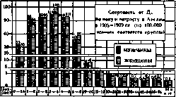
Figure 8.
Morbidity in the USSR. The incidence of D. in the USSR, Ukraine, Moscow and Leningrad for the period from 1886 to 1927 is given in table 15 (average annual morbidity per 10,000 population), as well as in fig. 12. In the USSR (Russia) in years free from epidemics, an average of about 15 cases of D. per 10,000 population were recorded annually, and in the pre-war period the morbidity rate never fell below 11.5. The first rise was noted in 1895-97 (up to 22 per 10,000 population). The second wave of D. begins in 1906, reaches its maximum in 1910 (43.1) and continues until 1915 inclusive, i.e. for 10 years. After this, there is a sharp decrease in D., and from 1919 (no data for 1917-1918) D. is recorded in unprecedentedly low figures. In recent years, table 18 (for 1903-1910 according to S.A. Novoselsky) gives a clear picture of this. N.I. Tezyakov, on the basis of data on the incidence of diphtheritis for 25 years (from 1886 to

Figure 9. In 1910, it was proposed to divide all the provinces of European Russia into 6 groups. The first 2 groups with the highest morbidity rates (from 40 to 54 per 10,000 population) included the former Yekaterinoslav, Kherson, Kharkov, Saratov, and Voronezh provinces. The 6th group with the lowest morbidity rates (less than 0.9 per 10,000 population) included all northern, northwestern, and northeastern provinces. Now such a distribution cannot be made: in the individual republics of the USSR, the morbidity rates in Ukraine (before the war - the highest) and Belarus (the lowest) are almost identical. Among individual provinces of the RSFSR, the lowest morbidity rates continue to remain in the northern provinces: Vologda, North Dvina, Novgorod, Zyryan, and in some national republics: Dagestan, Kirghiz, and Yakut. The difference in morbidity between other regions is small (see fig. 13 and 14). Morbidity by age and sex is illustrated in table 19. The largest number of diseases is observed in children under 10 years of age. In children under 5 years of age, morbidity among males is somewhat more frequent than among females, while in older ages, females show higher morbidity (fig. 15). 4. Spread of D. in cities and rural areas. Data on the spread of D. in cities and rural areas are different in different countries. In England for the period from 1905 to 1909, mortality from D. in children under 5 years of age was 99.6 in cities and 61.7 in rural areas per 100,000 population; in older age groups it was lower in cities than in rural areas: from 5 to 9 years - 47.8 and 56.4, from 10 to 14 years - 6.7 and 12.3, from 15 to 19 years - 1.2 and 3.2, etc. (the first figure shows mortality in cities, the second - in rural areas). In Russia, Serbia, and Austria, mortality from D. was significantly higher in rural areas than in cities, as seen from the following figures (table 20): Table 20. Mortality from D. per 100,000 population in cities and rural areas. Table 21. Number of registered patients per 10,000 population. Countries Russia . . . Serbia . . . Austria . . Years Cities Rural areas 1906-1910 44.6 72.0 1897-1906 34.0 123.0 1909-1910 19.3 27.7 In S.-A. S. H. mortality from D. is higher in cities than in rural areas (Vaughan). As for the morbidity of D., in the RSFSR the number of registered patients in cities is significantly higher than in rural areas (table 21); this is probably partly explained by better registration in cities. 5. Seasonality of D. spread. Figure 16 shows the monthly distribution of D. cases for 1925-27 in Denmark, France, RSFSR, and Ukraine (in absolute numbers), and fig. 17 for the Moscow and Leningrad provinces for comparison - the monthly distribution of D. in European Russia and some foreign countries before the world war. In Denmark, the rise of the monthly curve begins in September, the maximum is observed in November, December or January; the lowest figures are noted in June-August. In France, the rise of the curve begins in October, the maximum morbidity falls on December or January, the minimum figures occur in August-September. In the RSFSR and Ukraine, the rise of the monthly curve begins in September, the maximum of cases falls on October; in some years a second, less pronounced rise is observed in March.
I. Dobreyzer. Pathological Anatomy.
In the path.-anat. picture of D., one can distinguish: 1) local phenomena arising in the area of primary fixation of the pathogen, and 2) changes in the entire organism due to the action of the toxin circulating in the blood. As for local phenomena, the most frequent (one can say classical) form of tissue reaction to the introduction of the diphtheria bacillus is fibrinous inflammation in the form of diphtheritis or croup. This process usually occurs on the tonsils and the adjacent part of the pharynx [see separate table (p. 423-424), fig. 2]; in more severe cases it can spread from one side to the mucous membrane of the pharynx, then the esophagus and even the stomach, from the other side to the floor of the oral cavity, gums and tongue (the mucous membrane of the cheeks is almost never affected). Very often, especially in certain epidemics, lesions of the respiratory tract (nasal cavity, larynx, trachea and even bronchi) are also observed, in which the last three organs are rarely affected in isolation, but mostly in connection with D. of the pharynx, throat or nose [see separate table (p. 423-424), fig. 1]. Besides these usual localizations of D., one occasionally has to observe the same process on various wound surfaces of the skin (in children most often on excoriations of the upper lip, formed due to the corrosion of the skin by the nasal secretion in diphtherial rhinitis), on the conjunctiva of the eyes, in the external auditory canal and on the genital organs (mainly in girls in the area of the vulva and vagina, occasionally in boys in the preputial sac). In all these cases, infection usually occurs through scratches, injuries or rubbing of infectious material by infected fingers, which of course is much more frequent when there is already a lesion of the pharynx or nose (although in exceptional cases it can occur n
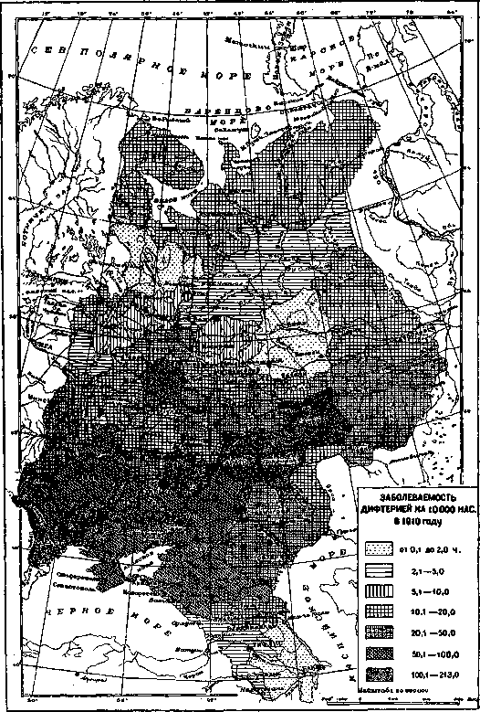
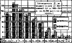
Figure 15.
which characterizes the mildest forms of the disease. Such catarrhs in the pharynx and throat are relatively rare, but in the nose (especially in small children) they occur more frequently and, if not in time g 1.......g..... N , | 1925 g. / -1925 J927 7г 19? // /^ \ 1926, /г 'A i Nr/ -'/' -J и ш Х1Г with ordinary catarrh, damage to the local vascular system. This damage to the vessels (in the form of their paralysis, creating blood stasis, and a significant increase in the porosity of capillaries and small veins, leading to edema and hemorrhages) is one of the typical features of diphtheritic intoxication and appears with greater intensity the more severe the disease. Impregnation of the fibrinous deposits with blood and significant edema of the surrounding parts are rightly considered a very bad prognostic sign. In some cases of so-called "hypertoxic D." (D. gravissima), mostly ending in death within 1-2 days, no inflammation may develop at the site of primary localization of the pathogens, but only a deep necrosis of the tissue and its impregnation with blood, while the inflammatory reaction is then found only at the demarcation line. Usually these cases are also accompanied by hemorrhages in other organs (hemorrhagic form). It should be noted, however, that such severe damage is observed almost 1 1 1 340» | .... 1925 g. 1927 g. г- \ \ , / "~ ">. -ч i / ^ \. к \ ^~ P925- 1 1 1 im \ 1926- \ / к \ / \ _____________ 1925 g. л \ \ 1! 28 g. 27 g. if ф У / \ Ио / i Я8п \v\ // яоо /У ?во \^ V/ 1 1 1 / / / ' 1 "-Ч £"*, / \ / / \> A > 1S 1925 g. ] 926 g. 927 g. ^ V/ П III 1750 1625 1500 1376 1250 1125 100» 875 750 626 500 Figure 16. Monthly distribution of D. cases for 1925-1927 (absolute numbers). recognized, may give rise to the spread of infection. A characteristic (though not obligatory) feature of diphtheritic rhinitis is the bloody nature of the discharge, which indicates a deeper involvement than that in D. of the pharynx alone. Isolated D. of the respiratory tract does not give hemorrhagic forms, and in general the changes associated with general intoxication (see below) in it are never significant. Correspond
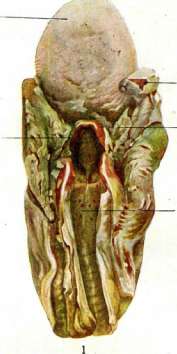
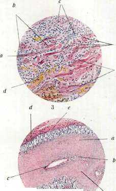
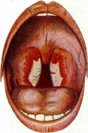
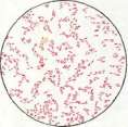
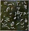
5 c Figure 1. Diphtheritis of the larynx, pharynx and their extension into the larynx: a-inflamed epiglottis with a fibrinous coating; b-inflamed tonsils (a) and pharynx (d). Figure 2. Diphtheritic myocarditis. (From Pfanndler-Schafer) Figure 3. Myocarditis in diphtheritis: a-preserved muscle bundles of the myocardium (cross section); b-state of cloudy swelling and granular degeneration; c-inflammatory infiltrate in place of dead muscle bundles; d-blood vessels. Figure 4. Changes in the adrenal glands in diphtheritis: a-deeper part of the adrenal cortex; b-modules of cortical substance; c-central vein of the adrenal gland; d-preserved parts of the cortical surface; e-capsule of the adrenal gland; f-vessels of the capsule. (Figs 1, 2 and 4-after illustrations of the Path.-anat. institute of Moscow State University) Figure 5. Diphtheritic bacilli: a-staining according to Löffler; b-double staining according to Neisser; c-staining according to Gram (after Kolle and Wassermann) Fig. 6. Diphtheritic bacilli. (After Schlossmann) To the section. Diphtheritis, Diphtheritis. Accordingly, death in lesions of the respiratory tract occurs either from asphyxia-mainly in cases of extensive spread of fibrinous inflammation along the bronchial tree to its small branches (the so-called 'descending croup')-but mainly (in children in more than 70% of all fatal cases) from complications with pneumonia. The latter usually has the character of catarrhal bronchopneumonia and is considered a complication on the grounds that in its etiology the diphtheria bacilli apparently play only a secondary role and may not play any role at all; its origin is most often due to secondary infection. Local manifestations in D. must also include lesions of the regional lymph glands and the collateral inflammatory-toxic edema of soft tissues adjacent to the site of primary localization. The first is expressed by an increase in the size of the glands (in severe cases very significant), which is based initially on severe hyperemia, inflammatory edema and often hemorrhages, and subsequently-proliferation of cellular (mainly reticulo-endothelial) elements. In addition, in highly toxic forms of D., in the regional glands (as well as in the tonsils in D. of the pharynx), a greater or lesser number of whitish or yellowish necrotic foci are always found on section, which together with hemorrhages sometimes penetrate the gland (resp. tonsil) so much that almost no normal lymphoid tissue remains in it. Regarding the collateral edema of adjacent tissues, it should be noted that its development proceeds quite parallel to the degree of intoxication, thanks to which in practice it is often used to determine the severity of the disease. In severe pharyngeal diphtheritis, it is extremely pronounced on the one hand on the soft palate and arches, often leading here due to the simultaneous increase in tonsils to almost complete closure of the isthmus of the fauces, on the other hand-in the intermuscular and subcutaneous tissue of the neck, from which it sometimes spreads downward below the clavicle line. The characteristic relief of the neck completely disappears in this case, and the head seems to pass directly into the trunk.
As for the changes caused by the general diphtheritic intoxication of the body, among them the most profound and constant are changes in the adrenal glands, the peripheral nervous system and the cardiovascular system. Lesion of the adrenal glands is particularly prominent in experimental animals (guinea pigs) that died from poisoning with diphtheritic toxin. In them, these organs are greatly enlarged and have a dark red color, which microscopically corresponds to extremely sharp hyperemia and numerous hemorrhages both in the cortical and medullary substance. The chrome reaction is either completely absent or more or less significantly weakened. If death does not occur too quickly (on the 2-3rd day or later), then extensive destructive changes in the parenchyma (necroses) are added to this. In humans with natural disease, the lesion, although not so bright, retains the same character. In cases of death from diphtheritic intoxication, significant hyperemia, hemorrhages and weakening or even disappearance of the chrome reaction are always found in the adrenal glands, clearly indicating a decrease (resp. cessation) of adrenaline production. The cells of the medullary layer are at this time more or less strongly vacuolized. In the cortical layer, a decrease in lipoid content and also vacuolization of cells are noted, often reaching necrosis (see separate table, fig. 4). In the nervous system, the main changes in diphtheritis are concentrated 1) in the peripheral nerves and 2) in the sympathetic and autonomic ganglia. In the first, the picture of changes is very well defined by the term 'multiple toxic parenchymatous neuritis' and consists on the one hand of phenomena of edema, circulatory disorders and small focal infiltration of nerve sheaths, but mainly-of phenomena of dissolution and disintegration of myelin. Processes of this kind can reach very great intensity, and then many nerve fibers are completely bare of myelin sheaths. The axis cylinders suffer less, but still a certain part of them (both in medullated and non-medullated fibers) dies with successive phenomena of swelling, deformation, fragmentation and disintegration. At the same time, in places of death of myelin and axis cylinders, proliferation of Schwann's nuclei is almost always noted. Despite the fact that diphtheric pareses and paralyses are usually discovered quite late (mostly around the third week of the disease), all the above-mentioned changes in nerves can be observed in persons who died from diphtheritic intoxication, already starting from the 3-4th day of the disease, and they are most pronounced in those nerves which are closest to the site of primary lesion (in D. of the pharynx-nn. glosso-pharyngei et vagi, to a lesser degree-nn. phrenici). This latter position is also justified in relation to the sympathetic and autonomic ganglia, of which the 2 upper cervical sympathetic nodes and ganglion nodosum n. vagi usually suffer most severely. Here, besides vascular and infiltrative phenomena, which are often not significant, one can find degenerative fatty changes, vacuolization and deformation of ganglion cells, displacement, pallor or, conversely, pyknosis of their nucleus, death of the nucleolus, decrease or disappearance of Nissl's granularity-in short, all gradations of gradual karyo- and cytolysis. On the part of the amphicytic sheath, these changes are accompanied either by proliferation of satellites or, conversely, degenerative processes in them, often leading to their death and to partial or complete exposure of nerve cells. The frequency of disorders from the cardiovascular system in D. can be largely explained by the lesion of the adrenal and nervous system. Indeed, a decrease in adrenaline production and changes in sympathetic nodes and nerves cause profound paralysis of blood vessels and a drop in blood pressure, which creates enormous difficulty for the work of the heart. On the other hand, most fibers of both cardiac plexuses originate from the vagus nerves and from the cervical part of the sympathetic trunks, i.e. from the most affected elements of the nervous system in D. of the pharynx, and of course damage to these fibers in connection with damage to the autonomic nodes of the heart itself must extremely strongly disrupt its innervation. It is quite understandable that under these conditions the heart becomes the place of least resistance of the body, on which the effect of the toxin is manifested with the greatest force and constancy. Changes in the heart muscle in diphtheritic intoxication can be either purely degenerative or inflammatory in nature. The first are more often found in cases of early death (within the 1st week) and consist of parenchymatous and fatty degeneration of muscle fibers, their vacuolization and the so-called granular disintegration (a variety of Zenker's degeneration, see separate table, fig. 3). Inflammatory phenomena usually develop from the 7-12th day of the disease and give a picture either predominantly of parenchymatous or of interstitial myocarditis. In the latter case, degenerative changes recede into the background, while hyperemia, hemorrhages, edema and focal or diffuse accumulations of various cellular elements (mainly histiocytes and lymphoid cells, to a lesser degree-neutrophils, eosinophils and plasma cells) in the intermuscular tissue predominate.
As for the topography of the process, the walls of the left ventricle are most severely affected in most cases, especially in the layers closest to the endocardium, as well as the papillary muscles. The atrioventricular bundle almost always participates in all the mentioned changes, although on the other hand, damage to it is rarely very deep. Both with severe myocardial degeneration and especially with a sharply expressed inflammatory process, the heart already acquires a very characteristic appearance macroscopically. It greatly expands and becomes so flabby that it often loses its normal shape. Its apex is smoothed out, and the entire organ takes the form of a bag with greatly expanded cavities and a rounded bottom. In the cavities, voluminous fibrinous clots are usually present, and if it is a matter of an inflammatory process, then on a section of the muscle made parallel to the surface, dark red stripes and spots of various sizes clearly stand out against a dull grayish or yellowish background.-As for the blood vessels, as has been said, a paralytic state is characteristic of diphtheritic intoxication, which is evidenced on the corpse by the external appearance of all organs with their congested venous and capillary network and often found hemorrhages.-Speaking of the state of the cardiovascular system, one cannot but also mention the thrombotic processes that often occur in D. The latter is very favored on the one hand by the toxic effect of diphtheritic poison on the inner lining and the slowing of blood flow due to weakening of cardiac activity and paralysis of the vessels, on the other hand by the increased coagulability of blood under the influence of diphtheritic toxin. The favorite places for thrombus formation here, in addition to the venous system (veins of the soft meninges, mesentery, vena azygos, hemiazygos, etc.), are the intertrabecular spaces near the apex of the left ventricle of the heart and the area of the left auricle. The presence of thromboembolisms, of course, often gives rise to embolisms and the development of infarcts. Of the remaining organs, the spleen, lymph glands, liver, and kidneys deserve mention. The spleen reacts weakly in D. In pure, uncomplicated by secondary infection cases, its increase is insignificant and occurs mainly due to plethora and hyperplasia of the follicular apparatus. At the same time, in the center of the follicles, one can almost always see more or less large collections of large ret.-endo. cells, often with signs of karyorrhexis in them. Similar changes are often also found in the follicular apparatus of other parts of the body (follicles and Peyer's patches of the intestines, lymphatic glands of the mediastinum and abdominal cavity). In the liver in toxic D., in addition to plethora and significant degenerative changes, miliary foci of necrosis are sometimes encountered.-The changes characteristic of D. in the kidneys belong to the category of nephroses and amount to degenerative processes in the epithelium of the cortical layer and the appearance of various kinds of cylinders in the tubules. However, in severe cases from the 4th-5th day of the disease, a true inflammatory process with corresponding changes in the interstitial tissue or glomeruli can often be observed.-In conclusion, it should be noted that death in D. can occur not only in the acute period of the disease, but also at later dates, 1-2, even 3 months after the onset of the disease, already among apparent health. This is the so-called death from late diphtheritic paralyses. It is connected with deep damage to the vagus or other respiratory nerves and can occur at any moment of the time during which the destructive (and partly reparative) period of neuritis extends. Since the vagus nerves suffer more and more often from other respiratory nerves in D. and since damage to the vagus nerves is usually aggravated by damage to the myocardium, death from late paralyses in the vast majority of cases is cardiac death. On autopsy, when examining the organs with the naked eye, no changes are found in such corpses as a rule, and only microscopic research reveals in the nerve trunks (especially in the vagus nerves) the picture of parenchymatous neuritis described above, while in the myocardium- for the most part, the consequences of the acute myocarditis in the form of diffuse sclerosis of the cardiac MUSCLE.
M.
Skvortsov. Clinics. The diphtheria bacillus can penetrate the human body through the mucous membrane of various organs and through the skin. Depending on the location of the process, we distinguish between D. of the pharynx, nose, larynx and generally the respiratory tract, eye, ear, genital organs and skin. The most common of these is D. of the pharynx. It is this form that is mainly meant when speaking of diphtheria. Diphtheria of the pharynx. Classification. Clinical forms and course of D. of the pharynx are extremely diverse. If we take as the basis of classification on the one hand the size and extent of the plaques (local distribution of the diphtheria toxin), and on the other hand the degree of general intoxication (general distribution), then all the diversity of clinical forms of D. can be reduced to the following four or even three main types. -I. Localized D. The diphtherial process is limited to the pharyngeal cavity: the plaques usually do not go beyond the tonsils, uvula, and arches. Symptoms of general intoxication are absent or moderately expressed. In its course, this form is the most favorable, well responsive to the action of serum. - II. Spread-out D. In this form, the plaques are not limited to the pharyngeal cavity: they extend to the nasopharynx and nasal cavity, and may also go into the oral cavity, but more often, especially in small children, they descend downward to the larynx and trachea (croup). General intoxication often runs parallel to the spread of the local process, however, not always (in croup, for example, despite the extensive spread of films, general intoxication is sometimes not strongly expressed). - III. Toxic D. (d. toxica), another generally accepted name: "severe D." (d. gravis). The plaques are sometimes limited to the pharyngeal cavity, but more often extend to the nasopharynx, soft and hard palate. From a clinical point of view, this form is characterized not by the appearance and extent of the plaques, but by the edema of both the soft tissues of the pharynx and the cervical cellular tissue. This edema is analogous to that observed in a guinea pig at the site of subcutaneous injection of diphtheria toxin. It appears already in the first days of the disease and is the earliest sign indicating the beginning of general intoxication. In this form, the consequences of general intoxication (nephrosis, paralyses, cardiovascular disorders, lesions of the vegetative-endocrine apparatus) can always be observed to a greater or lesser degree. Some call this form, on the proposal of Rauchfus, phlegmonous D., and French authors - false phlegmonous (Aviragnet). Rauchfus himself subsequently recognized this name as unsuccessful. Indeed, phlegmons and a sequential abscess do not occur here, and the edema of the pharynx depends on the diphtherotoxin-induced changes in the vessels. Another, more common, although also incorrect name is septic D. It gives an incorrect idea of the pathogenesis of toxic D. Sepsis, i.e., general infection with microbes, does not usually occur in toxic D. The French school, since the time of Roux, held the view that severe D. results from simultaneous infection with the diphtheria bacillus and the streptococcus - strepto-diphtheria of Sevestre and Martin. However, in its symptoms, so-called strepto-diphtheria essentially does not differ from that which is called toxic D. (edema of cellular tissue, hemorrhages, paralyses). The research of other authors showed that streptococcus cannot be found in the blood and organs. Thus, the pathogenesis of severe D. is reduced to intoxication by the poison of diphtherial bacilli alone. To this same view, French authors (Aviragnet) have also recently inclined. It is true that in a certain number of cases of D., the streptococcus plays a very important role in the pathological process, but exclusively as a microbe of secondary infection. In these cases, it causes the same phenomena that are generally characteristic of streptococcal infection: suppuration of the cervical glands, purulent otitis, pleurisy, etc.; streptococcus can always be isolated from the pus. Such cases are more correctly called D., complicated by streptococcal sepsis; the course of the disease is not always severe. Toxic D. is difficult to respond to the action of serum and requires very large doses. Very recently (1928), some German authors, in view of the frequent finding of hemolytic streptococcus in toxic D., again begin to attribute to it a major role in the genesis of this form and recommend combined treatment - with antidiphtheritic and anti-streptococcal sera. -As a variety of severe D., one can also distinguish malignant D. (d. maligna s. gravissima). The clinical picture and pathogenesis may vary. In some cases, malignant D. proceeds as a fulminating or hypertoxic form, when the child dies within 1-3 days with signs of rapidly progressing intoxication. In other cases, the disease proceeds more slowly, with the phenomena of hemorrhagic diathesis (hemorrhages under the skin, from mucous membranes, into internal organs) being prominently featured in the overall picture of severe intoxication. Finally, in the third category of cases, gangrenous decay develops in the pharynx, when the diphtheria bacillus is joined by a mixed infection with various microbes leading to gangrenous decay of tissues. Common to all three categories is the malignant course of the disease and the almost complete ineffectiveness of serum. Localized D. Typical form. The incubation period, as in other forms of D., is from 3 to 10 days. The disease begins either acutely, suddenly, or gradually, with inconspicuous symptoms. In the first case, the temperature immediately rises to 38°-39°, headache, general weakness and malaise appear. In the second category of cases, the patient notices poor appetite for several days, a slight increase in temperature (37.5-38°). Often even older children do not complain of sore throat, or it is insignificant. On objective examination, some acceleration of the pulse is found, the cervical glands are usually enlarged on one side and painful on pressure. The tongue is coated, the tonsils (both or mostly one of them) are swollen and reddened. On the reddened tonsil, one can see a plaque which in the first hours of the disease resembles a slight burn of the mucous membrane or a dense cobweb; it can sometimes even be removed with a cotton swab, but at this place a new, non-removable film appears extremely quickly. By the end of the first or beginning of the second day, the plaque takes on the characteristic properties of a diphtherial film: its color is dirty or yellowish, it is rather thick, protrudes above the surface of the mucous membrane, and cannot be removed without bleeding. If serum has been injected, sometimes no significant change in the patient's condition occurs within the first day, and the plaques may even increase. But after 24 hours, a sharp improvement occurs: the temperature sometimes drops to normal, the patient becomes cheerful and lively, appetite appears, the cervical glands decrease, the plaques change their appearance - they become more loose, as if raised above the mucous membrane, a sharply defined redness appears at the edges, the spread of the plaques stops. During the following days, a significant part of the plaques slough off, and in 2-3 days the pharynx is completely cleansed. Consequences of intoxication (paralyses, heart weakness), if serum was not injected late, usually do not occur. Without treatment with serum, in most cases the disease progresses, the plaques occupy both tonsils, may extend to the uvula, posterior wall of the pharynx; the cervical glands are enlarged and painful, but there is no edema of the cellular tissue. In the urine, traces of protein. In the blood, leukocytosis to varying degrees. The temperature of the remittent or irregular type persists for 7-12 days. The plaques slough off slowly, leaving no ulcers or defects of the mucous membrane. The disease ends by the 7th-15th day. Sometimes isolated paralyses (paresis of the soft palate) and moderately expressed cardiac disorders are observed. From this typical course, there can be various deviations. Rudimentary forms. The local process in the pharynx can be very weakly expressed in the form of pointy or patchy plaques - point D., or in the form of one hyperemia of the tonsils, with the fibrinous exudate completely absent - catarrhal D. The temperature is subfebrile or normal, general condition is good. Such forms are observed in adults and older children during an epidemic in the family or household. Recognition can be established on the basis of the simultaneous existence of typical cases in the family, and mainly with the help of bacteriological research (so-called bacteriological D.).-In a serious form, the process shows a tendency to spread to the uvula, soft palate, nasopharynx and posterior wall of the pharynx. The voice takes on a nasal tint, a sour-sweet odor from the mouth, salivation. The cervical glands are greatly enlarged, but there is no edema of the cellular tissue. The face is pale, appetite is absent. Temperature 38-39°. Serum rather quickly stops the process, although repeated injections are required. Such cases are transitional to the spread-out and toxic forms.-Protracted form. Sometimes the local process in the pharynx drags on for 2-4 weeks and longer. In these cases, the plaques, having disappeared from the tonsils, reappear in the same place or in other parts of the pharynx and persist for weeks, not responding to serum treatment.
The temperature is slightly elevated or even normal, the glands are almost not enlarged, the general condition is good. Such a prolonged course is occasionally observed both in primary D. and especially in secondary D., when it developed after measles. Apparently, in these cases, it is a matter of weakening of local resistance from the side of the pharynx in the presence of a well-expressed general immunity. Localized diphtheria occupies first place in frequency - 50-60% of all cases of diphtheria. Mortality is small: 1-2 percent. Widespread diphtheria. D. pharynx + D. nose. The transition of the process from the tonsils to the nasal cavity is observed no earlier than the 3-5th day of the disease and is accompanied by a new rise in temperature and worsening of the general condition; special symptoms appear, indicating damage to the nasopharynx and nasal cavity. The voice takes on a nasal tint, the mouth is open, the tongue is dry, covered with crusts, first a mucous, and then a bloody discharge appears from the nose, which corrodes the skin around the nostrils and on the lips. On the neck, not only the submandibular lymphatic glands swell, but also those located near the m. sterno-cleido-mastoidei. If the process has gone far and has involved the front parts of the nose, the films can be seen with the naked eye, after clearing the entrance to the nose from the purulent discharge and crusts. The spread of D. to the nose worsens the prognosis, as it is accompanied by an intensification of the phenomena of general intoxication. - Diphtheria of the pharynx + D. mouth. With the spread of the process to the oral cavity, dirty-gray, tightly adherent films appear on the palate, the mucous membrane of the lips and cheeks, as well as on the tongue. Clinical symptoms: profuse salivation, odor from the mouth, painfulness of chewing and swallowing, severe swelling of the submandibular lymph glands. The films come off slowly, leaving long-unhealing ulcers. - D. pharynx + D. throat. From the pharynx, the process can spread to the larynx, either directly spreading along the walls of the pharynx or, bypassing the latter, can be directly localized in the larynx. Diphtheritic damage to the larynx and respiratory tract is known under the name of croup. Under 'croup' in the clinical sense is understood any acute inflammatory disease of the upper respiratory tract, occurring with certain characteristic symptoms (rough cough, stenotic breathing). True and false croup are distinguished. True croup or simply croup is called diphtheritic damage to the larynx and trachea; in the vast majority of cases, the presence of a fibrinous exudate is observed on the mucous membrane. False croup differs from true croup etiologically: it is caused not by D., but by influenza, measles and other infections; fibrinous exudate is usually absent in it, only inflammatory hyperemia and edema are found. Croup (true, diphtheritic) can be secondary if it develops following damage to the pharynx or nose, and primary - with primary localization of the process in the larynx. But in a large number of cases of so-called primary croup, the initial damage to the nose and nasopharynx remains unrecognized; thus, compared to secondary, primary croup is not so common. In symptoms, course, and outcome, these two forms of croup do not show any significant difference. D. larynx (croup). The course of croup can be divided into three stages (Rauchfus). 1st stage - croupous cough. The first sign indicating the beginning of damage to the larynx is a sharp, loud cough, which very soon becomes rough, barking, at the same time the voice becomes hoarse, unclear, and then silent (aphonia). This period lasts 1-2-3 days and passes into the 2nd stage - the stage of stenosis. Breathing becomes difficult, unclear, with each inhalation a sawing or whistling sound is heard. Another sign of narrowing of the larynx - the retraction of the yielding places of the chest (supraclavicular and jugular fossae, intercostal spaces, subcostal region). At first, the child copes satisfactorily with the lack of air and remains calm. Then oxygen starvation begins - the child tosses in bed, jumps up, grabs the bed with its hands, asks to be taken in its mother's arms, throws its head back; the auxiliary respiratory muscles (mm. sterno-cleido-mastoidei, scaleni) are noticeably strained. If the disease is left to its natural course, in rare cases, when the film comes off, improvement occurs; in the vast majority of cases, the disease passes into the last stage - the stage of asphyxia. The child becomes quieter, drowsy, lies listlessly in bed. Breathing becomes frequent, but superficial, the retractions are not so noticeable. The lips, tip of the nose, and nails turn blue, the face is pale, sweat often appears on the forehead. The extremities are cold, the pulse is very frequent, thread-like, sometimes paradoxical. At times, attacks of acute suffocation occur, sometimes death occurs during such an attack; in other cases, the child dies after a more or less prolonged agony. Under the influence of serum, the described course of croup can change sharply - the disease can stop at any stage. The causes of stenosis are diverse: edema and fibrinous exudate, mechanically hindering the access of air, spasm of the laryngeal muscles, etc. There are cases when the fibrinous exudate from the larynx descends to the trachea and bronchi. These cases are known under the name of descending croup. The condition of the patient is extremely severe; neither serum nor intubation or tracheotomy eliminate the stenotic phenomena. Descending croup is observed in small children up to 2 years old and gives an extremely poor prognosis. There is also ascending croup, when the process begins in the bronchi and trachea and rises upward to the larynx. But the existence of this form is questionable. The frequency of croup is about 30% of all cases of D. Mortality 15-30%. Death occurs either from suffocation or, more often, from the resulting pneumonia. Toxic D. (d. toxica). This form sometimes develops from localized D. with the spread of the process to the nasopharynx and oral cavity, more often it arises as such from the very beginning. In the latter case, the disease begins suddenly, more violently than localized D. The temperature immediately rises to 39-40°, headache, repeated vomiting, pain on swallowing. The pulse is frequent (140-160), the face is pale, general weakness, sometimes excitement. The submandibular glands are enlarged, painful, under the angle of the jaw there is a flabby, doughy edema of the cellular tissue; sometimes the edema appears only on the second day. The mouth is open, a strong cloyingly sweet odor. The voice has a constricted, nasal tint. On examination of the mouth - the tongue is dry, coated, the pharynx is dark red, swollen; on one (usually) tonsil there is a dirty-gray coating, which is not removed by a cotton swab. This coating extremely quickly, within a few hours, occupies the entire tonsil, passes to the uvula, sometimes to the soft palate. On the second or third day, the disease reaches its full development, and it is not difficult to diagnose severe D., even without resorting to bacteriological research. Despite large doses of serum, the disease continues to progress in the first days after injection. However, 2-3 days after injection, the spread of the process stops, the temperature drops, the edema of the pharynx and cervical cellular tissue decreases. The pharynx, although slowly, clears up, and in place of the films, ulcers with dirty-gray bottoms remain, which heal very slowly. In the period of convalescence, the consequences of intoxication are always observed: paralyses, weakness of the heart, from which patients sometimes die in the 3rd-4th week and even later. Without serum, the process progresses uncontrollably: the films sometimes occupy the entire hard palate up to the teeth, go to the posterior wall of the pharynx and to the larynx, the edema of the cellular tissue occupies the entire neck and descends to the chest. Already due to the severe swelling of the pharynx, breathing becomes difficult (stenosis of the pharynx). Despite the spread of the process, the temperature very often falls on the 4-5th day of the disease, and in the future the severe picture of the disease continues to develop with subfebrile or even normal temperature. Recovery is an extremely rare exception; paralyses and weakening of cardiac activity usually kill the patient at the end of the 2nd or 3rd week. Toxic D. most often affects older children and adults; up to 2 years it is observed very rarely, but mortality in small children is very high (80% according to Egiz). According to the data of the Children's Clinic of 1st Moscow University in 1901-08, toxic D. constituted 2-8% of all cases of diphtheria. During the epidemic of 1908-12, its frequency increased to 17%. Mortality was 34%. In children's hospitals of Moscow during the same epidemic, mortality reached 50%. Malignant D. (d. maligna). The characteristic features of toxic D. (edema of the pharynx and cervical cellular tissue) are always observed in this form. But in hypertoxic D., the local process, compared with the extremely rapidly developing general intoxication, may turn out to be relatively moderate. Death occurs within 3 days from weakening of cardiac activity. This form needs detailed study both in relation to the virulence of the bacillus and in relation to the constitutional peculiarities of the organism. In hemorrhagic D., to the usual picture of toxic D., the phenomena of hemorrhagic diathesis are added.
The first signs of it are bruises at the site of serum injection and the hemorrhagic nature of the coatings; these signs are sometimes found from the first days of the disease, but more often in the later period. The pathogenesis is due to the deep lesion of the vessels and hematopoietic apparatus by the toxin. The number of blood platelets is reduced, clot retraction is insufficient, for which reason this form is considered as secondary thrombopenic purpura (Benjamin).-Gangrenous D. often joins the hemorrhagic one; hemorrhages into the tonsil apparently create favorable conditions for gangrenous decay of tissues under the influence of putrefactive bacteria. The frequency of malignant D. is 1-2%. The prognosis, with rare exceptions, is absolutely bad. - Other localizations of diphtheria. D. of the nose is one of the most frequent localizations of D. after the pharynx and larynx. Primary D. of the nose is especially common in newborns and infants; due to the underdevelopment of the tonsils, it occurs in them more often than diphtheria of the pharynx (according to Morozov, diphtheria of the pharynx in children in the 1st year of life is 9.3%, diphtheria of the nose is 41.8%). Symptoms: snuffling, discharge first serous, then sanguineous, usually from one half of the nose; the child refuses to take the breast; attacks of suffocation during sucking and during sleep. Temp. 38-39°; pallor, general restlessness. Under the influence of serum-rapid improvement. Without serum, an unexpected transition, bypassing the pharynx, to the larynx is not uncommon. In children in the 1st year of life, diphtheria bacilli are sometimes found in the nasal cavity without causing clinical manifestations of the disease. In older children the disease proceeds sluggishly: stuffy nose and sanguineous discharge from one half of the nose may last 1-2-3 weeks without causing signs of general intoxication.-D. of the eye is rare, usually as a secondary disease in D. of the pharynx and nose. A distinction is made between the superficial, or croupous, and the deep, or diphtheritic form. In addition to the conjunctiva, the process in most cases also affects the cornea, as a result of which vision is impaired to a greater or lesser degree. D. of the external genital organs: a very rare localization, especially in boys, arises most often as a secondary disease, but sometimes in girls it proceeds in the form of a subacute catarrhal form and is recognized only by bacteriological examination of the discharge from the mucous membrane of the vagina. Sometimes extensive coatings form with edema of the cellular tissue, which extend to the skin; a fatal outcome has been observed with signs of general intoxication.- D. of the skin and wounds. A distinction is made between D. of damaged and undamaged skin; the latter is extremely rare, and some (Adler) dispute its existence. During the war and famine years the frequency of D. of the skin sharply increased, according to Lande, up to 20%. The clinical picture is diverse; in addition to typical cases with films on the surface of the skin, cases have been described that proceeded under the appearance of pustules, impetigo, ecthyma, as well as under the appearance of phlegmons and panaritia. Similarly, in D. of wounds, films are sometimes absent, only the poor appearance and prolonged course of the wound attract attention; in the discharge-diphtheria bacilli (Lande). Complications of D.-Kidney. The usual form-nephrosis; glomerulonephritis is rare, apparently in combination with streptococcal infection. Time of appearance-acute (anginous) stage of the disease. Slight albuminuria is sometimes observed even with localized D. In severe D. albuminuria is a constant symptom; the amount of protein reaches 2-8%, with an abundant sediment consisting of epithelium, leukocytes, cylinders, salts. Blood is scarce. Blood pressure is not elevated. The functional capacity of the kidneys is slightly altered. Uremia is rare, edema also. The course is generally favorable. - Nervous system (paralysis). The pathogenesis is due to the lesion of the motor neuron by the toxin (toxic parenchymatous polyneuritis), and subsequently the gray matter of the spinal cord is also involved in the process (polyneuromyelitis). The diphtheria poison acts both through the blood and apparently also directly on the branches of the nerves in the area of the infectious focus. Diphtheritic paralyses have all the properties of peripheral paralyses (see Polyneuritides): they are flaccid, with muscle atrophy, with disappearance of tendon reflexes. Time of appearance: 3-4th week of the disease; in especially severe cases they appear early, before the disappearance of the coatings. The more severe D. and the later the serum treatment is started, the earlier the paralyses appear and the more severe they are. In the pre-serum period they were very common (20%), at present they are also not very rare (8% according to Jochmann). In D. of the pharynx paralyses develop with a typical sequence. First of all, paralysis of the soft palate appears (nasal voice, liquid food flows out through the nose). On examination of the pharynx the soft palate hangs down and remains immobile during phonation. These phenomena last 2-3 weeks and disappear without a trace. The next in time of appearance-paralysis of accommodation (c. ciliaris). Older children complain that they cannot distinguish objects at close range, cannot read. Sometimes strabismus (p. abducens), ptosis (p. oculomot.) are also observed. If the paralyses show a tendency to spread, paralyses of the extremities appear (ataxia, disappearance of knee reflexes). In severe cases, paralyses of the muscles of the neck and trunk join. The condition of the patient becomes extremely dangerous when paralysis of the larynx, respiratory muscles and diaphragm joins. The patient may die from suffocation or from pneumonia.-Cerebral paralyses are much rarer, exclusively in very severe D. during the period of cardiac disorders. Genesis-embolism of one of the branches of art. fossae Sylvii. The source of embolism is apical thrombi of the heart. The onset is sudden-a picture of apoplexy with subsequent hemiplegia. Recovery is rarely observed. Organs of circulation. Diphtheritic paralysis of the heart-the most important complication both in frequency and in view of the life-threatening character. The intensity of the lesion is extremely varied: from insignificant changes in the pulse (slowing, arrhythmia) to a sharply expressed decline in cardiac activity, from which the patient dies within 1-3 days. It is observed even with localized D. with delayed injection of serum, but as a constant phenomenon-in toxic D. A distinction should be made between circulatory disorders in the early period-on the 2-6th day of the disease, the so-called early paralysis of the heart, and disorders that arise after the disappearance of the films, on the 2-4th week, sometimes even later-late paralysis of the heart. Early paralysis is observed in very severe D., especially in the hypertoxic form. On the 3-4th day, sometimes already on the 2nd, simultaneously with the rapid spread of the process in the pharynx, the symptoms of cardiac decline sharply appear in the clinical picture: the skin is pale, cold, the pulse is very frequent, easily compressible, soon becomes threadlike, blood pressure is very low; the heart is dilated, the tones are dull, sometimes a systolic murmur, gallop rhythm or embryocardia. The liver is enlarged, vomiting, pains in the abdomen. Sometimes when attempting to sit or change position the patient as if suddenly dies, in other cases death occurs after a brief agony. Late paralysis is much more common. In severe D. at the end of the 2nd or beginning of the 3rd week, when the films in the pharynx have disappeared, no improvement in the general condition of the patient occurs: there is no appetite, the face is pale, the patient is apathetic. Urine is scarce, it contains much protein and cylinders. Paralysis of the soft palate. The pulse is usually slowed, sometimes drops to 60-40 per 1 min., the size of the heart increases, the tones are dull, doubling of the tones, blood pressure is low, the liver is enlarged. In not too severe cases such a condition lasts for some time (1-2 weeks), and the patient recovers. In more serious cases one after another three ominous symptoms appear. The first symptom-nausea, which turns into persistent vomiting. The second symptom, the origin of which remains unexplained-pains in the abdomen. The appearance of a disturbance of circulation-joining the described picture-gallop rhythm or embryocardia-should be considered especially dangerous. When reaching such a condition, the patient as a rule dies. In very rare cases, despite repeated attacks of collapse, recovery occurs. It is much rarer to observe on the 3-5th week a suddenly occurring paralysis of the heart in patients who before this did not show noticeable signs of cardiac disturbance.-Pathogenesis. The origin of paralysis of the heart in diphtheria is not yet fully elucidated. The diphtheritic toxin is an intense poison for the circulatory apparatus as a whole, but individual parts of the apparatus are apparently affected in a definite sequence. The first to be affected are the adrenal glands, in particular the chromaffin or adrenalin substance (Molchanov, Abramov), as well as the sympathetic system (Abricosov, Mogilnitsky). Much later changes in the vagus nerve and heart muscle are found. Along with diffuse myocarditis, more or less deep changes in the atrioventricular system-in the bundle of His and Tawara-have sometimes been found; in such cases during life complete transverse dissociation and ventricular automatism are observed.
However, the condition of the bundle of His and Tawara in most cases corresponds to the condition of the rest of the myocardium, and one cannot see the cause of heart paralysis in the isolated lesion of this bundle. Thus, early heart paralysis is caused primarily by the lesion of the adrenal glands and the sympathetic system; in late paralysis, the predominant role apparently belongs to the changes in the heart muscle and the vagus nerve. Other complications. Pneumonia is quite common in croup, as well as in severe D., accelerating the fatal outcome. The genesis in very rare cases is the direct action of the diphtheria bacillus itself, usually diplo- and streptococcus. Purulent complications (lymphadenitis, otitis, pleurisy, etc.) are not characteristic; they depend on the streptococcus. Combination of D. with other acute infectious diseases. Measles creates an increased predisposition to D.; the course is severe (in Moscow in 1923-27, 194 cases of this kind were autopsied in city hospitals out of 520 other diphtheritic autopsies); very frequent is the transition to the larynx, and the differential diagnosis from measles croup presents great difficulties. It is also often difficult to recognize in combination with scarlet fever (cases of fatal combinations 'scarlet fever-D.' in 1923-27 in Moscow, 165 were autopsied out of 2,303 cases of other scarlet fever autopsies). Recognition in many cases (especially in rudimentary forms) is possible only with the help of bacteriological research. But the cultivation research continues for 24 hours, besides in undoubted cases of D., at the first investigation, the answer is sometimes negative. Meanwhile, the injection of serum should be carried out as soon as possible; in toxic D., to delay it for 12-24 hours means to risk the life of the patient: the physician must remember that the responsibility for correct diagnosis and timely treatment lies with him, and not with the laboratory. Therefore, he must be able to make a diagnosis based on the clinical picture. The following signs are characteristic of D. of the pharynx: 1) thick films of dirty-gray color, rising above the level of the mucous membrane and tightly seated on it, 2) the tendency of the films to spread beyond the tonsils, 3) a large swelling of the neck glands, especially with edema of the neck tissue, 4) simultaneous lesion of the nose or larynx. In case of lesion of the pharynx with such a clinical picture, it would be a mistake to wait for the result of bacteriological research. In the differential diagnosis, the following diseases should be kept in mind: lacunar frog, follicular, ulcerative (or Vincent's angina, in the smear-Vincent's rods), phlegmonous or abscess of the tonsils (inability to open the mouth due to spasm of the masticatory muscles), pseudodiphtheritic (Hoffmann's rods), streptococcal and scarlet fever (see) without rash (diffused redness of the entire pharynx and soft palate), gangrenous frog, syphilis, as well as thrush. In the recognition of croup, false croup should be kept in mind above all. For the latter, the sudden onset (usually at night), rapid (within a few hours) development of stenosis and equally rapid improvement with a tendency to repetition of attacks are characteristic; the discrepancy in the development of individual symptoms (e.g., absence of aphonia in sharply expressed stenosis). In primary croup, diphtheria bacilli are often absent in the mucus from the pharynx. In doubtful cases, serum injection is mandatory. Less often do they cause errors: retropharyngeal abscess, aphthous laryngitis, tumors of the larynx, trachea or located nearby-papilloma, tbc of the tracheo-bronchial glands, etc. In primary D. of the nose in newborns and children of the first months, congenital syphilis should be remembered (other manifestations of this disease). Prognosis. The general mortality in the pre-serum period in Russia was 30% and higher; after the introduction of serum -14% (Rauchfus). The prognosis depends primarily on the form of D. (see above), then on age: the prognosis is the worse, the younger the child. Mortality in infancy is especially great: in croup-40% (Colley), in toxic D.-80% (Egiz). Further, the prognosis depends on the preceding disease and the general condition of the patient (especially poor prognosis when D. develops after measles). Finally, the prognosis is the worse, the later serum treatment is started. According to the resolution of the Scientific Medical Council of the People's Commissariat of Health, children and adults serving in children's institutions who have been in contact with the patient are considered non-infectious if there are no acute catarrhal processes in their pharynx and nose after a single bacteriological examination; if examination is impossible-after a week from the isolation of the patient. If the patient remains at home, these persons are not admitted to institutions and are considered infectious until the final recovery of the patient. The patient himself is considered non-infectious after two negative examinations of the discharge from the pharynx and nose, performed with a two-day interval after the disappearance of the clinical manifestations of D., but not earlier than 2 weeks from the onset of the disease; without bacteriological research-after 2 weeks after the complete disappearance of the clinical symptoms of diphtheria. Treatment. Non-specific treatment, both general (care, nutrition) and local (disinfection of the oral cavity, pharynx, nose, etc.), and with serum retains its significance. The diphtheria patient remains in bed as long as there are deposits in the pharynx and elevated temperature; as soon as the pharynx clears up and the temperature becomes normal, the patient can get up, if there are no complications. At the slightest hint of disturbance of cardiac activity or beginning paralysis, he must be put back to bed. Food in the acute period is liquid and semi-liquid (milk, soups, broths, cereals, fruit juices, etc.). With extensive deposits and large swelling of the pharynx, swallowing even liquid food often causes severe pain, as a result of which patients completely refuse to drink and eat; in such cases, it is useful to give patients to drink through a bent glass tube, which makes swallowing less painful. After the disappearance of the deposits, solid food (including meat) is allowed, regardless of the presence of protein in the urine. During recovery, especially from severe D., sufficient and varied food is of great importance; such patients are recommended to be given food in concentrated form: cream, meat powder, etc. In complete absence of appetite, in persistent vomiting, one has to resort to nutrient enemas. Local treatment in the vast majority of cases is reduced exclusively to gargling with weak solutions of hydrogen peroxide (1-2 tablespoons per glass of water) or boric acid (2%). With widespread deposits and abundant secretion of thick mucus, spraying with lime water (1 part Aq. Calcis + 2 parts water) or a 2% solution of soda is prescribed. Piocyanase, yatren, ichthyol have no special advantages. Local remedies should be used in the form of gargling or spraying. Smearing the pharynx in the acute period is inadvisable and even harmful due to the trauma inflicted. In case of lesion of the nose-1% yellow mercury ointment. Specific treatment-antitoxic serum-was proposed in 1894 almost simultaneously by Behring in Germany and Roux in France. Serum is obtained from horses, to which small, gradually increasing doses of toxin are injected over several months. Before releasing the serum from the institute for therapeutic use, it is subjected to control or testing for antitoxin content (each vial is labeled with how many units of antitoxin it contains). It is conventionally accepted that one unit of antitoxin (AE) is the smallest amount of serum that can neutralize 100 units of toxin. For treatment, sera are used that contain in 1 cm3 at least 200, usually 300, 500 AE and even more. According to the generally accepted view, the therapeutic strength of serum depends exclusively on the amount of antitoxin it contains; its concentration is of no importance, although Kraus, Schwoner and Barykin attach importance to the concentration of serum; in their opinion, a 500-fold serum acts weaker than a 300-fold one, since in sera of weak and medium concentration the affinity (Aviditat) of antitoxin to toxin is more pronounced. The essence of the action of serum is that it quickly and easily neutralizes the toxin present in the focus of infection at the moment of its formation (in statu nascendi), as well as the toxin freely circulating in the blood for a short time; but it cannot or can only to a limited extent displace the toxin from its combination with the body's cells (nerve, heart muscle, adrenal glands). This is possible if only a short time has passed since the combination of the toxin with the cells, and the dose of antitoxin is sufficiently large. But often by the time the serum is injected, almost all the toxin is already bound by the cells; thus, serum has a limit to its action: it cannot save those patients in whom a large amount of toxin, exceeding the lethal dose, is already bound by the body's cells before the start of treatment.
In order to more widely utilize the action of the serum and achieve the best therapeutic result, it is necessary to be guided by the rules developed from experiments on animals and observations in the clinic when administering it. The success of serum treatment depends on 1) the time of administration, 2) the dose of serum, and 3) the method of administration. The first and most important condition is the earliest possible administration. According to Kossel, the mortality from D. if the serum is administered on the 1st day of the disease is 0, on the 2nd day - 3%, on the 3rd - 13%, on the 4th - 23%, on the 5th - 40%, on the 6th - 50%. Approximately the same figures were obtained in the Children's Clinic of 1st Moscow State University (Molchanov). These figures show that the mortality from D. is the higher the later serum treatment is started. Therefore, when dealing with a diphtheria patient, one must not delay the administration of serum; this especially applies to toxic cases, where a delay of several hours in administering serum may prove fatal for the patient. There is no precisely established dosage. The dosage followed by most clinicians was developed empirically, based on clinical experience. Modern doses are significantly greater than those used in the first years of serum treatment, but still less than those recommended, for example, by American authors (100,000 units). When prescribing a certain amount of serum in each individual case, one must be guided mainly by 1) the form of the disease, 2) the time of administration, and 3) the age of the patient. For the localized form, 4,000-5,000 units are administered; repetition of the administration is rarely necessary. In cases with a clear tendency of the deposits to spread, but without swelling of the neck tissue, 6,000-7,000 units are administered and the same dose is necessarily repeated after a day, or in case of improvement - half of it. For the sharply spread form, for croup, especially for toxic D., 8,000-10,000 units are administered immediately and the same dose is repeated after 12-24 hours. Further administrations should continue, but in smaller doses, until the diphtheritic process visibly begins to decline. Usually, 30,000-100,000 units are used to treat a toxic case (Molchanov). English, American, and Danish physicians administer 80,000 units or more at once for toxic D. As for the time of administration, it can generally be said that the later treatment begins, the more antitoxin must be administered relatively. Age is given comparatively less importance in relation to dosage. However, for children under 1 year, a quarter of the dose is administered, and from 1 to 2 years - half the dose adopted for older children. The generally accepted methods of administration are subcutaneous, intramuscular, and intravenous. Subcutaneous administration is now almost entirely abandoned by everyone, since absorption occurs very slowly with it. Antitoxin is absorbed much faster with intramuscular administration. According to Morgenroth, the concentration of antitoxin in the blood with intramuscular administration is 10-20 times greater than with subcutaneous. Administration can be into the thigh muscles or the buttocks. For severe cases, the ideal method is intravenous, but it also has disadvantages: with it, the antitoxin is excreted from the body very quickly; therefore, after making the first administration into the vein, subsequent administrations must already be into the muscle. It is not always easy to find the cubital vein in small children. To avoid anaphylaxis, intravenous administration should not be done on patients who have previously been administered serum. In general, with any method of administration, for patients who have previously received any kind of serum in their history, it is recommended to use the Bezredka method (first administer 1-2 cm³ of serum under the skin or, better, into the muscle, and after 2-3 hours the full therapeutic dose). With timely and sufficient administration of serum, in most cases, after 12-24 hours, a turn is observed first of all in the general condition of the patient (improvement in well-being, disappearance of weakness, decrease in temperature, etc.), and then also in the local process: the spread of deposits ceases, the films swell and fall off more or less quickly (depending on the severity of the case). That the action of the anti-diphtheria serum is specific and depends on its antitoxin content can now be considered firmly established. In 1918, Bingel tried to prove that the action of the anti-diphtheria serum depends on the content of non-specific substances inherent in any foreign serum. However, Bingel's data were refuted by Feer, Birk, and others. Treatment of complications. Nephrosis does not require special treatment. Since uremia and edema are rarely observed, there is no need, especially with poor appetite, to prohibit meat and salt. For paralyses, adequate nutrition of the patient is of great importance, but it is often difficult to carry out. For paralysis of the soft palate, food should be semi-liquid (porridge, puree, etc.); for a severe form of paralysis, the patient has to be fed through a tube or through the rectum. Massage and electricity in small children with general weakness should be used with great caution (due to the excitement caused by these procedures). Of pharmaceutical preparations - strychnine orally or under the skin. Large doses of serum (30,000 units), recommended for spread paralyses (Comby, Heubner), in Molchanov's cases showed no noticeable influence on the course of the paralyses. For heart weakness, expressed more or less noticeably, strictest rest is on the first place. Such patients should not only not get up, but also sit in bed. Of medicinal preparations - caffeine, camphor, digalen, adrenaline [dosage of adrenaline: 0.25 - 0.5-1.0 Sol. Adrenalini (1:1,000) + 20.0 physiological solution, under the skin 1-3 times a day under control of blood pressure examination]. - Treatment of croup. Immediate administration of a large dose of serum: 8,000-10,000 units intramuscularly or intravenously; this dose is repeated within 24 hours. Further, half doses are administered until noticeable improvement. Air, humidified with steam, alleviates cough and promotes the secretion of mucus and films. This is most easily achieved with a steam atomizer; most pediatricians have recently abandoned special steam rooms. Schlossmann widely uses fresh cold air, but the applicability of this method in our climate has not yet been tested. Orally, mainly sedatives (bromine, codeine, adalin), and German pediatricians recommend Narcophin. Chloral hydrate and morphine should be avoided. With this method of treatment, about half of the patients (40-55%) recover without surgical intervention. Operation is necessary when the stenosis has reached a high degree of development and the child is visibly struggling with suffocation (agitation, sharp and constant retractions of the flexible parts of the chest, beginning cyanosis of the lips and skin). This moment corresponds to the boundary between the 2nd and 3rd stages of croup. The operation should not be postponed until complete asphyxia occurs (the 'expectant' method of Schlossmann), since the child's strength in the struggle with stenosis is exhausted, cardiac activity weakens, and pneumonia often develops. Of the two operations - tracheotomy and intubation - in childhood, intubation is used almost exclusively (SM.).
V. Molchanov. Prevention. General sanitary measures are of great importance in the fight against D.; such are early recognition of the disease, isolation, disinfection, and specific treatment (the latter-as a means of checking the course of the disease), explaining that reduction in mortality from D., which is noted in all countries since the discovery of the causative agent of D. and especially the introduction of serotherapy into practice (1894). Mortality, which was 50 per 100,000 population before 1894, gradually fell to 10 and even somewhat lower (see above - statistics); however, further reduction in mortality from D. ceased, and mortality remained for a long time at the same level, despite the widespread use of serotherapy. This serves as proof that general sanitary measures in the fight against D. have only limited significance: the main source of infection is not only patients but also those who have recovered from D. and healthy bacillus carriers, while isolation of bacillus carriers is for obvious reasons completely impracticable. Serotherapy gives limited results due to the late introduction of therapeutic serum, which however sometimes, in particularly severe and rapidly progressing cases, proves powerless even with its early introduction. Therefore, the idea of specific prevention of D. attracted general attention from the time of discovery of the causative agent. Immunization. Passive immunization. To obtain immunity, it is sufficient to introduce 1,000-1,500 antitoxic units of antidiphtheritic serum. In this case, the organism receives protective bodies 'passively' with the serum of an animal, previously hyperimmunized with the corresponding antigen (in this case diphtheria toxin). Therefore, passive immunity occurs in a short time after serum administration, most likely when introduced into the blood (into a vein), later - into muscles and even later - into the subcutaneous tissue. Thus, in the latter case, antitoxins appear in the blood 15-30 minutes after injection, but reach their maximum concentration only after 2-3 days. The main disadvantage of passive immunity is its short duration: it lasts only 3-4 weeks, since the antitoxin, bound to foreign serum protein, is broken down along with the latter and excreted from the body. If antitoxin is introduced with serum of the same species (for example, a guinea pig is given serum from an immunized guinea pig or a human from a human), then passive immunity lasts much longer. Repeated administration of antidiphtheritic serum to prolong immunity does not achieve the goal, since foreign protein on repeated administration is broken down much more quickly and the antitoxin disappears from the blood already after 5-7 days. Moreover, the first parenteral administration of foreign protein sensitizes the organism to its repeated administration, and therefore one should fear anaphylactic shock. Therefore, the widespread use of antidiphtheritic serum for prophylactic purposes is contraindicated, and passive immunization is used only when there is reason to fear that the child has already been infected and is in the incubation period of the disease. But after injection of antidiphtheritic serum, it is recommended to immediately begin active immunization. Active immunization. Since immunity in D. has an antitoxic nature, then as material for vaccinations in active immunization, it is necessary to use diphtheria toxin; however, due to the extreme sensitivity of humans to diphtheria toxin, introducing it into the human body is very dangerous. The first attempts at immunization with toxin were made by Dzhezhgovsky (Leningrad), who applied the toxin to the nasal mucosa using cotton swabs. Immunization with toxin could become widespread only from the time when methods were found for using detoxified diphtheria toxin for introduction into the human body. This goal is achieved mainly in two ways: either by adding antitoxic antidiphtheritic serum to the toxin (Behring) or by treating the toxin with formalin (4%0) at t° 40-42° (Ramon). Behring (1913) was the first to carry out active immunization against D., using as vaccinal material a mixture of diphtheria toxin with antitoxin (antidiphtheritic serum), taken in strictly defined ratios so that only a small part of the toxin remained unneutralized by the antitoxin. The war of 1914-18 interrupted Behring's work, which was continued by Park, Zinger (Park, Zmgher) and their colleagues in the U.S.A. Since 1913. The composition of Behring's mixture is unknown. Park proposed his mixture and his method of immunization, namely: initially his mixture contained in 1 cm3 approximately 3Lt + 3.5 AE; more precisely the mixture was titrated on guinea pigs in such a way that out of 5 guinea pigs receiving 5 cm3 of the mixture each, 2 died with more acute phenomena, and three survived 15-35 days (even if with paralysis phenomena); this mixture was injected subcutaneously to people in 1 cm3 doses three times with weekly intervals (regardless of age). Since this mixture gave a significant reaction, from 1924 it was replaced by another, containing in 1 cm3 only 0.1Lt, but more toxic, so that guinea pigs died from 5 cm3 of the mixture in 5-6 days, and from 1 cm3 - in 18-25 days. Park's mixture, besides America, is used in the Ukraine. In Moscow and in some other areas of the RSFSR, Korsun's neutral mixture is used; in its preparation, one starts from the so-called L0 mixture of Ehrlich (1 AE of serum + L0 dose of toxin should not cause the slightest infiltrate at the site of subcutaneous injection in a guinea pig). If the h0 mixture consists of 0.25 cm3 of toxin + 0.002 cm3 of antidiphtheritic serum, then both these doses are increased any number of times, and thus the so-called neutral mixture is prepared. Example: (0.25 cm3 of toxin + 0.002 cm3 of serum) × 1,000 = 250 cm3 of toxin + 2 cm3 of serum. To the measured amount of toxin, the required amount of serum is added with a pipette while constantly shaking. The mixture is left for a day at room temperature, and if it was prepared correctly, a significant loose precipitate (Ramón flocculation) falls out. The precipitate is not filtered, but carefully distributed by gentle shaking in the liquid before its use. 5 cm3 of the mixture, when injected into a guinea pig, causes a significant infiltrate at the injection site, which resolves within 4-5 days. The guinea pigs survive, but sometimes die after 25-30 days with paralysis and cachexia phenomena. For the first injection, 0.2 cm3 is taken; for the second - 0.4 cm3, and for the third - 0.6 cm3 (regardless of age). Immunization is carried out under the control of the Schick reaction (see). A successful result of immunization is judged by the transition from a positive to a negative reaction. The third method of immunization, finding increasingly wide application, is the subcutaneous injection of anatoxin (toxoid) in amounts of 0.5 cm3 for the first time and 1.0 cm3 for the next two. The second injection is made after a week, and the third after 2 weeks. The results obtained when using either mixture or anatoxin are equally favorable. Indeed, the positive Schick reaction turns negative in 92-97%, and morbidity and mortality are significantly reduced. The duration of immunity after using mixtures in 90% cases exceeds 6 years (the maximum observation period). It should be assumed that once acquired immunity remains lifelong in those persons who are generally capable of developing immunity. The presence of nephritis, disturbance of cardiac compensation, and acute infections are contraindications for active immunization. Anaphylactic phenomena are very rarely observed in the vaccinated, apparently due to idiosyncrasy to certain kinds of peptone used in the preparation of the toxin. Sometimes very severe symptoms (vomiting, cooling of the extremities, weakening of cardiac activity, clouded consciousness, erythematous rash) quickly disappear upon injection of adrenaline solution (1:1,000) - 1 cm3 to an adult and 0.5 cm3 to children under 5 years. Simultaneous injection of camphor is useful. The neutral mixture of toxin with antitoxin is stable, not changing even with prolonged storage under proper conditions (in a dark and cool place). Korsun.
Related articles
Mentioned in
Cite this page
“Diphtheritis.” Soviet Medical Encyclopedia. English translation of Bolshaya Meditsinskaya Entsiklopediya, 1st ed. (Moscow, 1928–1936), ed. N. A. Semashko. https://sovietmedicalencyclopedia.pages.dev/article/diphtheritis/