Labyrinthitis
Historical document, translated for reference. It reflects medical knowledge of the 1920s–30s and is not medical advice.
Summary
Labyrinthitis refers to inflammatory lesions in the semicircular canals and vestibule, often affecting the cochlear apparatus as well. This article classifies labyrinthitis into non-specific and specific types, describes various pathways of infection, and details symptoms and outcomes of different forms of the disease.
Encyclopedia article (1928–1936)
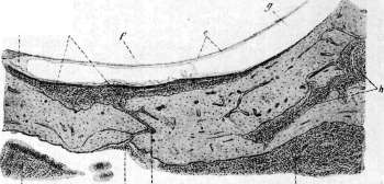
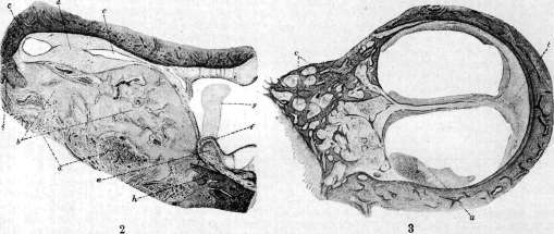
LABYRINTHITIS (labyrinthitis), inflammatory lesions in the area of the semicircular canals and vestibule. Often the cochlear apparatus is also affected in such cases. In these situations, one must speak of a disease of the entire inner ear (otitis interna). Labyrinthitis is divided into non-specific types, caused by pus-forming bacteria, and specific types, caused by tuberculous and syphilitic infection. Specific labyrinthitis arises primarily hematogenously, while non-specific types occur through the spread of infection from an inflamed middle ear (tympanogenic labyrinthitis) or from the cranial area (meningogenic). By course, acute labyrinthitis is distinguished, which can be caused by acute or subacute otitis media, and chronic labyrinthitis. By the extent of the inflammatory process, 1) limited labyrinthitis (labyrinthitis circumscripta) is distinguished, in which the inflammatory process is observed in one part of the labyrinth (oval window, round window, one of the semicircular canals); patho-anatomically, they have a proliferative character; 2) diffuse (labyrinthitis diffusa), which affects the entire labyrinth. These can be serous, sero-fibrinous, and purulent. The spread of infection from the middle ear to the labyrinth occurs through different pathways: for example, in otitis media, in which exudative phenomena predominate, infection spreads to the labyrinth through the windows. In proliferative inflammation, observed primarily in chronic middle ear lesions, the labyrinth becomes infected through the bony capsule. Destruction of the bony capsule occurs most easily and quickly in the area where the vascular canals are most developed (ampullae, area of the oval window, and facial nerve). The infectious process proceeds along the bony canals of vessels connecting the middle ear with the labyrinth; granulation tissue forms there, which expands the bony passages and thus gradually establishes direct communication between the middle ear and the labyrinth (Fig. 1). The mechanism of infection through the windows proceeds differently. In this case, infection of the labyrinth initially occurs while the integrity of the membranes is preserved through the direct passage of bacteria through the membrane. Subsequently, the membrane swells, is infiltrated by round-cell elements, then the tissue breaks down and ruptures. Tympanogenic labyrinthitis is observed in acute otitis media in 0.1% of cases, and in chronic cases in 1%. The largest number of labyrinthitis cases in chronic diseases occurs in cases with cholesteatoma (74%). Non-specific tympanogenic labyrinthitis. Limited labyrinthitis (fistula) (labyrinthitis proliferans circumscripta). Most often observed in chronic purulent otitis media with and without cholesteatoma and in tuberculosis of the middle ear. This disease is strictly limited. It is most often observed in the area of the semicircular canals, less often in the cochlea. The course is chronic, creeping. Often a sero-purulent labyrinthitis, caused by an exacerbation of chronic otitis media, joins the limited proliferative labyrinthitis (Fig. 1). In cases of recovery, which usually occurs after removal of the primary focus in the middle ear, the proliferative mass becomes scarred (Fig. 2). Symptoms. In addition to complaints of dizziness, occurring with rapid turns and tilts of the head, with relatively good hearing, a pressor nystagmus (fistular) can be elicited. When air pressure in the external auditory canal is increased, a horizontal nystagmus toward the diseased ear is obtained; the same can be achieved by pressing a finger against the tragus of the diseased ear. If the defect is located in the area of the promontory, the nystagmus is directed toward the healthy side (atypically). When air is rarefied in the auditory canal, nystagmus occurs in the opposite direction. Despite the presence of a defect in the bony capsule of the labyrinth, the fistular symptom cannot sometimes be elicited. This is explained by the fact that the defect is sometimes covered by dense granulation and cholesteatoma mass, which prevents the transmission of pressure to the perilymphatic and endolymphatic spaces. Examination of the labyrinth for rotational irritation reveals normal excitability, decreased excitability, or absence of excitability. Serous diffuse labyrinthitis is caused by the penetration from the middle ear into the labyrinth not of bacteria, but of toxins. The basis of the patho-histological process is collateral edema (hydrops labyrinthi). The penetration of bacteria, however, can cause sero-fibrinous exudate. At the same time, inflammatory hyperemia of the soft parts of the labyrinth is noted. Symptoms: decreased conductive-type hearing (decreased hearing, especially for low tones, prolongation of bone conduction), caused by disease of the middle ear, can be intensified due to involvement of the cochlea. General phenomena, such as headaches, temperature, may be more pronounced, but cannot be considered pathognomonic. Dizziness is considered pathognomonic: the patient feels that objects are moving toward the diseased or healthy ear; at the same time, the patient has a sensation of 'sinking,' 'falling' from the bed. These sensations are sometimes accompanied by vomiting, nausea, and are intensified with the slightest turn of the head. From the side of the eyes, spontaneous nystagmus is initially observed toward the healthy side, and later toward the diseased ear, which is usually caused by the increased excitability of the affected labyrinth. During this period, the typical reaction of deviation of the upper limbs toward the slow component of nystagmus can be noted. If the process progresses, the function of the diseased labyrinth will gradually fade, and then the direction of nystagmus changes toward the healthy ear. Examination of the caloric reaction in such cases reveals loss of function. Dizziness gradually passes, and if the labyrinthitis has a tendency not to go beyond its limits, then the spontaneous nystagmus toward the healthy ear also completely disappears over time. Purulent diffuse labyrinthitis. The inflammatory exudate consists of multinuclear leukocytes, the soft parts of the labyrinth are hyperemic and infiltrated with leukocytes, sometimes to the point of tissue dissolution (Fig. 3). Symptoms are the same as in serous labyrinthitis, but the course is more acute: sudden dizziness, almost complete loss of hearing, coarse horizontal-rotatory nystagmus, in contrast to the fine horizontal nystagmus in serous labyrinthitis, toward the healthy ear. Complete non-excitability of the diseased labyrinth. The outcome can be twofold. 1) The purulent focus gradually dissolves and organizes into a connective tissue-osteoid scar (Fig. 4); at the same time, subjective as well as objective (nystagmus) symptoms gradually disappear; the excitability of the inner ear is not restored, and over time, the same duration of nystagmus after rotation in both directions is observed (compensation). 2) The process ruptures through the internal auditory canal and the cochlear and vestibular aqueducts into the cranial cavity and causes intracranial disease (meningitis, epidural, subdural, cerebellar, cerebral abscesses). Such labyrinthogenic intracranial complications are often caused by labyrinthitis acuta serosa in subacute, prolonged otitis media caused by Streptococcus mucosus. In the tympanic cavity, the process usually ends but continues to spread to the mastoid process and posterior cranial fossa, where it causes great destruction. Usually with these otitis media, labyrinthitis develops 1-2 months after the onset of the disease. Labyrinthitis diffusa purulenta is observed much more frequently in chronic otitis media with cholesteatoma. This is explained by the fact that the cholesteatoma process causes gradual destruction of the bony labyrinth capsule. It has been repeatedly noted that cholesteatoma masses grew into the labyrinth. Cholesteatomatous otitis media, often causing fistulous labyrinthitis and maintaining the labyrinthine disease by its process, gives mainly chronic labyrinthitis. Thus, the function of the labyrinth can gradually fade, and then labyrinthitis proceeds without special subjective phenomena (latently). Only examination of labyrinthine function for caloric irritation reveals absence of nystagmus. Rotational reaction, if Figure 1. Bony vascular canal connecting the middle ear cavity and the capsule of the horizontal semicircular canal, and contributing to the formation of proliferative inflammation (d) in the perilymphatic space in purulent cholesteatomatous chronic inflammation. Granulation tissue (a) on the surface of the bony semicircular canal in the middle ear (b) penetrates into the vascular canal (c), blocked by round-cell infiltrate and passing into the perilymphatic inflammatory focus (d). The latter slightly compresses the wall of the endolymphatic canal (e); d-endolymphatic space; f-perilymphatic space; h-granulation focus that has destroyed the bony basis of the semicircular canal capsule, obtained as a result of expansion of the bony vascular canals. Figure 2. Healed limited inflammatory process in the area of the horizontal semicircular canal. The entrance (fistula-b) is closed by newly formed bone tissue (b), in which a granulation mass rich in blood vessels (a) is noted. The newly formed bone tissue completely closes the fistula and protrudes into the middle ear cavity and semicircular canal. In the perilymphatic space (d), in addition to proliferated bone tissue, a newly formed connective tissue mass is noted.
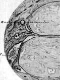
The endolymphatic canal of this last one is sharply deformed. The crista ampullaris with its constituent elements (cupula) is well preserved despite the prolonged inflammatory process that had been near it; postmortem changes in the area of the epithelium (spaces). Figure 3. Acute purulent labyrinthitis (section through the second turn of the cochlea). Bloody pus in the perilymphatic space (a). Abundant vascular engorgement and hemorrhage with round-cell infiltration in the ligamentum spirale (b). In the bony canals of the modiolus (c), all lymphatic spaces around the vessels and nerves are filled with a dense network of fibrin penetrated by pus. Figure 4. Healed purulent labyrinthitis with formation of bone tissue. Both cochlear staircases (scala vestibuli et scala tympani-a) and the contents of the lamina spiralis ossea (b) after preliminary death of nerve elements are filled with osteoid tissue. Newly formed bone tissue is noted in the well-preserved lig. spirale (c). The ductus cochlearis (d) is preserved in a round shape with a thickened wall of the membranae Reissneri (e). Complete absence of traces of the organ of Corti. The gangl. spirale (/) is comparatively well preserved. Figure 5. Longitudinal fracture through the upper wall of the auditory canal parallel to the axis of the pyramid and fracture of the apex of the pyramid (view from above). (Figures 1-4 according to Zange, fig. 5 according to Passow.) (For illustration of the article Labyrinth. Labyrinthitis.)
(
Ь c
\ &- See article Labyrinth, Labyrinthitis. The process is chronic, and the duration of nystagmus after rotation in both directions is equal (compensation). Traumatic tympanogenic L. is caused either by activation of the infectious agent present in a latent state in the middle ear, or by the introduction of infection from the outside with direct damage to the middle and inner ear. As a result of trauma, all types of labyrinthitis are observed with the above-mentioned symptom complex. Treatment of tympanogenic L. At the slightest suspicion of the appearance of L., it is strictly necessary to avoid the slightest shaking of the head, which could cause changes in both blood and general hydrostatic pressure in the skull; such a change may be caused by turning the head or the entire body, or by increased use of the abdominal press during defecation. Therefore, complete rest and monitoring of regular bowel movements (enemas, calomel) are recommended. For acute serous, and sometimes even purulent diffuse L., associated with acute purulent otitis, carefully performed conservative treatment with the application of cold to the ear area and nervina, urotropin internally, in a number of cases leads to recovery without the use of surgical intervention. However, in such cases, attention should be directed to detecting the slightest signs of developing meningitis or cerebellar abscess. It is necessary to verify a doubtful clinical diagnosis serologically (lumbar puncture) with attention to the number of leukocytes (from 1 to 5 in 1 mm³ is still considered normal). The general condition must be taken into account, for example, a decrease in headaches, a decrease in temperature (not above 38°) even with leukocytosis (up to 10) should still delay surgery on the labyrinth until the next puncture. In the presence of doubtful meningeal phenomena, deterioration of the condition of the cerebrospinal fluid gives grounds to proceed with surgery on the labyrinth. An indication for surgery is also an abscess of the cerebellum. The cardinal symptom of a cerebellar abscess is a large horizontal-rotatory nystagmus toward the diseased ear with loss of function on the side of the diseased labyrinth (Neumann's symptom) and spontaneous misplacement of the index finger on the side of the diseased ear. These symptoms appear earlier than the usual pathological cerebellar ones. Too cautious and restrained approach to surgical treatment of the labyrinth sometimes leads to the operation being delayed. But it is not excluded that sometimes surgical intervention can disrupt the anatomical relationships that have developed as a result of inflammation in the form of the formation of a protective barrier preventing further penetration of infection. Therefore, establishing indications for labyrinthotomy requires a careful analysis of the clinical picture. If acute otitis, which caused serous or even purulent L., is complicated by mastoiditis, then first of all, antrotomy should be performed and then one should wait. Similarly, one should proceed in cases of limited L. caused by chronic purulent otitis, with the only difference that in most cases a radical operation has to be performed. However, in cholesteatomatous processes, the following should be kept in mind: if a cholesteatoma penetrates into a necrotic labyrinth, being deprived of its source of nutrition in the middle ear (removed by surgery), it will not grow further and will not pose a great threat in terms of intracranial complications. Consequently, in such cases, there are no indications for labyrinthotomy. If, however, a cholesteatoma has penetrated into a still reactive labyrinth and thus still has the possibility of developing on the basis of the vessels present in it, then removal of the primary focus from the middle ear is not sufficient to stop the growth of the cholesteatoma. Then there is an indication for careful removal of all pathological tissue from the labyrinth. Operations. Of the many methods proposed, those of Neumann and Ruttin are used more often than others. Neumann, after radical trepanation and exposure of the boundaries of Trautmann's triangle, reaches the posterior semicircular canal and opens the horizontal semicircular canal at the site of its posterior crus. Then follows the opening of the vestibule from behind and the opening of the cochlea by removing the promontory from the lowest anterior edge of the oval window downward and forward. In Ruttin's method, the path to the labyrinth is made from behind not through the posterior semicircular canal, but through the lateral semicircular canal. From the front, as in Neumann's method. Meningeal L. The free communication that exists between the subarachnoid space and the inner ear through the cochlear aqueduct and the internal auditory canal sufficiently clearly explains the frequency of complications from the inner ear in various diseases of the meninges. Among them, in terms of frequency, infectious processes are in first place, with spinal meningitis, and among non-infectious ones - tumors and chronic serous meningitis in the posterior cranial fossa. Almost half of all deaf and deaf-mute people owe their deafness precisely to spinal meningitis. Pathological changes are found exclusively in the cavities of the labyrinth with completely normal or slightly changed eardrums and middle ear cavities. Specific L. Tuberculous L. (tuberculosa) patho-anatomically manifests as 1) proliferative L. with the formation of tubercles and abundant tumor-like growths, and the process involves not only the bony but also the membranous labyrinth; 2) serous L.; the serous process is primary, and to it is joined a proliferative one; the process proceeds slowly, asymptomatically, with a gradual extinction of function; 3) purulent L.; it is extremely difficult to clinically establish the tuberculous nature of L., as it is rarely possible to find tubercle bacilli in serous and purulent secretions. Sometimes, based on the course of the process (asymptomatically, slowly) and the nature of the otitis that caused L., it is possible to make a presumptive diagnosis. A distinctive feature of tuberculous L. is that they much less frequently lead to meningitis than L. associated with acute purulent otitis. Tuberculous L. can heal spontaneously, which most often occurs in the serous form after removal of the focus in the middle ear. Treatment of tuberculous L. Chronic serous tuberculous L. very often respond to conservative treatment under the influence of climatic and light therapy (heliotherapy). Acute purulent L. in tuberculosis of the middle and inner ear require immediate opening of the labyrinth, as in these cases there are few chances that during the course of the disease, protective connective tissue growths can form on the path to the cranial cavity. Syphilitic lesions of the VIII pair (most often the process involves the meningeal covering of the nerve with its vessels - meningo-vasculitis) depending on the extent of the process, are considered either as L. or as neuritis and finally as poliencephalitis. Since clinically it is often impossible to differentiate these diseases, it is practically advisable to treat them as neuro-labyrinthitis. These lesions are observed in different stages of luetic disease: in secondary, tertiary syphilis, tabes, early and late congenital syphilis. In secondary syphilis, neuro-labyrinthitis often occurs simultaneously with acute purulent syphilitic otitis. The otoscopic picture in this case (slight hyperemia of the eardrum, scanty secretions) does not correspond to the data of functional examination. Hearing research indicates a lesion of the cochlear apparatus (shortening of bone and air conduction for high tones, spontaneous nystagmus in both directions, reduced excitability to caloric irritation, and sometimes extinction with preservation of excitability to rotational). Both branches of the n. acustici n. vestibularis are more often affected than the n. cochlearis. In contrast to these tympano-labyrinthine syphilitic lesions in secondary syphilis, neuro-labyrinthitis are much more common. Usually they are accompanied by symptoms of specific meningitis. In general, in most cases they are caused by meningo-neuritis of the VIII nerve. Symptoms can manifest in three forms: acute, severe, subacute. 1. Suddenly, in complete health, without any prodromes, the patient experiences severe noise and ringing in the ears, nausea, vomiting, dizziness and completely loses hearing. If specific treatment is not started immediately, deafness becomes permanent. 2. Initially, the patient notes intermittent noises in one or both ears, then accidentally when turning the head, experiences dizziness. Subsequently, hearing gradually decreases, and dizziness intensifies. This continues for 2 to 3 weeks, after which hearing further deteriorates and may completely disappear if specific therapy is not urgently applied; dizziness gradually disappears. 3. Sometimes the process proceeds covertly. The patient cannot accurately determine the time of onset of the disease. Minor hearing impairments may last for several weeks or months, but with a tendency to increase. Attacks may take an intermittent course. Latent luetic L. are also observed: hearing loss and disappearance of vestibular function may occur unnoticed by the patient.
Such latent lesions of the labyrinth sometimes occur together with symptoms of paresis or paralysis of the facial nerve. Lesions of the labyrinth are observed in an average of 15-20% of cases of secondary syphilis. The prognosis is favorable with immediate and energetic specific treatment. The symptoms of neuro-labyrinthitis in the 3rd stage begin to appear in the 3rd year from the start of the infection. Of all the luetic diseases of the labyrinth, 7/10 fall on diseases of the 3rd period. The symptoms are the same as in diseases in the 2nd stage. The basis of syphilitic neuro-labyrinthitis can be three phenomena: meningo-neuritis of nerve VIII, primary cellular lesions of labyrinth centers, and lesions of nerve endings in the labyrinth. Early congenital syphilitic oto-labyrinthitis. Children with such lesions give a high percentage of deaf-mutes. At its basis lies either a dystrophic process - atrophy of the auditory nerve and anomalies of development up to cortical centers, or specific - lesions of the labyrinth, meningitis involving nerve VIII, spirochetes in the auditory and facial nerves with a healthy labyrinth, or degenerative neuritis without disease of the membranes, endarteritis in the labyrinth, changes in the ganglia of the cochlear and vestibular nerves. Neuro-labyrinthitis in late congenital syphilis is significantly easier to diagnose than in early syphilis. The disease begins at an age of 6 to 33 years and proceeds sometimes slowly, sometimes quickly. In general, it can develop over a period from several days to 10-18 years. At a certain stage of the disease, the process can become stationary or intensify or weaken. Symptoms: bone conduction is almost normal (or slightly shortened or prolonged), as well as in the Schwabach test; Rinne-negative; hearing for conversational speech is sharply reduced; reaction to caloric irritation is partially or completely preserved while there is no reaction to rotational irritation; galvanic reaction is disturbed (nystagmic reaction is normal, static is absent); fistula symptom is positive [when pressing on the tragus - nystagmus toward the opposite ear, and it can take different forms: horizontal, rotatory, diagonal (Nepbert's symptom)]. The prognosis is unfavorable. At its basis lies a primary lesion of the bony capsule, peri- and endolymphatic spaces, destruction of nerve elements. Treatment of all the above-mentioned manifestations of syphilis, both osteoperiostitis and neuritis and meningo-neuritis, comes down to the use of mixed specific therapeutic agents (see Syphilis). Contrary to the opinion that the sometimes observed during treatment with neosalvarsan sharp decrease in hearing up to its loss is allegedly caused by arsenical intoxication neuritis, Ehrlich expresses the view that this phenomenon is a purely specific disease of the nerves after the application of salvarsan (neurorecurrence), caused by insufficient treatment. Therefore, to eliminate the neurorecurrence, it is recommended to repeat the specific treatment in a more intensive form. It is possible to distinguish neurorecurrence from arsenical neuritis as follows: the first appears several weeks after completed treatment with neosalvarsan, while the second - during treatment with it. Pilocarpin should also be included among the specific preparations. It facilitates the absorption of intra-labyrinthous exudate. Pilocarpin is administered subcutaneously after previously conducted specific treatment.
Related articles
Mentioned in
Cite this page
“Labyrinthitis.” Soviet Medical Encyclopedia. English translation of Bolshaya Meditsinskaya Entsiklopediya, 1st ed. (Moscow, 1928–1936), ed. N. A. Semashko. https://sovietmedicalencyclopedia.pages.dev/article/labyrinthitis/