Denervation
Historical document, translated for reference. It reflects medical knowledge of the 1920s–30s and is not medical advice.
Summary
Denervation, or sympathectomy, is the surgical removal or severing of sympathetic nerves to a body part, affecting vascular tone, glandular activity, or interrupting trophic, pain, or deep sensitivity pathways. The technique was first performed in 1883 by William Alexander for genuine epilepsy, but comprehensive development of the surgical anatomy, technique, and clinical application is credited to the Lyon school of surgeons led by Jaboulay.
Encyclopedia article (1928–1936)
Denervation, sympathectomy, denudation and decortication (of arteries), the deprivation of sympathetic innervation of a particular area of the body. Corresponding to the physiological role of the sympathetic nerve (see Autonomic Nervous System), the operation of denervation affects vascular tone, or the activity of the corresponding gland, or interrupts the course of trophic, pain, or deep sensitivity nerves. Technically, the operation consists in the severing or excision of nerves; this is done in cases where a long-term or permanent result is desired; when it is only necessary to temporarily deprive an organ (or area) of sympathetic innervation, then medicinal substances that interrupt conductivity for a shorter period are administered. The first denervation operation was performed by the English surgeon William Alexander (1883); he removed the upper cervical sympathetic ganglion for genuine epilepsy. However, the comprehensive development of the surgical anatomy, technique, and clinic of denervation belongs to the Lyon school of surgeons led by Jaboulay. Operations on the sympathetic nerve are more often performed under general anesthesia (Jabouley).
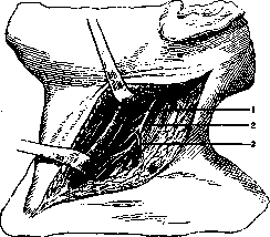
Figure 1. Approach to the cervical part of the sympathetic nerve: 1-internal jugular v.; 2-sympathetic n.; 3-inferior thyroid a. (From Biuning-Stahl).
For an operation on the cervical part, the patient is placed in a semi-sitting position (Jabouley, Chalier) with the head thrown back and the face turned to the opposite side. The incision of the skin is from the mastoid process to the clavicle along the anterior edge of the sternocleidomastoid muscle (if it is necessary to expose the upper part of the cervical sympathetic nerve) or along its posterior edge (for operations on the lower ganglia); to remove the entire cervical sympathetic nerve, either incision can be used; in the subcutaneous layer, the external jugular vein is ligated and severed. The aponeurosis is incised; the muscle is displaced either medially or to the side; along its entire course, the vascular-nerve bundle is isolated; it is lifted, and then the muscles lying on the vertebrae and the aponeurosis covering them are exposed.
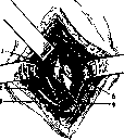
Figure 2. Right scalene-vertebral angle. Exposure of the stellate ganglion in the region of the vertebral artery: 1-intermediate ganglion; 2-thyroid artery; 3-phrenic nerve; 4-subclavian artery; 5-stellate ganglion. (From Leriche).
Immediately inside the anterior tubercles of the cervical vertebrae lies the sympathetic nerve (see Figure 1). Depending on the need, it is removed either entirely or partially; sometimes only its peripheral branches or connecting branches are severed. For cases requiring removal of one stellate ganglion, Leriche advises making an incision along the course of the sternocleidomastoid muscle fibers, then penetrating between its two legs to reach the depth and locate the a. vertebralis, which, according to Leriche, is the most important landmark: the stellate ganglion lies behind it, at the point of its origin from the subclavian artery (see Figure 2). The solar plexus is exposed by median laparotomy. Lyon surgeons performed its "retraction" by pushing away the surrounding tissues from the truncus coeliacus; injection of anesthetic substances into the area of the solar plexus is possible both by laparotomy (Czerny) and by posterior puncture under the XII rib according to Kappis. The lumbar and sacral parts of the sympathetic trunks are exposed by laparotomy below the navel along the median line in the Trendelenburg position; by incising the posterior leaf of the parietal peritoneum along the lateral surface of the vertebrae, the sympathetic nerve is exposed, and both its trunk and connecting branches become accessible for excision (Jonnesco). Jabouley at one time approached the sacral part of the sympathetic nerve through the perineum; by separating (decollement) the rectum from the sacrum, he tore any nerves encountered in his path; in other cases he removed the entire chain of ganglia at once. The connecting branches in the thoracic part are exposed by a longitudinal incision about 3 cm to the side of the line of spinous processes; the muscles are retracted, the transverse processes are severed at their base; the posterior branch of the intercostal nerve serves as a guide, along which one reaches the connecting branch by separating the intercostal muscles. If the operation is performed under local anesthesia, one can check oneself with electrodes before severing the connecting branch: touching them will cause the same pain for which the operation is undertaken. For denervation of peripheral arteries, two methods can be used: in the first, the nerves going from the mixed nerve to the artery accompanying them are severed; this is achieved either by visually cutting the nerve twigs that approach the artery at different levels - the operation of Prokin (1927), or by stripping the artery from its sheath for a distance of several centimeters (Jabouley's "denudation" operation). Leriche (1914) developed another method in which the outer sheath of the artery is removed for a length of 7-10 cm, along with the sympathetic nerves embedded in it; for this, the artery is freed from surrounding tissues as for ligation, then the outer sheath is lifted with two small forceps into a transverse fold and cut through to the muscular layer; the incision is continued downward for the required length, guiding with a grooved probe. Then the outer sheath is removed like a cuff from the entire circumference of the artery. However, this is not always successful; more often removal occurs in pieces. To find any remaining parts, it is recommended to pour hot physiological solution over the artery, from which the whitish remnants of the outer sheath become clearly visible. When the operation is properly performed, the area deprived of the outer sheath narrows significantly. The wound is sutured tightly. Razumovsky instead of removing the outer sheath proposes injecting 70% alcohol into it. Denervation is indicated, first, for severe pains of unknown cause (for example, neuralgia) or from intractable suffering (for example, inoperable cancer). Second, denervation is indicated for certain vascular disorders (vasomotor aura of epileptics, spasms of limb vessels in the pre-gangrenous period). Third, for diseases requiring activation of the process in the sense of enhancing arterial blood circulation, improving tissue nutrition, and enlivening granulations (for example, leg ulcers, trophic and gangrenous ulcers of the extremities, scleroderma). Operations on the cervical part of the sympathetic nerve, including denervation of the carotid artery, are performed for trigeminal neuralgia, for epilepsy (see), which begins with vasomotor phenomena, for Basedow's disease (see), for migraine with facial pallor, for glaucoma, for angina pectoris, for bronchial asthma. Anesthesia of the splanchnic nerves, according to Kappis, has been successfully applied in reflex anuria (Neuwirth). Excision of the sacral part of the sympathetic nerve is performed for sciatica, vaginismus, and other neuralgias of pelvic organs, for trophic disorders and spontaneous gangrene of the lower extremities. Severing of the communicating branches (rami communicantes) is performed for neuralgia, painful stumps, for "gastric crises" in tabetics, for traumatic aseptic edema of the extremities. Denervation of arteries is applied in nephritis dolorosa, for pains from inoperable cancers, for dysmenorrhea, for leg ulcers, for Raynaud's disease, for intermittent claudication, for spontaneous gangrene, for trophic ulcers, for bone and joint tuberculosis, and for fractures (with the aim of enhancing and accelerating callus formation). The result of denervation is the immediate disappearance of pain. Removal of the cervical sympathetic nerve, and sometimes also denervation of the carotid artery, causes the appearance of Horner's triad (pupil constriction, narrowing of the palpebral fissure, and enophthalmos); then they lead to a decrease in intraocular pressure, increased lacrimation, decreased sweating, and hyperemia of the corresponding half of the face. Severing of the communicating branches causes a change in "deep" sensitivity: the limb becomes weaker, heavier, and the previous confidence in movements is lost. Denervation of the vessels causes redness of the extremity after a few hours due to active hyperemia, which lasts for several weeks. Exudation from ulcers increases, granulations become more lively, and ulcers often heal quickly, and strangely enough - not only on the operated side but also on the opposite side, if any existed. But these favorable consequences are not permanent - usually after a few months, both pain and trophic disorders recur. According to Rubashov, denervation for causalgia gives 50% permanent recoveries, for leg ulcers - 25%, and for gangrenous phenomena - 20%. Apparently, regeneration of sympathetic nerves occurs; but perhaps other nerves take over the function of the removed ones. A number of severe complications have been described in connection with denervation; thus, thrombosis of arteries denuded of their outer sheath have been described, ruptures of operated arteries, suppurations; these complications most often lead to amputation. The complications just mentioned make it preferable to perform Jabouley's or Prokin's operation rather than Leriche's, as the first two are significantly less traumatic to the artery. Then, arteries with healthy walls, non-sclerotic, tolerate Leriche's operation better; it is known that the inner part of the arterial wall in young people is nourished by the blood flowing through its lumen; such an artery tolerates removal of the outer sheath successfully; in elderly people with sclerosis of the inner membrane, arteries are nourished more through the vasa vasorum, which develop abundantly in the outer sheath, whose removal in this case will cause obvious harm, and often death of the arterial wall.
Related articles
Cite this page
“Denervation.” Soviet Medical Encyclopedia. English translation of Bolshaya Meditsinskaya Entsiklopediya, 1st ed. (Moscow, 1928–1936), ed. N. A. Semashko. https://sovietmedicalencyclopedia.pages.dev/article/denervation/