Facial Nerve (facial nerve, the seventh pair of cranial)
Historical document, translated for reference. It reflects medical knowledge of the 1920s–30s and is not medical advice.
Summary
The facial nerve is the seventh cranial nerve that controls facial muscles and transmits taste sensations. This article describes its anatomical course, functions, and various pathological conditions including paralysis.
Encyclopedia article (1928–1936)
Facial Nerve, facial nerve, the seventh pair of cranial nerves, emerges from the posterior part of the pons Varolii. Together with the intermediate nerve of Wrisberg and the auditory nerve, it enters the internal acoustic opening of the temporal bone. Further, the facial nerve separates from the auditory nerve and enters the canal of the facial nerve (canalis n. facialis), called the Fallopian canal, located in the pyramid of the temporal bone. The course of the facial nerve in the Fallopian canal is winding. At its beginning, the nerve runs horizontally forward, then, after participating in the formation of the geniculate ganglion, it turns backward and passes along the inner wall of the tympanic cavity. Having passed beyond its limits, the facial nerve turns downward and exits externally through the stylomastoid foramen. Still within the bony canal, the facial nerve gives off anastomoses to the spheno-palatine ganglion (pterygopalatine ganglion) and to the otic ganglion (Arnold's ear ganglion). The most significant of the branches separating in the canal is the chorda tympani. After leaving the facial nerve, the chorda tympani penetrates into the tympanic cavity through a special bony canal and exits through the fissure of Glasser. Subsequently, having incorporated the fibers of the lingual branch of the trigeminal nerve, the chorda tympani branches in the submandibular and sublingual glands and in the anterior parts of the tongue. In the canal, the facial nerve gives off a small branch (n. stapedius) for the innervation of the muscle of the same name. After exiting the canal, the facial nerve gives off the posterior auricular branch, which innervates the muscles of the auricle, the stylohyoid nerve (n. stylohyoideus) for the muscle of the same name, and the digastric nerve (n. digastricus) for the posterior belly of the m. biventer. After giving off these branches, the facial nerve penetrates the parotid gland (fig. 4), passes through it, and forms in front of the external auditory meatus the greater goose's foot (pes anserinus major), from which branches depart for the mimetic muscles of the face. The largest branches of the n. facialis on the face are the temporal, buccal, labial, and marginal mandibular branches; for the innervation of the platysma myoides on the neck, the subcutaneous branch of the facial nerve descends (ram. subcutaneus colli n. facialis).- The main mass of the facial nerve consists of motor fibers. Secretory and possibly gustatory fibers go mainly in the chorda tympani, into which they enter from the intermediate nerve of Wrisberg and from the greater petrosal nerve. According to Hunt, general sensitivity fibers also pass in the facial nerve. In some animals, including fish, the facial nerve is undoubtedly a mixed motor-sensory nerve. In its trunk in humans, numerous fibers of the autonomic nervous system pass. The nucleus of the facial nerve (nucleus n. facialis) is represented in the pons Varolii by cells located in the covering of the pons Varolii near the nucleus of the abducens nerve. The nucleus of the facial nerve ends in the very anterior part of the medulla oblongata. At the height of its development, the nucleus consists of three groups of cells, of motor type according to Nissl's structure; Cells detached from the main nucleus are located dorsally from it and are united under the name of the accessory nucleus (nucl. accessorius n. facialis). In its phylogenetic development, the nucleus of the facial nerve undergoes displacement (neurobiotaxis according to Kappers): in lower vertebrates it lies dorsally and as the animal develops it shifts ventrally. The cortical center of the facial nerve is located in the lower quarter of the anterior central gyrus. The cellular groups for the frontal branches lie higher than for the oral branches. The axons of the cells of the cortical center of the facial nerve, descending to the pons, are located in the internal capsule somewhat in front of the fibers for the limbs. Partially not quite reaching the nuclei of the facial nerve in the pons, partially already at their level, the corticobulbar fibers of the facial nerve cross in the pons and approach the cells of the nucleus of the facial nerve of the opposite side. Part of the uncrossed fibers ends in the nucleus of its own side. The root of the facial nerve is formed from the axonal processes of the cells of its own nucleus, a very small part of the fibers enters it from the nucleus of the opposite side.- Through the facial nerve, most reflexes of the face are realized, both from mucous membranes and from skin-sucking, blinking, partly corneal, conjunctival, sneezing, nasolabial, etc. In intrauterine development, oral reflexes are the earliest. They are already elicited in a 2-month fetus, when others are still absent. Pathology. Lesion of the facial nerve is a component part of a huge number of syndromes of cortical, subcortical, and stem localization. Among the independent diseases of the facial nerve, the most common is its paralysis (syn. prosoplegia* syn. Bell's palsy). Among the paralyses of individual cranial nerves, facial nerve paralyses occupy second place after the oculomotor nerves. Among etiological factors, first place is taken by cooling, causing the so-called rheumatic paralysis of the facial nerve. The basis of rheumatic paralysis is acute infectious neuritis. There is a familial and individual predisposition to rheumatic paralysis of the facial nerve. The proximity of the facial nerve in its intrabony course to the organ of hearing sometimes leads to its paralysis as a complication of purulent otitis. Relatively rare are paralyses of the facial nerve in general acute infections; syphilis plays a much greater role in its origin. Inflammatory and neoplastic processes at the base of the brain often cause unilateral, more rarely bilateral, paralysis of the facial nerve. Its paralyses are also part of the syndromes of both diffuse and selective nuclear lesions of the pons Varolii in hemorrhages, bulbar paralysis, poliomyelitis, amyotrophic lateral sclerosis. Paralysis of the lower part of the facial nerve is a common component of capsular hemiplegia. Traumas of the base of the skull, temporal bone, parotid gland, and face also often lead to paralyses of the facial nerve. In military material of cranial nerve paralyses, the facial nerve occupies first place. Surgical intervention on the parotid gland and temporal bone, used mainly for purulent processes, often entails paralysis of the facial nerve. Congenital paralysis of the facial nerve is caused by the absence or underdevelopment of nuclear cells or a defect in the structure of the Fallopian canal. Birth paralysis of the facial nerve is most often caused by the application of forceps, and sometimes by pressure on the emerging face from the side of the normal or pathologically changed (exostoses) symphysis of the mother. Facial paralysis occurs predominantly between the ages of 20 and 24 years. Acutely occurring in children paralysis of the facial nerve with general infectious phenomena and elevation of temperature serves as a manifestation of pontine poliomyelitis, sometimes caused by the poliomyelitis virus. Paralysis of the facial nerve is as a rule a single disease. It recurs, according to Bernhardt, in 7% of all cases. Recurrent paralyses of the facial nerve are located somewhat more often on the side of the first lesion than on the opposite side. The recurrence usually occurs once, as an exception twice and up to four times. Sometimes recurrent paralysis of the facial nerve is a determining component part of a complex syndrome (see Recurrent paralysis) with phenomena of migraine-like character (Rossolimo). Usual- 593
However, peripheral paralysis of the facial nerve develops acutely. Paralyses of traumatic origin or developing on the basis of otitis or endogenous intoxication (diabetes, gout) immediately manifest as motor disorders. In rheumatic paralysis of the facial nerve, in half of the cases it is preceded by a sensory disorder in the form of pain in the ear and parotid region. It is not decided whether these pains depend on a simultaneous disease of the sensory nerves of the head (trigeminal, occipital) or they are caused by a lesion of the sensory fibers of the facial nerve itself. Objective changes in sensitivity in paralysis of the facial nerve occur, according to Donath, in 11%. Paralysis of the facial nerve of otitic origin, as well as compression in brain tumors, is sometimes preceded by mild twitching for hours and even days. Paralysis of the facial nerve caused by gross trauma, e.g. its severing, occurs immediately. Paralyses of other etiology, including rheumatic ones, usually develop within the first 24-48 hours.
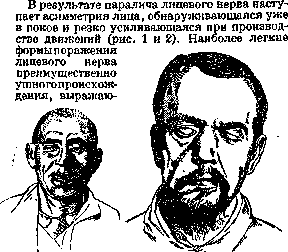
Figure 1.
Figure 2.
Figure I. Paralysis of the left facial nerve. Figure 2. Complete paralysis of the facial nerve. Appearance of the patient's face when closing the eyes; on the right—incomplete closure of the eyelids, manifested only by a slight asymmetry without gross motor deficits, are distinguished under the name of hemimimia (hemimimia). The forehead wrinkles on the paralyzed side are smoothed out, the nasolabial fold loses its depth, the palpebral fissure widens (lagophthalmus) due to paralysis of the orbicularis oculi muscle, and the corner of the mouth drops. Tears flow from the eye on the paralyzed side (epiphora), since the displacement of the opening of the tear duct occurs due to the paralysis of the orbicularis oculi muscle and Horner's muscle. When attempting to close the eye on the paralyzed side, the eyeball moves upwards, sometimes inwards. This phenomenon was named by Bernhardt in honor of the author who first described it, Bell's phenomenon. Essentially, Bell's phenomenon is a normal associated movement that is revealed during paralysis due to the non-closure of the palpebral fissure. On the paralyzed side, the ability to produce separate closing of NERVUS
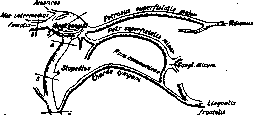
504 one eye disappears—Revilliod's symptom. The tip of the nose in facial nerve paralysis is pulled toward the healthy side, the nostril opening is narrowed on the paralyzed side. Toward the healthy side, even at rest, the corner of the mouth is also pulled, which is especially pronounced when showing the teeth. Protrusion of the lips like a trunk forward, raising and everting them on the paralyzed side is impossible. When evertive movements are attempted, paralysis of the usually associated contracting platysma is also revealed. Taking food is difficult, because it is not moved due to the loss or weakening of the buccal musculature; food often gets stuck between the teeth and the cheek. Motor disorders are sometimes joined by secretory and trophic ones due to damage to autonomic fibers. The production of tears and saliva changes in the direction of increase or decrease. The face on the paralyzed side is often puffy. In far-advanced cases, atrophy of the skin and even bones sometimes occurs on the paralyzed side. With bilateral facial nerve paralysis, the above-described symptoms are observed on both sides (see Diplegia facialis). Peripheral facial paralysis, as a general rule, has a flaccid character. The peculiarity of the mimic musculature—the small size of individual muscles, their location in the subcutaneous tissue—makes mild atrophy difficult to catch. Of particular importance as an indicator of flaccid facial nerve paralysis is the change in electrical excitability. Numerous facial reflexes disappear in peripheral facial nerve paralysis. Lesions of individual branches of the facial nerve on the face, mainly of traumatic origin, are expressed by paralysis of the corresponding musculature. The involvement of the nerve to the stapedius in facial paralysis is manifested by increased sensitivity to auditory stimuli, especially to low tones, on the paralyzed side (hyperacusis). Level diagnosis of facial nerve paralysis is based on taking into account both the features of the paralysis itself and the symptoms from other parts of the nervous system, resp. individual nerves. In supranuclear central paralysis, especially, and sometimes exclusively, the lower branch of the facial nerve is affected. Electrical excitability and reflexes do not change, sometimes pathological reflexes appear (Oppenheim, Toulouse, Bekhterev, Fuchs, and others). For topical diagnosis of peripheral facial paralysis, the regularities established by Erb are used. When a lesion occurs between points 1 and 2 (Fig. 3), paralysis of all facial musculature occurs with the preservation of taste, hearing, and innervation of the palate. When the lesion is localized between points 2 and 3, a taste disturbance on the anterior 2/3 of the tongue and salivation are added to the paralysis of the facial musculature due to damage to the chorda tympani. The localization of the disease between points 3 and 4 is sometimes characterized by the addition of the hyperacusis syndrome to the just outlined syndrome. For lesions of the geniculate ganglion, the complex Ghent syndrome is recognized as more or less characteristic: a combination of painful facial paralysis with hearing impairment and a herpes eruption on the auricle, and sometimes paresis of the soft palate. When the facial nerve is diseased above the geniculate ganglion, i.e., at the base of the brain, peripheral facial nerve paralysis is exclusively motor in character. It is also preserved with nuclear localization of the paralysis. As a difference from radicular facial nerve paralysis, nuclear ones are more characterized by a certain dissociation in favor of greater involvement of the lower part of the face. Hysterical facial nerve paralysis is rare. Sometimes
-r zygomatici ph-r. buccales-

Figure 3. Scheme of the facial nerve. Lines 1, 2, 3
and so on—for topical diagnosis of facial nerve disease. in organic paralysis of one side, hysterical develops on the opposite—Ziehen's automimetic paralysis. Peculiar phenomena in the pathology of the facial nerve are hypertonia and hyperkinesia. Sometimes they develop simultaneously with paresis in the same muscles, for example, in head tetanus. Sometimes hypertonia of some muscles goes along with paresis of others (Lazarev's syndrome in diseases of the base of the brain, Josephowicz's syndrome in temporal tumors). Hypertonia and hyperkinesia are especially frequent in the recovery period of facial nerve paralysis, where they are part of the concept of "secondary contracture" of the facial nerve. As a result, a peculiar asymmetry is formed on the side of the facial nerve lesion, opposite to that which characterizes the first period of paralysis. On the paralyzed side, the nasolabial fold becomes sharper, the palpebral fissure narrows. When innervating muscles of one level, the musculature of another level is synergistically tonically contracted. At rest on the paralyzed side, lightning-fast clonic twitchings in the chin muscles, in the circumzygomatic musculature (pommette—French), and in the orbicularis oculi muscle are not uncommon. Hypertonic phenomena are much rarer in central facial paralysis. In these cases, they are localized either on the side of the paralysis or on the opposite one. In the area of facial musculature, primary spasms, single and bilateral, are frequent (see Tics). Sometimes facial nerve paralysis is preceded by myokymia; sometimes it occurs with tonic forms of facial spasms. In generalized myokymia, the face is usually spared. Prophylaxis of facial nerve paralysis in subjects who have already suffered this disease once boils down to protecting the face from cooling. The prognosis in facial nerve paralysis is based on taking into account the underlying disease, the duration and severity of motor disorders, and the degree of changes in electrical excitability. Severe forms of the reaction of degeneration in the 3rd to 6th month of paralysis indicate the irreversibility of the process. Treatment of facial nerve paralysis boils down first of all to the therapy of the underlying disease (otitis media, syphilis, neoplasms, etc.). At the beginning of rheumatic paralysis, salicylates and local counter-irritants are appropriate. Subsequently, electrotherapy, massage, diathermy, mud therapy (surgical intervention on the nerve and cosmetic operations on paralyzed muscles—see below) are used.
M. Neiding. Injuries to the facial nerve and their surgical treatment. In cases where therapeutic treatment of facial nerve paralysis is unsuccessful or when there are large defects of the nerve (for example, when removing the parotid gland along with the nerve), where regeneration cannot be expected, surgical intervention is indicated, which is divided into three groups: 1) all kinds of plastics aimed at achieving a cosmetic result, 2) methods for reviving the facial muscles, 3) operations on the sympathetic nervous system. Operations of the first group include all kinds of lifting of the corner of the mouth and lower eyelid using Bush silk, thread, Momburg wire, or Stein fascia. Incisions for operations to lift the corner of the mouth follow the nasolabial fold (plica naso-labialis), from which the orbicularis oris muscle is dissected. To this, fascia is attached, fixed at the other end to the zygomatic arch, either by wrapping it around or pulling it through a hole made in the bone. Burk divides the fascia into four ends, suturing one to the wing of the nose, the second and third in a V-shape to the corner of the mouth, and the fourth to the triangular muscle of the lower lip. In all these operations, due to the subsequent drooping of the corner of the mouth, it is necessary to perform hypercorrection by 1-2 cm. To the operations of the second group, restoration of the facial nerve by means of suture should be primarily attributed. The shortness of the trunk of the facial nerve makes indication for this intervention very rare. Therefore, comparatively long ago, grafts from the accessory nerve and hypoglossus nerve were proposed. Suturing the peripheral end of the facial nerve to the central end of the XII pair is performed more often and gives better results than the same operation with the XI pair. After plastic surgery with the latter, a number of associated movements are often observed: grimaces are accompanied by movement of the shoulder girdle. The better results of suturing with the XII pair are explained by the proximity of the center of this nerve to the center of the facial nerve (see. Medulla oblongata). The negative side of this method is the impairment of speech, characterized by lisping. The technique of anastomosis with the XII pair - an arcuate incision from the mastoid process to the hyoid bone, convex downward. The subcutaneous muscle of the neck, the external jugular vein, and the facial vein are severed. The trunk of the facial nerve is visible after incision of the parotid-masseteric fascia (fascia parotideo-masseterica) and sharp upward traction of the parotid gland (Fig. 4). According to Taylor, removal of a small bone plate more widely opens the stylomastoid foramen. The hypoglossal nerve is easily found and isolated, as it forms the upper edge of Pirogov's triangle (see. Neck), and becomes visible when lifting and sharply pulling upward the submandibular gland. To isolate the hypoglossal nerve, the digastric and stylohyoid muscles are severed posteriorly. The anastomosis can be applied either by the method of lateral implantation or the central part of the completely severed hypoglossal nerve is implanted into the peripheral facial nerve. Results should not be expected earlier than six months. Haberland performs implantation of thin branches of the hypoglossal nerve directly into the mimetic muscles of the face (neurotization). These methods compete with muscle plastic surgery. Lexer proposed using the masseter muscle to replace the affected orbicularis oris muscle and the temporal muscle to close the eyelids, as these muscles receive innervation from the trigeminal nerve. Incisions along the nasolabial fold (plica naso-labialis) 1½-2 cm and along the lower edge of the lower jaw. From the first, the orbicularis oris muscle is dissected, from the second by sharp upward traction, the anterior edge of the masseter muscle is exposed, from which a flap with the upper limb is separated (Fig. 5). According to the anat.-clin. investigations of G. Richter, the muscle must be taken necessarily to the bone, as the branches of the trigeminal nerve run for a long distance on the posterior side of the muscle near the periosteum, giving branches into the muscular tissue. Both skin incisions are connected by a tunnel, through which the muscle is pulled and sutured after preliminary splitting to the orbicularis muscle of the upper and lower lip. To form a flap from the temporal muscle, the incision is made along the edge of the hairy part, the muscle flap about 2 cm wide is also taken to the bone and necessarily with the temporal fascia. With a second incision extending from the upper eyelid to the lower eyelid at the outer corner of the eye, the orbicularis muscle is exposed. Special attention must be paid to ensuring that the peripheral part of the flap is reinforced as far as possible to the middle part of the lower eyelid. For muscle plastic surgery, Jianu also proposed the digastric muscle without favorable results; Gomoiu for the same purpose proposed the sternocleidomastoid muscle, which was successfully performed by Hildebrand. Gersuny proposed transplantation of the orbicularis oris muscle from the healthy side. Leriche to combat lagophthalmos proposed transection of the sympathetic trunk under the superior cervical ganglion. In Leriche's results were excellent. Novikov in removal of a tumor of the parotid gland along with the facial nerve to mask the paralysis successfully resected the superior cervical ganglion. Albert, Jianu and Butsoianu remove the superior and middle nodes; Geymanovich recommends resection of the truncus sympathicus, etc. Hesse, in addition to removing the lower two-thirds of the superior ganglion, performs resection of the branches (ramicotomy C8-T1 or C6-C7). Leriche and Fontaine consider it recently necessary to remove the entire sympathetic trunk below the middle ganglion. The results obtained by most authors are good, but only with respect to the eye; improvement in eyelid closure is achieved due to the phenomenon caused by removal of the sympathetic nerve - Horner's syndrome: 1) recession of the eyeball due to paralysis of the smooth muscle fibers in Tenon's capsule, 2) narrowing of the pupil as a result of paralysis of the muscle that dilates the pupil (m. dilatator pupillae), 3) narrowing of the palpebral fissure due to paralysis of the so-called Müller's muscle fibers of the eyelid, 4) increase in blood supply of the corresponding half of the scalp and the appearance of some secretory phenomena. Most physicians at the present time perform intervention on the sympathetic nerve, combining it with muscle and plastic surgery of the remaining defects, mainly in the area of the corner of the mouth.
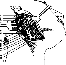
Figure 4. Operations on the facial part of the head: 1-gl. parotis; 2-n. facialis; 2-processus mastoideus; 4-mm. stylo-hyoideus and Biventer; 5- m. sterno-cleidomastoideus; 6-n. accessorius; 7-a. carotis externa; 8- a. maxillaris externa; 9-n. hypoglossus; 10-a. lingualis; 11-v. jugularis externa; 12-a. sterno-cleidomastoidea; 13-v. facialis.
to use the masseter muscle (m. masseter) to replace the affected orbicularis oris muscle and the temporal muscle to close the eyelids, as these muscles receive innervation from the trigeminal nerve. Incisions along the nasolabial fold (plica naso-labialis) 1½-2 cm and along the lower edge of the lower jaw. From the first, the orbicularis oris muscle is dissected, from the second by sharp
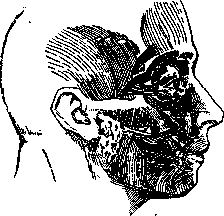
Figure 5. Muscle plastic surgery in paralysis of the facial nerve.
upward traction, the anterior edge of the masseter muscle is exposed, from which a flap with the upper limb is separated (Fig. 5). According to the anat.-clin. investigations of G. Richter, the muscle must be taken necessarily to the bone, as the branches of the trigeminal nerve run for a long distance on the posterior side of the muscle near the periosteum, giving branches into the muscular tissue. Both skin incisions are connected by a tunnel, through which the muscle is pulled and sutured after preliminary splitting to the orbicularis muscle of the upper and lower lip. To form a flap from the temporal muscle, the incision is made along the edge of the hairy part, the muscle flap about 2 cm wide is also taken to the bone and necessarily with the temporal fascia. With a second incision extending from the upper eyelid to the lower eyelid at the outer corner of the eye, the orbicularis muscle is exposed. Special attention must be paid to ensuring that the peripheral part of the flap is reinforced as far as possible to the middle part of the lower eyelid. For muscle plastic surgery, Jianu also proposed the digastric muscle without favorable results; Gomoiu for the same purpose proposed the sternocleidomastoid muscle, which was successfully performed by Hildebrand. Gersuny proposed transplantation of the orbicularis oris muscle from the healthy side. Leriche to combat lagophthalmos proposed transection of the sympathetic trunk under the superior cervical ganglion. In Leriche's results were excellent. Novikov in removal of a tumor of the parotid gland along with the facial nerve to mask the paralysis successfully resected the superior cervical ganglion. Albert, Jianu and Butsoianu remove the superior and middle nodes; Geymanovich recommends resection of the truncus sympathicus, etc. Hesse, in addition to removing the lower two-thirds of the superior ganglion, performs resection of the branches (ramicotomy C8-T1 or C6-C7). Leriche and Fontaine consider it recently necessary to remove the entire sympathetic trunk below the middle ganglion. The results obtained by most authors are good, but only with respect to the eye; improvement in eyelid closure is achieved due to the phenomenon caused by removal of the sympathetic nerve - Horner's syndrome: 1) recession of the eyeball due to paralysis of the smooth muscle fibers in Tenon's capsule, 2) narrowing of the pupil as a result of paralysis of the muscle that dilates the pupil (m. dilatator pupillae), 3) narrowing of the palpebral fissure due to paralysis of the so-called Müller's muscle fibers of the eyelid, 4) increase in blood supply of the corresponding half of the scalp and the appearance of some secretory phenomena. Most physicians at the present time perform intervention on the sympathetic nerve, combining it with muscle and plastic surgery of the remaining defects, mainly in the area of the corner of the mouth.
G. Richter.
Related articles
Cite this page
“Facial Nerve (facial nerve, the seventh pair of cranial).” Soviet Medical Encyclopedia. English translation of Bolshaya Meditsinskaya Entsiklopediya, 1st ed. (Moscow, 1928–1936), ed. N. A. Semashko. https://sovietmedicalencyclopedia.pages.dev/article/facial-nerve-2/