Sublingual Gland
Historical document, translated for reference. It reflects medical knowledge of the 1920s–30s and is not medical advice.
Summary
The sublingual gland is the smallest and most anterior of the three pairs of major salivary glands, located beneath the tongue. It is a mixed gland containing both serous and mucous cells, with multiple ducts opening into the oral cavity.
Encyclopedia article (1928–1936)
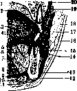
SUBLINGUAL GLAND, glandula sublingualis (fig. 1), the smallest and most anterior of the three pairs of major salivary glands; it has the shape of an olive, somewhat flattened in the transverse direction. Its main axis runs from back to front and from outside to inside, i.e., parallel to the body of the mandible. Figure 1. Vertical median section of the face: 1-hard palate, covered with mucous membrane {2); 3-tongue; 4-vessels; 5-ductus Whartoni; 6-ductus Bartholini; 7-sublingual gland; 8-m. genio-glossus; 9-m. mylo-hyoideus; 10-m. digastricus; 11-platysma; 12-lower jaw; 13-chin; 14 and 19-lower and upper folds of the oral mucosa; 15-1st premolar; 16-skin; 17-m. buccinator; 18-subcutaneous tissue; 20-BepxHHH4enrocib.(noTestut.) mandible. Its length is 25-30 mm, width 10-12 mm (vertical diameter), thickness 6-8 mm. Weight is 3-5 g, i.e., approx. 1/2 weight of the submandibular gland and approx. 1/10 of the parotid. Unlike the submandibular and parotid glands, it does not have a closed osteofibrous aponeurotic bed, but is as if entirely immersed in loose connective tissue, lying on the upper surface of m. mylo-hyoidei between imm. genio-hyoideus, genio-glossus and hyo-glossus on one side, and the medial surface of the body of the mandible on the other. Its anterior rounded end, pressing against the inner surface of the body of the mandible, forms on Hei-iiossa sublingualis. The medial surface of the gland is adjacent to m. genio-glossus and to ductus submandibularis. Here, at first almost parallel, and then crossing ductus submaxillaris, passes the P. nerve and P. vein (p. et v. sublingualis) (fig. 2). The posterior edge of the P. gland contacts the anterior edge of the submandibular gland. The upper surface elevates the mucous membrane of the floor of the oral cavity and forms on both sides of the frenulum of the tongue two protrusions, called carunculae sublinguales, s. carunculae salivales. In structure, the P. gland belongs to the complex glands: it consists of separate lobules,
which in turn break down into primary and secondary glandules-tubules. By function, it belongs to the mixed glands and contains (primary glandules) both dark-colored serous glandules and light-colored mucous ones, as well as mixed ones containing simultaneously mucous cells and half-moons of Januzzi. The terminal glandules, however, in the overwhelming majority, are mucous. They do not contain, according to Raube-4 py(Rauber),HHKa ких вставных тр^-5 бочек and salivary tubules. The dark half-moons of Januzzi are shaped like a cap or thimble over the terminal glandules. The excretory apparatus of the P. gland consists of intraglandular and extraglandular ducts; the first consist of intralobular and interlobular ducts;
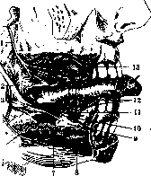
Figure 2. Right sublingual gland: 1-p. buccinatorius; 2-p. lingualis; 3-gangl. submaxilla-re; 4-rami communicantes cum n. linguali; 5-remains of submandibular gland; 6-n. mylo-hyoi-deus; 7-m. mylo-hyoideus; 8-m. digastricus; 9-glandula sublingualis; 10-ductus submaxil-laris Whartoni; 11-n. sublingualis; 12-rami linguales; 13-ductus parotideus Stenonis. (According to Spalteholz.)
the second, which are the excretory ducts of this gland, are very numerous. Some of them open along plica sublingualis in the amount of 20 to 30 into the oral cavity and are called ductus Rivini, s. sublinguales minores. Others open
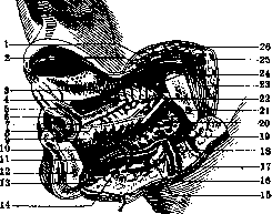
Figure 3. Sublingual gland from the side: 1 and 2-a. et v. facialis; 3, 4 and 5-a. et n. lingualis; b-openings of excretory ducts; 7-opening of Wharton's duct (8); 9-sublingual gland; 10 and 11-vascular branches to the sublingual gland; 12-lower jaw; 13-t. digastricus; 14-a. mentalis; 15-lymph gland; 16, 18 and 26-v. facialis; 17 and 19-a. facialis; 20-upper part of sublingual gland; 21-canal for vessels and nerve in bone; 22-p. lingualis; 23-t. masseter; 24-upper mucosal fold; 25-ductus Stenoni. (According to Testut.)
into the excretory duct of the submandibular gland (ductus submandibularis), near its termination. This gives reason to believe that both these glands represent only differentiated parts of one common gland (Gegenbaur). A certain number of excretory ducts unite into one large1 main--ductus salivalis sublingualis major, s. Bartholini. The latter goes to the caruncula sublingualis together with the excretory duct of the submandibular gland and opens either together with it or nearby in the oral cavity (fig. 3). The structure of the excretory ducts of the sublingual gland is as follows: the connective tissue wall, rich in elastic fibers, nerves, and blood vessels, is lined with two layers of epithelium, of which the deep layer is cuboidal and round-celled, the superficial layer is cylindrical with cilia directed into the lumen of the duct. The sublingual gland sometimes has "accessory glands". However, they should not be considered as fully formed independent glands, but as simple accumulations of glandular cells, scattered irregularly around the main gland. Each such accumulation of glandular cells has a special excretory duct, which opens independently on the floor of the oral cavity at the caruncula sublingualis or slightly medial to it. Testut calls them the ducts of Walther after the anatomist who first observed them in 1724 in humans. Their position is very variable. The openings are sometimes arranged in series in one line having the same direction as the upper edge of the gland, sometimes scattered without any order on the caruncula sublingualis or somewhere nearby. Their number is also variable: according to some authors 4-5, according to others 25-30. The sublingual gland is very rich with blood vessels and nerves. It receives arterial blood from two sources: a. lingualis and a. submentalis. The direction of the vessels and their branching is almost the same as that of the excretory ducts, with which they pass together in the interlobular septa. A dense network of capillaries surrounds the terminal glandules. The veins repeat the course of the arteries and carry their blood to the v. jugularis ext. The lymphatic vessels drain into the submandibular lymph glands. The nerves come from the n. lingualis (after anastomosis with the chorda tympani) and from the ganglion submaxillare. Injuries and diseases of the sublingual gland. Wounds of the sublingual gland due to its anatomical position are extremely rare. Gunshot wounds most often lead to the formation of an internal fistula, however, this has no practical significance. - Salivary fistulas may also result from sequestrating inflammation, ulceration in cancer, in mixed tumors, and in stones in the ducts, accompanied by inflammatory phenomena. - Foreign bodies can penetrate in penetrating wounds (metal fragments, bone fragments, teeth) and through the external opening of the main excretory duct (bristles, hairs, fruit seeds). However, this is extremely rare, since the external opening even of the main excretory duct is only 1 mm. Along with foreign bodies, bacteria also enter, and as a result of this, inflammation of the duct or the gland itself occurs, formation of stones around the foreign body, and obstruction of the lumen either by the foreign body or by a stone. Treatment consists in the earliest possible removal of the foreign body. - Salivary stones-see Salivary glands. Stones of the sublingual gland remain unnoticed for a long time. Patients learn about them only from so-called salivary colic or from a tumor appearing under the tongue during meals (tumor salivalis). Treatment--total removal of the gland together with the excretory duct, since its remnants may lead to recurrences. - Inflammatory processes, sialoadenitis et sialodochitis-:see Submandibular gland. Chronic inflammations of specific origin (tuberculosis, actinomycosis and syphilis) are extremely rare. Primary tuberculosis of the sublingual gland has not been described. Cysts of the sublingual gland and its excretory ducts are exclusively of retention type (see Submandibular gland, cysts) and are found mainly in the sublingual gland in the form of so-called ranula (see). Tumors of the sublingual gland are observed very rarely. Mixed tumors, so characteristic of other salivary glands, in the sublingual gland according to some authors (Bruning) occur as an exceptional rarity, according to others (Kuttner) have not yet been observed with certainty. From epithelial tumors, adenomas are occasionally encountered, which do not reach large sizes here, but nevertheless require total enucleation with the gland, as they cause disorders of swallowing, speech, and in some cases constant salivation. Carcinoma of the sublingual gland is more often described as carcinoma of the floor of the mouth. It occurs in the form of a medullary or scirrhous type. The first one is more malignant. Treatment--total enucleation of the gland. If the capsule is infiltrated, then enucleation is performed within healthy tissue. Operations on the sublingual gland. Complete removal of the sublingual gland can be performed either from the oral cavity side or from the outside. In the first method, the incision of the mucous membrane is made parallel to the edge of the lower jaw at a distance of г/2 or 1 cm from the bone edge. The incision should not be made closer to the bone, as it will then be difficult to suture the edges of the mucous membrane. Further from the bone-toward the midline-one may encounter the a. et v. sublingualis and the excretory duct of the submandibular gland. After opening the mucous membrane, the sublingual gland is easily enucleated by blunt dissection. It must be remembered that the posterior pole of the gland lies near the lingual nerve, which must be spared. With slight bleeding, the mucous membrane is sutured tightly: otherwise, a tampon or drain is introduced. In the second method, the skin incision is made, starting from the midline, parallel to the edge of the lower jaw, 2 cm behind it, to the middle of the submandibular gland. After separating the m. platysmae, the submandibular gland is displaced backward and the lower surface of the m. mylohyoidei is exposed (well freed); its fibers are separated by blunt dissection and the lower surface of the tumor or cyst of the sublingual gland is reached. The anterior belly of the m. digastricus, mm. geniohyoideus and genioglossus are retracted to the sides with blunt hooks: the a. et v. sublingualis are separated. The submandibular duct is spared as much as possible. Finally, the gland is separated from the mucous membrane of the oral cavity. Usually the mucous membrane is injured at this time, and then it is necessary to leave a drain for several days. The platysma and skin are sutured. The mucous membrane from the oral cavity side is also sutured.
Related articles
Mentioned in
Cite this page
“Sublingual Gland.” Soviet Medical Encyclopedia. English translation of Bolshaya Meditsinskaya Entsiklopediya, 1st ed. (Moscow, 1928–1936), ed. N. A. Semashko. https://sovietmedicalencyclopedia.pages.dev/article/sublingual-gland/