Submandibular Gland
Historical document, translated for reference. It reflects medical knowledge of the 1920s–30s and is not medical advice.
Summary
This article from the 1928–1936 Soviet Medical Encyclopedia details the anatomy, topography, capsule structure, and surrounding formations of the submandibular salivary gland.
Encyclopedia article (1928–1936)
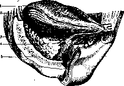
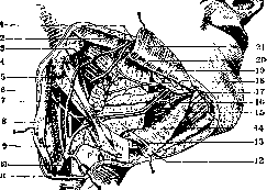
SUBMANDIBULAR GLAND (glandula submaxillaris, s. submandibularis), the second largest of the three large salivary glands. Weight from 7–8 to 15 g; length 2.5–3.5 cm; thickness 1.5 mm. Color at rest is grayish-yellow, when functioning it is grayish-pink, and sometimes more or less red. Shape is ovoid-flattened, somewhat resembling an irregular triangular prism, the major axis of which is directed from behind forwards and from without inwards, parallel to the lower jaw (Figure 1). One can distinguish three surfaces in the gland—outer, inner, and lower—and two ends—anterior and posterior. The submandibular gland lies in the trigonum hyomaxillare, bounded by the two bellies of the digastric muscle of the mandible and the edge of the lower jaw. Like the parotid gland, the submandibular gland is located in a closed osteofibrous bed (submandibular) (Figure 2). Its walls are: lower-outer, formed by the first cervical aponeurosis; upper-outer, formed by the inner surface of the body of the lower jaw, on which the gland forms a special depression (fossa submandibularis); upper-inner, exclusively muscular, covered, according to some authors, by the deep layer of the first cervical aponeurosis, according to others, by the fascia of the muscles forming this wall (mylohyoid muscle, posterior fibers of the hyoglossus muscle, and the posterior belly of the digastric muscle with the styloglossus muscle) (Figures 3 and 4). The first aponeurosis of the neck, lining
-9

Figure 3. Region of the submandibular gland: 1 and 17 - facial artery and vein; 2 - masseter muscle; 3 - superior pharyngeal constrictor muscle; 4 - stylohyoid muscle; 5 and 15 - digastric muscle; 6 - external carotid artery; 7 - hypoglossal nerve; 8 - sternocleidomastoid muscle; 9 - internal jugular vein; 10 - common facial vein; 11 - skin; 12 and 27 - mylohyoid muscle; 13 and 20 - hyoglossus muscle; 14 - lingual vein; 16 - genioglossus muscle; 18 - sublingual gland; 19 - lingual nerve. (According to Testut.)
the gland from below and in front, and the muscle fascia covering the gland from above and in front, form its capsule, with which, however, it does not grow together as tightly as the parotid gland. This capsule is dense on the outside and very thin on the inside. The submandibular gland is very easily enucleated from its bed, which communicates with neighboring organs through the following clefts: 1) between the hyoglossus muscle and mylohyoid muscle along the course of the lingual vein, hypoglossal nerve, and submandibular duct with the bed of the sublingual gland in the oral cavity; 2) along the course of the facial vein and external maxillary artery with the facial region. In the fascial bed, besides the gland, there is a certain amount of fatty tissue, lymph glands (3–6), and the external maxillary artery. By its outer surface, the gland is in contact with the medial surface of the lower jaw, and by the posterior edge of this surface with the internal pterygoid muscle. Along the lower edge of the lower-outer surface pass the mental artery and vein. Here also, in a single line, are located from 3 to 6 lymph nodes.
Figure 1. Submandibular gland from the side: 1 - tongue; 2 - lingual nerve; 3 - submandibular gland; 4 - Wharton's duct; 5 - lower jaw. (According to Testut.)
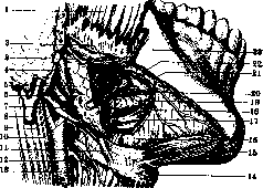
Fig. 4. Region of the submandibular gland: 1 - m. masseter, 2 - plexus sympathicus, 3 - a. facialis, 4 - m. digastricus; 5 - submandibular gland; 6 and 17 - n. hypoglossus; 7 - a. carotis ext.; 8 - v. jugularis ext.; 9 - a. carotis int.; 10 - v. lingualis; 11 - m. stylo-hyoideus; 12 - a. thyreoidea sup.; 13 - a. carotis communis; 14 - os hyoideum; 15 - m. digastricus; 16 - m. mylo-hyoideus; 18 - branch for the submandibular gland; 19 - anterior part of the submandibular gland; 20 - ductus Whartoni; 21 - v. lingualis; 22 - gangl. submaxillare; 23 - upper jaw. (After Testut.) (submandibular lymph glands). Although they more frequently lie above the aponeurosis, nevertheless they are in direct contact with the glandular tissue. The inner surface is in intimate relation with the posterior belly of the digastric muscle, accompanied by the stylohyoid muscle; it approaches particularly tightly to the tendons of these muscles at the place of their attachment to the hyoid bone and thus frequently lies adjacent to the hyoid bone itself. From the inner surface, two processes as it were depart from the gland: one—shorter and wider—runs backward and frequently reaches the internal pterygoid muscle; at this place the submandibular gland is in contact with the parotid gland; the other process—narrow and long—runs forward and, lodging in the cleft between the mylohyoid and hyoglossus muscles, passes into the oral cavity and there comes into contact with the posterior part of the sublingual gland. Sometimes the posterior part of the anterior process is undeveloped, and then this process gives the impression of a separate, independent, isolated lobule. Some authors, by analogy with the parotid gland, consider it an accessory submandibular gland (gland. submandibularis accessoria). However, the fact that this process sends its secretion to the mucosa of the oral cavity not through its own excretory ducts, but through the general excretory duct of the submandibular gland, suggests that this is not an accessory submandibular gland, but an accessory lobe of it. The lower surface, which is the most extensive, is covered by the lamina of the first cervical aponeurosis, the platysma myoides muscle, subcutaneous fatty tissue, and skin. Across this surface, obliquely from back to front, the gland is crossed by the anterior facial vein. This same surface has to be lifted to separate the muscle fibers during the ligation of the lingual artery. With its posterior end, the submandibular gland closely approaches the parotid, and both of them are separated from each other by a fibrous septum (called by some authors the stylomandibular ligament). At this place, the fatty tissue of the submandibular gland bed communicates with the tissue of the tonsillar region and the pharyngeal space. By its structure, the submandibular gland belongs to compound glands: it breaks down into lobules of the second order, simple lobules, and tubular glandules immersed in a connective-tissue stroma. By its function, it belongs to mixed glands and contains serous, mucous, and mixed glandules. Mucous glandules are fewer in it, but they are more voluminous, their tubules are longer and wider, and they are surrounded by mucous cells. Mixed glandules (tubules) consist of serous and mucous cells. The initial part of these tubules is generally mucous, the terminal part is serous. The serous parts of the gland have an alveolar character, the mucous ones—alveolar-tubular. On the blind ends of the alveoli are located "Gianuzzi's demilunes." These are short intercalated parts (Schaltstücke) lined with low cylindrical cells. There are various views on the nature of these demilunar bodies: according to some authors, they may originate from mucous cells that have freed themselves from their secretion; according to others, the demilunar bodies supply only a serous secretion (and never a mucous one). They differ both in morphological and physiological senses from mucous cells and stand in connection with them perhaps only as stages of development or as functional stages. The excretory apparatus of the submandibular gland consists of intralobular ducts, pouring their contents into 3–5 interlobular ducts, which unite into one main excretory duct of the submandibular gland—Wharton's duct, ductus salivalis, s. duct. submaxillaris Whartoni, opening into the oral cavity after passing beneath its floor. The length of the duct is 4–6 cm, the diameter from 2 to 5 mm. The narrowest place is the outlet opening. Immediately behind the outlet opening, the duct expands in a spindle-like manner, and foreign bodies easily get stuck here; the posterior two-thirds of the duct are again somewhat narrowed. The extensibility of the outlet opening is very insignificant in those cases when it needs to be expanded sharply, urgently (insufflation, injection, probing), whereas prolonged and systematic bougienage easily expands it. The structure of the main duct is as follows: it, like the other ducts of the submandibular gland, consists of a connective-tissue stroma rich in elastic fibers, nerves, and vessels, lined from the inside by two layers of epithelium: deep—cubic and round-cell—and superficial—cylindrical with cilia facing into the lumen of the channel. Unlike other excretory ducts, Wharton's duct contains in its wall between the two layers of connective elastic tissue a layer of smooth muscle fibers intersecting among themselves in various directions. Having passed a certain distance inside the gland itself, Wharton's duct together with the medial process of the gland penetrates through the cleft between the hyoglossus and mylohyoid muscles and appears on the floor of the oral cavity, where it lies under the mucous membrane along the medial margin of the sublingual gland, forming an elevation on the lower surface of the tongue called the sublingual caruncle (caruncula salivalis, s. sublingualis). It opens next to the outlet opening of the duct of the opposite side at the lower part of the frenulum of the tongue (frenulum linguae). On both sides of the frenulum, in its posterior part, a small tubercle rises with a round opening at its apex—this is the external opening of Wharton's duct—ostium umbilicale. This tubercle is called by some authors the inferior salivary papilla. Physiology of the submandibular gland—see Salivary glands. The submandibular gland is very rich in blood vessels and nerves. The submandibular gland receives its arterial blood supply from two sources: from the external maxillary artery and the submental artery, and according to Rauber, also from the lingual artery. In their direction and branching, vessels as a rule follow the excretory ducts of the gland, passing together with them in the layers of connective tissue separating the lobules of the gland. The terminal tubules are enwrapped by a dense network of capillaries. Some authors (Kovalevsky, 1885) distinguish arteries of the excretory tubes and arteries of the terminal parts by their length and by their resistance (they are richer in elastic fibers). Large veins repeat the course of the arteries and empty partly into the submental vein, partly into the anterior facial vein. The lymphatic vessels of the gland carry their lymph mainly into the submandibular lymph glands, and from the latter into the deep lymph glands of the neck. Nerves originate from the lingual nerve, which receives the chorda tympani, and from the submandibular ganglion. Wounds and diseases of the submandibular gland. Wounds of the submandibular gland are extremely rare thanks to its deep position. Salivary fistulae of the submandibular gland and foreign bodies...—see Salivary glands, Parotid gland. Salivary calculi (sialolithiasis) are a very rare disease (Fig. 5) (see Salivary glands, pathology). Parasites of animal origin are extremely rare. Isolated cases of the finding of cysticerci and echinococci have been described. Inflammatory processes (sialadenitis) are acute and chronic. Etiology: penetration of infection either from the oral cavity (mainly)—ascending infection along the excretory ducts or by a hematogenous path (metastatic inflammation)—in typhoid fever, gonorrhea, etc., or finally from neighboring organs. Acute inflammations of the submandibular gland can lead to the formation of phlegmon of the oral cavity (Ludwig's angina). Secondary acute sialadenitis of the submandibular gland in general infectious diseases or after operations occurs exclusively in infants. Acute inflammations are difficult to distinguish from acute inflammation of the submandibular lymph glands and from phlegmon of the floor of the oral cavity. Sometimes the inflammation is limited only to the region of the excretory duct (sialodochitis). A special form is fibrinoid sialodochitis (Kussmaul), in which fibrinous-purulent clots clog the excretory duct and cause salivary colic (pyorrhoea salivaris). They are frequently accompanied by chills and fever. Certain chronic poisonings, especially with lead, can also cause chronic inflammation of the submandibular gland, leading to strong proliferation of connective tissue. The incision in inflammation of the submandibular gland is made parallel to the lower margin of the lower jaw. It should be remembered
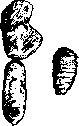
Figure 5.
regarding the submandibular artery passing here. Some authors consider it advisable in inflammation of the submandibular gland (Kuttner) to completely enucleate it, especially in its chronic forms, when it is difficult to differentiate an inflammatory tumor from a malignant neoplasm and even more difficult to be guaranteed against malignant degeneration of the tumor. Chronic specific inflammations. 1. Tuberculosis of the submandibular gland is an extremely rare disease. Treatment is the complete removal of the submandibular gland. The diagnosis of tuberculosis of the submandibular gland is difficult, especially when a general examination of the body yields no decisive data. In the presence of fistulas, the diagnosis should be differentiated by examining the pus for druses to rule out actinomycosis of the submandibular gland. 2. Syphilis and actinomycosis of the submandibular gland are also extremely rare. Enlargement of the submandibular gland, occurring with the simultaneous enlargement of all salivary and lacrimal glands, is known as Mikulicz's disease (see Mikulicz's disease). Cysts. Aside from rare echinococcal cysts, cysts of the submandibular gland are exclusively retention cysts. They are formed as a result of complete or partial closure of the lumen of its excretory ducts. This closure can be either congenital due to epithelial blockage—atresia—of the excretory duct before birth, or acquired as a result of inflammation and cicatricial bands. A cyst of Wharton's duct has the character of a roller-like or fusiform swelling with an axis parallel to the direction of this duct; complete blockage of its lumen ultimately leads to atrophy and obliteration of the submandibular gland. Cysts of the submandibular gland are less common than cysts of the submandibular duct. They can be unilocular, but can also be multilocular—upon simultaneous blockage of several small excretory ducts. They can reach the size of a chicken egg; they are spherical in shape. Treatment of cysts of Wharton's duct consists in creating a surgical communication between the dilated duct and the oral cavity. Treatment of cysts of the submandibular gland is its enucleation. Tumors. Sarcomas are found in all known forms and exhibit all kinds of transitional grades between relatively benign and highly malignant neoplasms, ranging from fibrosarcoma to small round-cell sarcoma and even melanoma. However, in general, as primary tumors they occur extremely rarely; more often they originate from mixed tumors, which are characteristic of the salivary glands (Fig. 6) (see Salivary glands, pathology). Of the epithelial tumors of the submandibular gland, adenomas are an extreme rarity; more often they are carcinomas in the form of medullary carcinoma or scirrhus. However, so far only 18 cases of carcinoma of the submandibular gland have been described in the world literature (Bruning). Treatment is exclusively surgical: total removal within healthy tissue. Operations on the submandibular gland. Complete removal of the submandibular gland. The incision is made a finger's breadth below the inferior
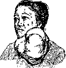
margin of the lower jaw, parallel to it. After dividing the fibers of the platysma muscle, the anterior facial vein is ligated. Then the capsule of the gland is exposed at its lower pole and the gland is dissected away from the capsule (if the tumor is neither malignant nor mixed), and the external maxillary artery and vein are ligated here. Gradually, by blunt dissection, having freed the gland, it is delivered from its bed. At its upper margin, the external maxillary artery and vein and the main excretory duct are ligated once more. A rubber drain is inserted into the wound, and it is closed in layers. For cosmetic purposes, it is necessary to specially suture the platysma muscle separately. During extirpation of the submandibular gland for benign tumors, or during its enucleation, one should adhere as much as possible to the capsule of the gland, as this method avoids injury to the lower (labial) branch of the facial nerve.
Fig. 7
Related articles
Mentioned in
Cite this page
“Submandibular Gland.” Soviet Medical Encyclopedia. English translation of Bolshaya Meditsinskaya Entsiklopediya, 1st ed. (Moscow, 1928–1936), ed. N. A. Semashko. https://sovietmedicalencyclopedia.pages.dev/article/submandibular-gland/