Parotid Gland
Historical document, translated for reference. It reflects medical knowledge of the 1920s–30s and is not medical advice.
Summary
The parotid gland is the largest of the salivary glands, located in the face behind the ascending ramus of the mandible. This article describes its embryology, anatomy, histology, and the structure of its duct system.
Encyclopedia article (1928–1936)
PAROTID GLAND (glandula parotis), the largest of the salivary glands, located on the face, in a deep depression behind the ascending ramus of the lower jaw, in the so-called retromandibular fossa (fossa retromandibularis). The shape of the gland is entirely adapted to the walls of this bed and has irregular, difficult to compare contours; with some stretch, it can be compared to a three-sided, vertically placed prism, one side of which faces outward, while the other two face forward and backward. The parotid gland is found in a rounded shape and flattened, extending far forward onto the cheek or downward along the sternocleidomastoid muscle to the level of the lower jaw. The greatest thickness is reached by the posterior half of the gland - about 1.5 cm. The color of the gland is grayish-yellow, close to the color of the surrounding fat, from which the gland differs by a more pronounced grayish tint, lobulation, and greater density. The volume of the gland varies considerably, the smallest gland relates to the largest as 1:5; the average weight of the parotid gland is 25-30 g. Embryology of the parotid gland. The first rudiments of the parotid gland are found in the eighth week of embryonic life. The primary form of this gland, as well as other salivary glands, is a cylindrical outgrowth of the oral cavity epithelium; the distal part of this outgrowth branches, serving as the basis for the formation of further elements of the gland; on cross-sections, solid epithelial cords are visible, in the center of which cavities form (future ducts). On the 15th week, the capsule of the parotid gland is formed. On the 12th week, the parotid gland lies very close to the bone rudiments of the lower jaw. Sometimes acini parotidis are seen among the cells of the periosteum of the lower jaw. At this time, the parotid gland also lies close to the rudiments of the eardrum. The canalization of the ducts, the formation of the terminal tubules of the parotid gland proceeds through their systematic separation and distribution. The cells of the parotid gland develop in the fifth month.
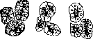
From an anat.-physiol. point of view, the parotid gland belongs to the tubulo-alveolar serous glands; its cells produce a watery secretion containing protein and salts. The parotid gland is a lobulated gland; individual lobes (primary) are formed as a result of grouping small tubulo-alveolar formations (acini); the connection of a certain number of such lobes gives larger lobes of the gland (secondary). Individual lobes are separated from each other by well-developed, fat-penetrated connective tissue. Acini have the form of blind elongated sacs, the cells of which (secretory epithelium) are located on a thin plate devoid of formed elements (membrana propria). The epithelium consists of small cubic or conical cells with a nucleus located in the lower third of them and basophilic protoplasm filled to one degree or another with highly light-refractive granules (Langley's granules) (fig. 1). In the interlobular connective tissue there are cellular elements
a
b
c
of fig. 1. Acini of the serous gland in three different physiological states: a - in a state of rest; b - after a short period of work. The granules have disappeared from the main part of the cell; c - after intensified work. Granules are present only at the apex of the cell,
u
p
a
s
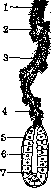
light as1SH - individually or in groups, forming true lymphatic nodules. In the connective tissue septa, vessels, nerves, and excretory channels of the gland-ducts pass. ? Upon exit from the sac (acinus) of the gland, saliva successively passes through the intercalated (fig. 2), intralobular, and interlobular ducts in order to enter the main excretor of the parotid gland, Stenon's duct (ductus parotideus Stenonis). The intercalated, intermediate canal (Schaltstucke of German anatomists, canal of VoP'ya) extends from the sac of the gland to the intralobular canal, consists of its own membrane covered with low prismatic epithelium with homogeneous, highly light-refracting protoplasm. In the intralobular canals are formed as a result of the fusion of several intercalated canals, pass in the thickness of the lobes themselves; their wall is built of thin connective tissue and prismatic epithelium with a centrally located nucleus rich in chromatin and protoplasm having longitudinal striation. These cells produce part of the saliva, namely- lime salts. The walls of the interlobular canals consist of connective tissue containing elastic fibers, smooth muscles, vessels and nerves, and are lined in two layers with epithelium-deep, flatter and superficial, prismatic.
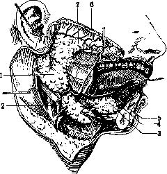
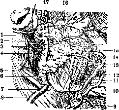
Stenon's duct excretes the saliva of the parotid gland into the mouth; the duct begins on the anteromedial surface of the gland near the anterior edge, at the border of the lower and middle thirds of it. The method of formation of Stenon's duct from the interlobular ducts varies: either by the fusion of two converging ducts with an almost equal lumen, or the duct penetrates deeply into the substance of the gland, going obliquely downward backward, receiving on its way from 6 to 14 lateral channels from above and below. After exiting the gland, the duct goes obliquely upward forward, not reaching 15-20 mm to the zygomatic arch, it turns forward and goes horizontally along the outer surface of the masseter muscle in the company of the transverse artery of the face (art. transversa faciei), located slightly above the duct, and branches of the facial nerve, which pass some over Stenon's duct, others under it (fig. 3 and 4). Further, the duct bends inward in front of the masseter muscle, pierces the fatty layer of Bishai, piercing obliquely the buccinator muscle (m. buccinator), goes 5-6 mm under the mucous membrane and opens into the vestibule of the mouth, corresponding to the upper second
Fig. 3. Salivary glands of the right side: 1-gl.parotis; 2-submandibular gland; 3-sublingual gland; 4-Barton's duct; 5-duct of the sublingual gland; 6-Stenon's duct; 7-continuation of the parotid gland forward.
large molar tooth (fig. 5) in the form of a narrow slit; sometimes this opening is located on an elevation in the form of a papilla. The entire length of the duct varies from 15 to 40 mm with a lumen diameter of up to 3 mm. On the masseter muscle, an accessory parotid gland (glandula parotis accessoria) adjoins the duct; the duct of which opens into Stenon's duct; therefore it should be considered not as an additional independent gland, but as an additional lobe of the parotid gland. The projection of Stenon's duct onto the skin passes by a line from the tragus of the auricle to the angle of the mouth. The wall of Stenon's duct consists of strong connective tissue rich in elastic fibers, vessels and nerves, and epithelium lining the lumen of the canal; the epithelium consists of two layers-deep
Fig. 4. Parotid gland, denuded of the layers covering it: 1-mastoid process; 2-styloid process; 3-facial nerve; 4-posterior belly of the digastric muscle; 5-sternocleidomastoid muscle; 6-parotid gland; 7-interglandular septum; 8-external jugular vein; 9-submandibular salivary gland; 10-lower jaw; 11-masseter muscle; 12-external maxillary artery; 13-buccal muscle; 14-buccal glands; 15-Stenopov's duct; 16-zygomatic arch; 17-superficial temporal artery and vein.
cubic and superficial cylindrical; at the place of opening into the mouth, the epithelium of the duct takes on the character of the oral mucosa epithelium. The parotid gland is rich in vessels and nerves; its arteries take origin from many sources: aa. auricularis post., transversa faciei, carotis externa; all these vessels give the richest arterial network, the capillaries of which approach the proper capsule of the gland, without coming into contact with the secretory epithelium of the gland. The veins pass in the interlobular septa, carrying blood to the external jugular ve
Fig. 5. Stenon's duct on a horizontal section of the face (right side, lower segment)
from above): 1-parotid gland; 2-lymph vessels of the gland; 2-upper jaw; 3-medial pterygoid muscle; 4-pterygoid process; 5-lower jaw; 6-masseter muscle; 7-Stensen's duct; 8-buccal muscle; 9-mucous membrane of the mouth. It is penetrated by numerous vessels of various calibers, which also pass through the septa of the lobules; the lymph vessels are represented by endothelial slits devoid of valves; they carry lymph to the lymphatic glands of the parotid. The nerves of the parotid gland receive from 3 sources: from the auriculotemporal nerve (n. auriculo-temporalis), the great auricular nerve (n. auricularis magnus), and sympathetic branches. All these nerve branches branch out in the interlobular connective tissue of the gland, breaking down into myelinated and non-myelinated fibers, forming around the primary lobules plexuses (plexus peri-lobularis), the fibers of which penetrate into the lobules themselves. Some of these branches are true vasoconstrictors, others are secretory; the latter pass between the acini and form a second plexus of nerves (plexus perialveolaris); a third type of fibers ends in the walls of the excretory ducts of the gland, the method of their termination is not yet clarified. The true secretory nerve of the parotid gland is the auriculotemporal nerve (Claude Bernard), its removal in case of parotid fistula leads to cessation of salivation in humans, as well as in animals (Leriche). The sympathetic nerve decreases or stops the secretion of the parotid gland.
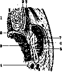
The bed and aponeurosis of the parotid gland. The bed of the parotid gland is, for the most part, lined with a thin layer of areolar tissue, in some places thicker, taking on the character of an aponeurosis. The parotid gland, like all glands, is surrounded by a connective tissue sheath, a true capsule. The capsule, covering the gland with a thin sheet, sends partitions into the depth of the gland and thereby divides it into separate lobules. Around the capsule are aponeurotic formations of the neighboring musculature: on the outside, the superficial (or first) aponeurosis of the neck, behind it the prevertebral aponeurosis, and inside the stylopharyngeal aponeurosis and the vascular sheath. Usually this series of aponeuroses is described as one whole, aponeurotic sheath of the gland, distinguishing in it the superficial (external) and deep (internal) layers (figs. 6 and 7). The superficial layer of the aponeurosis of the parotid gland is a continuation of the aponeurosis of the outer surface of the sternocleidomastoid muscle and passes onto the face, attaching to the angle and the posterior edge of the ascending branch of the lower jaw, partly to the aponeurosis of the masseter muscle (fascia parotideo-masseterica) and to the lower edge of the zygomatic arch.
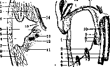
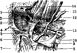
The deep layer, separating from the previous one at the anterior edge of the sternocleidomastoid muscle, goes to the lateral walls of the pharynx, successively covering the posterior belly of the digastric muscle, the styloid process and the ligaments and muscles attached to it; then the aponeurosis covers part of the posterior surface of the medial pterygoid muscle (m. pterygoideus internus) and at the posterior edge of the ascending branch of the lower jaw merges with the superficial layer. Below (fig. 8) both layers pass into each other in a narrow space between the angle of the lower jaw and the sternocleidomastoid muscle, thereby creating a strong partition between the bed of the parotid gland and the bed of the submandibular gland. At the top (fig. 7) the superficial layer is attached to the lower edge of the zygomatic arch and to the
Fig. 6. Schema of the bed of the parotid gland of the left side (horizontal section): 1-masseter muscle; 2-lower jaw; 3-medial pterygoid muscle; 4-deep layer of the aponeurosis of the parotid gland; 5-styloid process; 6-superficial layer of the aponeurosis; 7-skin; 8-subcutaneous tissue; 9-sternocleidomastoid muscle; 10-pharyngeal opening of the parotid gland; 11-digastric muscle; 12-internal jugular vein; 13-internal carotid artery; 14-pharynx. Fig. 7. The same bed in a vertical transverse section (left side, anterior segment): 1-superficial layer of the aponeurosis of the parotid gland; 2-external carotid artery; 3-internal jugular vein; 4-internal carotid artery; 5-external jugular vein; 6-fusion of the two layers of aponeurosis; 7-muscles of the styloid process; 9-deep layer of the aponeurosis; 10-position of the pharyngeal opening; 11-inferior surface of the skull; 12-external auditory canal. Fig. 8. The two beds of the parotid and submandibular glands: 1-parotid gland, lower pole cut off; 2-its bed; 3-fibrous partition between the glands; 4-bed of the submandibular gland; 5-anastomosis between the external jugular vein (7) and the facial vein (13); 6-sternocleidomastoid muscle; 8-sublingual muscles; 9-cervical aponeurosis; 10-hyoid bone; 11-anterior belly of the digastric muscle; 12-external maxillary artery; 14-masseter muscle.
cartilaginous part of the external auditory canal. The deep layer at the base of the styloid process fuses with the periosteum of the inferior surface of the temporal bone. Some parts of the capsule of the parotid gland are very strong (e.g., on the outer surface of the gland and at its lower pole), others, on the contrary, are very thin (e.g., the part adjacent to the pharynx and to the external auditory canal). Due to the processes of the capsule penetrating into the depth of the gland, it is possible to separate the gland from the capsule only with great difficulty, and it is especially difficult to separate the outer part and the anterior edge of the gland; conversely, the gland is easily dissected out near the external auditory canal, near the masseter muscle, near the muscles of the styloid process and the digastric muscle, and near its lower pole.
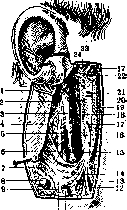
The bed of the parotid gland (figs. 9 and 10), freed from its contents, i.e., from the parotid gland and other organs, represents a three-sided depression with the greatest vertical dimension. The outer surface of the bed exists only when the parotid aponeurosis is intact; but when it is removed, an opening in the form of a vertical slit is obtained, the anterior edge of which is the posterior edge of the ascending branch of the lower jaw. Posterior
10 AND
the margin of the opening is formed by the mastoid process and the sternocleidomastoid muscle. Movements of the head, as well as of the lower jaw, change the size of the entrance to the bed. The upper margin of the entrance is formed by the temporomandibular joint and the external auditory meatus; the lower margin forms a partition between the bed of the parotid gland and the submandibular gland. The anterior surface of the bed is formed by the ascending ramus of the lower jaw and the masticatory muscle covering it on the outside and the pterygoid muscle on the inside; between the latter and the parotid gland passes the sphenomandibular ligament. The posterior surface of the bed is formed by the posterior belly of the digastric muscle, the styloid process with its two ligaments and three muscles, and the stylopharyngeal aponeurosis. The lower, cervical foundation of the bed is formed by the interglandular septum (septum intermaxillo-parotideum) (fig. 8). The upper, temporal foundation of the bed (fig. 7) is formed by two slopes: the posterior one - the external auditory meatus and the anterior one - the temporomandibular joint; thus the dome of the bed constitutes the base of the skull along the course between the spine of the sphenoid bone and the base of the styloid process. Thus the bed has bony-muscular-aponeurotic walls. Besides the parotid gland, the external carotid artery and external jugular vein, the facial and auriculotemporal nerves, and lymphatic vessels pass through this bed. The syntopy of the parotid gland is complex both with the organs lying outside the gland's bed (external syntopy) and with those inside the bed (internal syntopy). External syntopy. The parotid gland, repeating the shape of its bed, also has three surfaces (external, anterior, and posterior) and two bases. The external surface of the gland is slightly convex. The skin of this area is thin, mobile, smooth in women and children, and partially covered with hair in men. The subcutaneous tissue (except in obese individuals) is thin and adherent to the skin. Deeper lies the superficial fascia; in this layer pass some bundles of the platysma muscle of the neck and the risorius muscles, as well as small vessels and nerve branches originating from the cervical plexus. Even deeper is located the parotid aponeurosis (part of the parotidomasseteric fascia). The posterior surface of the gland is adjacent to all those organs and tissues that constitute the posterior surface of the bed of the parotid gland. Occasionally the parotid gland gives off a process between the sternocleidomastoid and digastric muscles. The anterior surface of the gland fills all the depressions of the anterior surface of the bed, occasionally giving off a process (retromandibular process of the parotid gland) between the medial pterygoid muscle and the lower jaw and often along the external surface of the masticatory muscle, without quite reaching its anterior edge; in this case the gland with its protruding forward edge covers its excretory (Stensen) duct and masks its origin. Between the gland and the constantly moving ascending ramus of the lower jaw, a serous bursa is often found. The upper part of the parotid gland covers a significant portion of the capsule of the temporomandibular joint and fuses with it. Medial to this joint, the gland is adjacent to the cartilaginous and bony parts of the external auditory meatus, into which an abscess often opens in purulent parotitis. With its lower pole, the parotid gland borders the bed of the submandibular gland. With its inner margin, the parotid gland is turned toward the pharynx, often reaching its wall formed by the superior constrictor of the pharynx (m. constrictor pharyngis superior) (see Carotis arteria, fig. 19). Here are located the mandibular nerve and its branches, the otic ganglion, the chorda tympani, branches of the maxillary artery (a. maxillaris int.), and the ascending palatine artery (a. palatina ascendens); deeply above passes the terminal part of the Eustachian tube. By means of a weak fibrous septum, the so-called 'pharyngeal wings' (see Carotis arteria, fig. 19), the posterior surface of the parotid gland is separated from the vascular-nervous bundle of the neck. Internal syntopy of the parotid gland. Besides the parotid gland, its bed contains arteries, veins, nerves, lymphatic vessels, and lymphatic glands. The main artery of the bed is the external carotid artery (a. carotis externa), which penetrates the anteromedial part of the bed, first passes between the aponeurosis and the gland, then deepens into the substance of the gland, having a slightly oblique direction toward the neck of the articular process of the lower jaw; occasionally the external carotid artery passes outside the gland, between it and the pharynx (Testut). In the gland, the external carotid artery gives off branches: the posterior auricular, superficial temporal, and maxillary (fig. 10). Somewhat lateral to the external carotid artery, from top to bottom, runs the external jugular vein, leaving the gland at its lower pole; as the vein passes through the gland, the transverse facial and posterior auricular veins flow into it; the trunk of the vein in turn is formed by the superficial temporal and maxillary veins. The parotid bed is penetrated by numerous large lymphatic vessels coming from the skull and face and flowing into the lymphatic glands of the parotid gland. The lymphatic glands of the parotid gland are divided into superficial and deep; the former are located under a thin layer of the external surface of the gland and collect lymph from the skin of the face, the external surface of the auricle, the external auditory meatus, and the tympanic cavity; the deep lymphatic glands, which are very small, lie along the course of the external carotid artery and internal jugular vein; to them flows lymph from the external auditory meatus, the soft palate, and the posterior half of the nasal cavity. Lymph from the glands of the parotid gland goes partly to the glands located near the exit of the external jugular vein, and partly to the glands under the sternocleidomastoid muscle. Of the nerves passing through the substance of the parotid gland, the most important are the facial and auriculotemporal (nn. facialis et auriculo-temporalis). The facial nerve, upon exiting the skull through the stylomastoid foramen (foramen stylo-mastoideum), immediately enters the substance of the parotid gland, running obliquely from behind forward, from medial to lateral, and slightly from above downward; at first the nerve lies deeply and, moving forward, approaches the external surface of the gland, always lying lateral to the external carotid artery and external jugular vein. At the posterior edge of the ascending ramus of the lower jaw, sometimes earlier, still within the substance of the gland, the nerve divides into its main branches. The auriculotemporal nerve separates from the mandibular nerve most often by two branches, surrounds the middle meningeal artery, passes between both pterygoid muscles above the maxillary artery, and behind the articular process of the lower jaw penetrates the parotid gland, where the nerve divides into a number of trunks; the first one turns upward and goes along and behind the superficial temporal artery; this branch anastomoses with the facial nerve; the second short trunk gives in its peripheral part a thickening in the form of a plate, from which numerous finest twigs emerge; some of them enter the skin of the auricle and external auditory meatus, anastomose with the sympathetic plexus of the external carotid artery and its branches, while others in the form of numerous finest twigs enter the parotid gland; they anastomose with each other and with the branches of the facial nerve, thus forming a whole nerve network on the deep surface of the gland, from which terminal branches go into the substance of the parotid gland. a. sirotkin. Diseases of the parotid gland.-Congenital malformations. Absence or abnormal position of the parotid gland is rare; Schreiber described a case of right-sided aplasia with simultaneous dystopia on the left. In one case of Gruber, the parotis was not found in its normal place, but instead on the posterior border of the region of the cheek there was a very large gland corresponding in its position to the parotis accessoria and simulating a tumor. The opening of the duct is not always in its place. In one case in a little girl, two openings as thin as a hair were observed, located several centimeters from the sternum along the inner edge of the sternocleidomastoid; during chewing, saliva was secreted from them. A. sirotkin also reports on congenital salivary fistulas.-Injuries.
Greater significance than deformities are injuries to the parotid gland. It should be noted that of all the salivary glands, almost only the gl. parotis is subject to injury due to its more superficial location. These injuries are more often observed in wounds from cold steel weapons. Injuries to the parotid gland are also possible during operations, but simultaneous injuries to large nerve and blood vessels are so prominent that they completely overshadow injuries to the parotid gland. Diagnosis of parotis injuries is not always easy in cases with minor bleeding. The presence of characteristic glandular tissue in the wound quickly resolves the question; otherwise, diagnosis is sometimes made post factum based on the discharge of saliva from the wound. It follows that with any kind of wound in the area of the parotid gland, careful examination of the wound is mandatory. With any injury, suturing of the wound with careful stitching of the glandular tissue is necessary. With subsequent discharge of saliva from one or another part of the wound, the application of a pressure bandage for 5-6 days usually eliminates this complication. More serious are injuries to the duct of the gland, although they occur much less frequently; in diagnosis, one must be guided by the direction of the duct's course (see above); with any wound crossing this line, one must keep in mind the possibility of duct injury. Diagnosis of the injury is not difficult; it is usually made based on the discharge of saliva from the central segment of the duct. Kuttner recommends placing a piece of sugar in the mouth, having previously dripped a little diluted acetic acid on it, to enhance the reaction. The prognosis is quite favorable, although it was previously believed that such an injury must necessarily result in a fistula. According to König, duct injuries are often overlooked, and instead of restoration of duct patency, there occurs a secretly progressing fatty degeneration of the gland, as Rolando established in his experiments in the 19th century. The method of treatment is primary suture with approximation of the ends of the severed duct, and drainage of the duct with a bristle or thread is irrational, as it eliminates all chances for primary tension. Making an opening into the mouth is done only in the presence of a crushed wound when primary tension cannot be expected. In cases of penetrating wounds into the mouth, the mucous membrane should be left unsutured so that saliva can make its way into the oral cavity. The formation of an oral salivary fistula does not cause any functional disorders; Salivary fistulas. The presence of a fistula is determined by the discharge of saliva at an abnormal location on the mucous membrane or on the skin. Usually, external fistulas are distinguished if saliva is discharged through the skin, and internal ones when discharged through the mucous membrane into the oral cavity. Further, parotid gland fistulas and duct fistulas are distinguished. Fistulas represent a very unpleasant suffering, mainly due to constant wetting of the skin with subsequent eczema, as well as the fact that food is not sufficiently moistened with saliva. Besides injuries, etiological factors include abscesses, neoplasms (cancer), lues, tbc, etc. A parotid gland fistula is easily recognized: it usually has a short course (except in cases of a fistula of the accessory part) and goes directly into the gland tissue; the discharge of secretion is much less than with a duct fistula, since only one part of the gland is affected; this small secretion discharge is one of the characteristic differential signs between a gland fistula and a duct fistula. Usually, a small opening is observed on the top of a granulation papilla, discharging clear saliva. Between meals, no discharge of secretion is observed, but during meals, secretion is discharged in drops and even in a stream. Duct fistulas are of great importance, being much more difficult to treat, and those fistulas in which the skin and mucous membranes have fused in the form of a lip-like fistula, and the peripheral part of the fistula facing the oral cavity is closed, are the most persistent. Diagnosis presents no difficulties, but it is necessary to consider the signs that distinguish a parotid gland fistula from a duct fistula. One of these signs is mentioned above. Further, the location of the fistula opening in front of the chewing muscle will always indicate a duct fistula (it is necessary to remember about the accessory part), and finally, the absence of secretion discharge from the external opening of Stensen's duct indicates a duct fistula. Corresponding to the three parts of the duct, glandular, masticatory, and buccal fistulas are distinguished; buccal fistulas respond best to treatment. Treatment can be conservative or operative. Usually, conservative treatment is started first. Cauterization with silver nitrate, thermocautery, tincture of iodine, and subsequent pressure bandages are applied. Some authors proposed closing the fistula with tafta and pouring collodion over it, then prescribing a liquid, non-irritating diet. Kausch introduced paraffin around the scar. Conservative methods of treatment should also include treatment with appropriate doses of X-rays, due to which the parenchyma of the parotid gland atrophies so much that all secretion from it ceases.--There are more than 55 methods of operative treatment. The ideal operation is the restoration of duct patency by suturing the freshly cut central and peripheral segments. To avoid narrowing or closure, the suture is made over a silver wire or a catgut thread passed with the help of a needle from the central segment to the peripheral one. Such a suture of freshly cut ends gives too many failures and is therefore almost never used. Similarly, operations aimed at restoring the missing part of the duct by plastic reconstruction using skin (Kuttner), epidermal, mucous membrane flaps or a piece of facial vein have been abandoned. These operations require larger incisions and, besides being unreliable, leave extensive disfiguring facial scars. The most deserving of attention are implantation methods, in which the central end is sutured into an opening in the mucous membrane. According to Langenbeck, the central end of the duct is dissected from the fistula to the mucous membrane, in which an opening is made, and the duct is sutured into this opening. The freshened skin opening is sutured. Opel further simplified this operation by not looking for the central end, but suturing the fistula opening of the duct into the opening in the mucous membrane. For this purpose, an elliptical incision is made in the skin around the fistula, with the fistula opening located at the posterior pole of the ellipse. Then this flap is dissected from the underlying tissues to the posterior pole, where the fistula opening is located; under the flap, tissues are punctured obliquely from front to back with a knife, and the skin flap together with the fistula tract is passed into the oral cavity and sutured here to the mucous membrane. The skin wound is sutured. Sometimes the goal is achieved by simply puncturing the cheek from the fistula opening with a trocar or thermocautery, followed by drainage of the newly formed opening into the oral cavity until epithelialization and healing of the skin opening occur. With fistulas of the gland itself or duct fistulas located near or in the gland tissue, it is more difficult to achieve good results with the methods described. In this respect, the methods of Hesse, Buchman, and Sapozhkov are the most reliable. With Hesse's method, the fistula tract is excised to the gland tissue, the edges of which are well mobilized and sutured with a purse-string suture around the fistula opening, which is submerged into the gland tissue. This place is additionally covered by a flap created on a pedicle from fascia. The skin wound is sutured tightly.--According to Buchman, the fistula opening is excised to the fascia. On both sides of the fistula opening, two oval incisions are made in the fascia, and a duplication is formed over the fistula opening from the fascial layers, sutured with wire. Above them, the edges of the incised fascia are drawn together and sutured with wire. The skin wound is sutured tightly. The wire sutures are brought out to the surface.--The simplest is Sapozhkov's method. According to this method, the fistula opening is excised to the superficial fascia (fig. 11). Then, 1 cm above and below the skin wound, two punctures are made into the skin to the fascia, and through one puncture into the other on a needle, a strong silk thread is passed, which is tightly tightened, tied, and cut (fig. 12 and 13). Thus, the fistula tract is ligated subcutaneously (fig. 14). The fistula tract can be curetted before applying the ligature. The skin wounds are sutured tightly. If a cyst forms after such ligation, it can be punctured from the mucous membrane side to form a fistula into the oral cavity. If all operative interventions fail, removal or physiological destruction of the gland is resorted to. For this purpose, injection of sterile oil, alcohol into the gland, ligation of Stensen's duct and the system
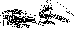
According to Buchman, the fistula opening is excised to the fascia. On both sides of the fistula opening, two oval incisions are made in the fascia, and over the fistula opening a duplication is formed from the fascial layers, sutured with wire. Above them, the edges of the incised fascia are drawn together and sutured with wire. The skin wound is sutured tightly. The wire sutures are brought out to the surface.--The simplest is Sapozhkov's method. According to this method, the fistula opening is excised to the superficial fascia (fig. 11). Then, 1 cm above and below the skin wound, two punctures are made into the skin to the fascia, and through one puncture into the other on a needle, a strong silk thread is passed, which is tightly tightened, tied, and cut (fig. 12 and 13). Thus, the fistula tract is ligated subcutaneously (fig. 14). The fistula tract can be curetted before applying the ligature. The skin wounds are sutured tightly. If a cyst forms after such ligation, it can be punctured from the mucous membrane side to form a fistula into the oral cavity. If all operative interventions fail, removal or physiological destruction of the gland is resorted to. For this purpose, injection of sterile oil, alcohol into the gland, ligation of Stensen's duct and the system
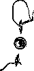
Fig. 13
Fig. 13
mechanical compression of the gland, leading to its atrophy. Finally Leriche proposed neurexaeresis n. auriculo-temporalis, the fibers of which are torn out for the parotid gland, thanks to which secretion ceases and the fistula closes. Neurexaeresis does not always achieve its goal, which is why the operation did not receive general recognition. This is explained by the presence of an autonomous nervous system in the gland itself (Lukomsky, Herzen). Foreign bodies (bristles, grains) are rarely encountered and primarily cause phenomena of stagnation, then enlargement of the gland and the appearance of shooting pains. Sometimes with the addition of infection, an acute Figure 14.
inflammation with spread to the gland and subsequent suppuration. Stones of the duct and gland are also a rare phenomenon. The etiological factors are considered to be foreign bodies t phenomena of stagnation, presence of infection. Clinically they manifest as a tumor at the site of location7of the stone, enlargement of the gland due to stagnation, pains that increase with food intake* due to increased secretion and obstruction to secretion, especially when the stone is located in the duct. Stones within the gland substance sometimes cause no subjective symptoms besides swelling. Path.-anat. changes can be significant. Besides atrophy due to compression, inflammation, pressure sores, ulcers with subsequent scarring may be observed. Treatment is surgical. Operation when a stone is present in the duct is performed from the oral cavity side. The mucous membrane and duct wall are incised over the stone and the latter is removed. The wound is sutured. A stone of the gland is removed through a skin incision with the gland capsule. Inflammations of the P. g. Acute inflammations-see Parotitis. Chronic non-specific inflammations of the P. g. are extremely rare; usually there is diffuse swelling without change in the external coverings, painless or slightly painful on pressure, almost always strictly limited. Sometimes inflammation of the duct, its obstruction, swelling and presence of granulation tissue at its external opening are observed. Küttner described very interesting inflammatory tumors. These descriptions concerned the submandibular gland. Histologically there is proliferation of connective tissue and small-cell infiltration. Küttner considers it very probable that such tumors also occur in the P. g. Only biopsy reveals the true nature of the tumors. Chronic inflammatory swellings are usually treated with iodine, potassium iodide internally. If there is growth of the tumor and ineffectiveness of therapeutic treatment, it should be removed, since there is suspicion that we are dealing with a malignant form. Chronic specific inflammations. Actinomycosis of the P. g.-usually of secondary origin, although primary involvement through the excretory ducts is not excluded. In far-advanced cases it is difficult to determine whether we are dealing with primary or secondary involvement; only in the initial stages by the emergence of typical grains from the duct can we judge what we are dealing with. Treatment-internal potassium iodide, radiotherapy. If the latter is ineffective, then extensive surgical intervention.-Tuberculosis of the P. g. is rare. It should be noted that of all the salivary glands, the P. g. is affected by tuberculosis most frequently. Usually there is diffuse or limited dense swelling with several or one area of softening. Sometimes the skin cover is slightly inflamed, usually unchanged. Sometimes facial neuralgias are described, due to involvement of nerves passing through the gland. Diagnosis is difficult, the process is often mistaken for a tumor, and only histological research reveals the true nature of the disease. Treatment-quartz, heliotherapy, general strengthening; if unsuccessful, operation is indicated, with only the affected parts of the gland to be removed.-Syphilis of the P. g. in the early period can give symptoms of acute parotitis, and only the presence of other signs of early syphilis clarifies the picture. In the gummatous period there is diffuse dense (interstitial, fibrous form) swelling or gummatous neoplasms, which can be distinguished from true neoplasms often only with the help of biopsy. Treatment-specific anti-syphilitic. A special disease described by Mikulicz (see Mikulicz's disease) is rarely encountered. Treatment-general strengthening, arsenic, x-ray. The prognosis is relatively favorable.-Cysts of the gland and duct are not uncommon; usually with a cyst of the gland there is retention of secretion due to obstruction of the duct and subsequently interstitial inflammation. A duct cyst is usually recognized by a ridge palpable along the course of the duct. Treatment-dilatation, if unsuccessful-operation. Echinococcal cysts (Subbotin) are extremely rare. Clinically they manifest as swelling in the area of the P. g. For differential diagnosis, Casoni reactions should be performed (see Echinococcus). Treatment consists in enucleation of the cyst or echinococcotomy, with washing with a 1/2% solution of formalin and suturing tightly. Tumors of the P. g. can be divided into connective tissue, mixed and epithelial. Angiomas are extremely rare (they should not be confused with subcutaneous angiomas located in the area of the P. g.) and are clinically characterized by a tendency to increase rapidly in volume. Treatment-surgical (profuse bleeding) or electrocoagulation. Lipomas are true only those that originate from the interlobular connective tissue; they are opposed to lipomas located inside the gland capsule but not in its substance. Treatment-surgical. Sarcomas occur in all known forms and show all possible transitional forms between relatively benign and highly malignant, from fibrosarcoma to small-cell sarcoma and melanoma. Treatment-radiotherapy and surgical. - The most characteristic for salivary glands are mixed tumors, consisting of derivatives of two embryonic layers. In understanding their nature there are three directions: 1) supporters of the endothelial theory (Volkmann, Steinhaus, Bogolyubov) believe that these tumors originate from the endothelium of blood vessels and should be classified as tumors of the connective tissue type, 2) supporters of the epithelial theory (Kaufmann, Aschoff and others) believe that the parenchyma of the tumor consists of various types of epithelium, 3) supporters of the theory of mixed origin (Landsteiner, Lubarsch) believe that here there can be both connective tissue and epithelial cells. Clinically these tumors usually have a nodular form, less often a smooth surface, and the consistency depends on the predominance of one or the other tissue. The location of the tumor depends on which part of the gland the growth comes from: they can be located on the masseter muscle or under or in front of the auricle; usually solitary. Sometimes the gland is delimited from the tumor, sometimes it completely or partially merges with it. In a number of cases pains are observed, depending on compression of nerves by a large tumor. All these tumors are considered path.-anat. benign, but clinically they can have a malignant course, since recurrences are possible, and therefore treatment should be only surgical-resection, and sometimes also extirpation of the gland. Of epithelial tumors, adenomas are rarely observed, carcinomas are significantly more common, appearing in the form of cell-rich, rapidly growing medullary tumors or cell-poor, wrinkling scirrhous tumors. In the latter, paresis and paralysis of the facial nerve soon occur, the skin is drawn in a radial pattern. In the medullary form, ulcers develop, lymph glands are affected. In both forms, pains, loss of hearing, disturbance of respiration, chewing, swallowing are observed. Prognosis-unfavorable. In the early stage they can be mixed with tumors of the inflammatory-specific type. Recurrences almost as a rule. Treatment-surgical. Operation for a tumor consists in enucleation of the tumor or its removal together with the P. g., depending on malignancy. During the operation it is necessary to avoid damage to the facial nerve, and only if it is impossible to preserve it due to infiltration by the tumor, it should be sutured after resection (or in case of accidental injury). Therefore, for enucleation of the tumor, the incision is made horizontally along the course of the facial nerve. As far as possible, all small branches are preserved to avoid subsequent paralysis of the muscles. The tumor is bluntly enucleated together with the capsule from the gland tissue. Often on the posterior surface the tumor is intimately fused with the gland tissue, and blunt dissection here is difficult. To prevent recurrence of the tumor, partial resection of the adherent part of the gland is recommended. Then the gland tissue together with the capsule is sutured. If a large cavity is present, a drain is inserted. To remove the P. g., a vertical incision is made from the level of the upper attachment of the auricle and 4-5 cm below the angle of the mandible. For a large tumor or when the P. g. is infiltrated, an additional horizontal incision parallel to the zygomatic arch is recommended. The gland in the capsule is enucleated from the lower end. Here together with it, the lymphatic glands are also removed if they are affected. To prevent bleeding in extensive involvement, a temporary ligature can be applied to the a. carotis comm. When the tumor infiltrates blood vessels, sometimes it is necessary to ligate the a. carot. ext. and even the a. carot. int. Razumovsky performed ligation of the a. carotis comm. four times. In most cases this ligation does not lead to serious consequences, because due to gradual compression of the vessels by the tumor, collateral circulation has already developed. After freeing the lower pole, the gland is carefully enucleated from above and from the sides. If the gland is fused with the sternocleidomastoid muscle, the latter is partially excised.
Usually ligated are the vv. jugularis ext., facialis ant. et post., a. transversa faciei, a. auricularis post., aa. occipitalis, tempor. superficialis, zygomatico-orbitalis, maxillaris int. The most difficult moment is the separation of the Parotid Gland from the proc. styloideus and the crossing of the fascia going to the pharynx, as damage to the a. carotis int. and v. jugularis int. with the n. vagus passing here is possible. After removal of the gland, a drain is inserted into the resulting cavity for 1-2 days. The skin is sutured. To avoid damage to the cornea in case of damage to the n. facialis during the operation, the eye must be kept closed in the postoperative period.
Related articles
Mentioned in
Cite this page
“Parotid Gland.” Soviet Medical Encyclopedia. English translation of Bolshaya Meditsinskaya Entsiklopediya, 1st ed. (Moscow, 1928–1936), ed. N. A. Semashko. https://sovietmedicalencyclopedia.pages.dev/article/parotid-gland/