LACRIMATION
Historical document, translated for reference. It reflects medical knowledge of the 1920s–30s and is not medical advice.
Summary
Lacrimation refers to the secretion and drainage of tears, involving the lacrimal gland which produces tear fluid and the lacrimal drainage system which carries this fluid to the nose. The article describes the anatomy, physiology, innervation, blood supply, composition, and pathological conditions related to tear production.
Encyclopedia article (1928–1936)
LACRIMATION. The lacrimal organs consist of two completely isolated topographically and different in purpose departments, namely: the apparatus that secretes a special secretion called tear fluid or tears, and the apparatus that carries this fluid from the conjunctival sac into the nose. The function of the first is performed by the lacrimal gland (gl. lacrimalis), located in the bony depression of the anterior part of the orbit, immediately behind the upper outer part of the lacrimal gland; 2-h J
palpebral part of it; 3-
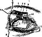
Figure 1. Displacement of the lacrimal gland: 1 - orbital
orbital margin. By the tendon of the t. levator
skin with t. orbicularis; 4 - septum orbitali; 5 - cartilage toriS palp. Super. Of the upper eyelid; 6 - conjunctival surface of the eye; 7 - the tendon divides into two unequal parts, m ievator palpebrae superi-considered by some authors as oris; 8 - the lateral part of this tendon, dividing-independent glands, with-g,глтм1тлл-тт nnmn-^n,™ т, Щaya °be parts of the lacrimal gland; S'-lateral attachment of this tendon to the edge of the orbit and to the deep-coverings; 9- edge of the orbit. The upper pole, bity. ^ - fat lobule. by form and size resembling a bean, lies directly under the periosteum of the orbit and is called the "orbital lacrimal gland" (pHC.l)(gl. lacrim. or-bitalis); its transverse size varies from 20 to 25 mm, longitudinal-from 12 to 14 mm, thickness is approximately 5 mm. The lower pole is significantly smaller in size than the upper and is called the "palpebral lacrimal gland" (gl. lacrim. palpe-bralis); it lies directly under the first, above the upper fornix of the conjunctiva, so that when the upper eyelid is everted and the eye is turned inward and downward, it protrudes as a yellowish bumpy mass. According to Figure 2. The lacrimal gland in its anat. structure to the lacrimal glands I have great similarity with SALIVARY GLANDS and CONSIST OF MANY tubular glands, collected in the thickness of the conjunctiva, especially along its upper transitional fold, there is also a whole series of small glands with an identical purpose and known under the name of Krause's glands. The excretory ducts of the orbital gland, numbering from 3 to 5, pierce the palpebral gland and together with its own open through a series of holes on the mucous membrane of the upper fornix near its outer edge. From here the tear fluid, due to its gravity, flows down the anterior surface of the eye, moistening the latter, and along the lacrimal groove (rivus lacrima-lis), formed by the posterior edge of the lower eyelid and the eye itself, collects in the inner corner of the palpebral fissure in a depression called the "lacrimal lake", from where the second part of the lacrimal apparatus, so-called lacrimal drainage pathways, begins. The innervation of the lacrimal gland is very complex and moreover not yet completely clarified; it is assumed that in it participate the I and II branches of the trigeminal nerve, the facial, as well as sympathetic fibers from the cervical plexus. The first carries out its participation through one of the terminal branches of its I branch, namely through the n. lacrimalis; the latter before entering the gland divides into two branches, of which the lower anastomoses with the n. zygomatico-temporalis, a branch of the II branch. The secretory fibers for the lacrimal gland are supplied by the facial nerve, with some assuming that these fibers begin from the nucleus of the n. glossopharyngei, upon exit from which they through the portio intermedia Wrisbergi join to the trunk of the n. facialis, and then through the n. petrosus superficialis major reach the ganglion sphenopalatinum, from where through the n. maxillaris sup. and some other anastomoses connect with the n. lacrimalis and together with it enter the lacrimal gland. The fibers of the sympathetic nerve enter the gland together with its blood vessels, and whether they also have an auxiliary secretory effect on the gland is not yet decided. The lacrimal gland is supplied with blood from the a. lacrimalis, a branch of the a. ophthalmicae. Tears represent a sterile, transparent, slightly alkaline and somewhat opalescent liquid, consisting of 99% water and approximately 1% solid parts of organic and inorganic nature, mainly sodium chloride, as well as sodium and magnesium carbonate, and calcium sulfate and phosphate. Their purpose is first of all to, by moistening the anterior surface of the eye, especially the cornea, maintain the transparency of the latter, absolutely necessary for normal vision, and then to wash out of the conjunctival sac all kinds of foreign elements of both organic and inorganic nature and carry them away into the nose. In addition, according to the research of some authors, tears to a certain extent also possess bactericidal properties, perhaps due to the presence in them of "lysozyme" (see Lysozyme), and also take some part in the nutrition of the cornea due to the content of lipoids. During sleep, tear secretion completely ceases. The normal amount of tear fluid secreted by the glands per day is negligible and amounts to approximately 1/2 cm3. Deviations from the norm in this respect may manifest themselves in the direction of decreased secretion - hyposecretion, as well as in the direction of its strengthening, i.e. hypersecretion. Hyposecretion of tear fluid is a relatively rare phenomenon and therefore has relatively little practical interest, unless it is a consequence of scarred wrinkling of the conjunctiva and obliteration of the excretory ducts of the glands as a result of severe diseases of the mucous membrane, e.g. trachoma. Apart from such cases, decreased secretion is observed in paralysis of the facial nerve, especially in the area of the ganglion geniculi, as well as in paralysis of the trigeminal nerve, further in some mental diseases, e.g. in melancholy, in certain poisonings, e.g. in botulism, and sometimes in severe infectious diseases with high temperature. Much more often one has to deal with hypersecretion of tears, and it can manifest itself in two forms: either in the so-called psychic crying, caused through special still unknown centers by various emotional disturbances (grief, anger, tenderness, joy, etc.), or it is purely reflex in nature and due to the close connection of the trigeminal nerve with the secretory tear fibers is especially often caused by all kinds of irritations in the area of the I and II branches of the n. trigemini. This includes first of all all inflammatory processes of the anterior part of the eye itself, such as: cornea, sclera, iris and ciliary body, as well as eyelids and conjunctiva, and in such cases hypersecretion of tears is usually accompanied by photophobia, blepharospasm and dilation of vessels; further especially abundant secretion of tears is observed when foreign bodies-dust, fall on the cornea or mucous membrane, and then when the eye is irritated by poisonous substances in vapor or gaseous state, etc. This kind of increased secretion of tear fluid should be regarded as a protective reflex aimed at mechanically removing from the eye a substance foreign to it. This should also include increased secretion in neuralgia of the trigeminal nerve, as well as its irritation from the side of the meninges, further in various manipulations in the nose in the form of cauterizations, lubrications, etc. The sensitive reflex point for the lacrimal gland is also the lacrimal sac, diseases of which are usually accompanied by increased secretion of tears; the latter significantly decreases or even completely ceases after its extirpation. As a rule, increased secretion of tears caused by reflex from the trigeminal nerve is always unilateral and moreover on the same side from which the reflex comes. On the contrary, bilateral hypersecretion of tears, besides psychic crying, is observed e.g. in light irritation of the optic nerves, and also by some authors is noted as an initial symptom of tabes dorsalis in the form of temporary attacks resembling the so-called crises, and then during pregnancy, in Basedow's disease, myxedema, migraine and hysteria. There is an assumption that the increased secretion of tears in psychic crying occurs specifically through the strengthening of the function of the orbital gland, while the palpebral gland accordingly reacts to all kinds of irritations of a reflex nature.-Under ordinary conditions all the tear fluid produced by the glands goes unnoticed into the nose, causing no subjective sensations, in case of hypersecretion it, even with normal condition of the lacrimal pathways, does not have time to enter the drainage system, causing in patients a very distressing condition called "tearing". In addition to disorders of the functional activity, the lacrimal glands are also subject to organic diseases, which in general come down to inflammation

Figure 3. Displacement of the lacrimal gland.
to inflammatory processes (see Dacryoadenitis), displacements of the gland from its normal position, and to new growths. Displacement usually affects only the orbital glands, and due to relaxation of the ligamentous apparatus, the gland is not held in place but gradually descends along the anterior surface of the cartilage, bulging forward the tars-orbital fascia, and in severe cases, ultimately ends up completely under the skin of the outer third of the upper eyelid. In such cases, the latter produces the impression of a sagging bag (Fig. 3), in which the displaced lacrimal gland lies freely and can be easily palpated, and is easily reducible to its place, under the orbital rim, from where it again begins to appear upon removal of pressure. As a rule, the disease is not accompanied by an inflammatory reaction, and the gland itself remains painless and retains its normal size. The condition can be either unilateral or develop simultaneously on both sides, and it is characteristic predominantly of young age. Treatment is surgical and consists of attempts to fix the gland in its place, and if this is not possible, to its complete extirpation. Tumors of the lacrimal glands are characterized by an increase in their volume, sometimes to very significant sizes, a firm or fluctuating consistency, the absence of an inflammatory reaction, painlessness, and sometimes also a forward or downward displacement of the eye. Among benign tumors, cysts should be noted, which develop either in the excretory ducts (dakryops) or in the gland itself as a result of impaired secretion outflow, caused by concrements or scar strictures. Solid tumors of the lacrimal glands are quite rare and usually belong to the so-called mixed tumors (Mischgeschwulste), i.e., new growths constructed from the most varied cells and tissues, such as: epithelial, connective tissue, glandular formations, hyaline cartilage, bone, etc.; tumors of this kind are characterized by relative benignity. Among malignant tumors, which are even rarer, adenocarcinomas, adenosarcomas, and epitheliomas are characteristic for this organ. Therapy for new growths of the lacrimal glands can only be of a surgical nature, with small isolated tumors of the palpebral gland being removed from the conjunctival side under the eyelid, limited tumors of the orbital gland through a skin incision in the area of the outer half of the eyebrow; with b. or m. significant spread into the orbital cavity, it becomes necessary to resort to a temporary resection of its outer wall (Krenlein's operation), and if the tumors are malignant, the question may also arise of the complete exenteration of the ORBIT.
Related articles
Mentioned in
Cite this page
“LACRIMATION.” Soviet Medical Encyclopedia. English translation of Bolshaya Meditsinskaya Entsiklopediya, 1st ed. (Moscow, 1928–1936), ed. N. A. Semashko. https://sovietmedicalencyclopedia.pages.dev/article/lacrimation/