Slavyansky
Historical document, translated for reference. It reflects medical knowledge of the 1920s–30s and is not medical advice.
Summary
This article provides a biography of Kronid Fedorovich Slavyansky (1847-1898), a prominent Russian gynecologist of the late 19th century. It details his academic career, contributions to gynecology, and his work in pathology and histology of the female reproductive system.
Encyclopedia article (1928–1936)
Slavyansky Kronid Fedorovich (1847-1898), an outstanding Russian gynecologist of the second half of the 19th century. After graduating from the Medico-Surgical Academy, S. was retained at the clinic of Krassovsky for further training, as while still a student he had published a series of works that attracted attention. In 1870, S. defended his dissertation: "On the normal and pathological histology of the human Graafian follicle" (St. Petersburg). In 1871, he was accepted as a privat-docent at the Academy and was sent abroad for study. Abroad, he worked in Vienna, Berlin, Leipzig, Edinburgh, London, and Paris, wrote several works, and returned to Russia as a broadly educated specialist with good training in the then-emerging "new" gynecology. In 1876, S. was elected professor at Kazan University, but already the following year he returned to the Academy, where he first conducted the propaedeutic obstetrics clinic, and from 1883 until his death, the hospital clinic. During his professorship, S. expanded intensive scientific and teaching activities, which resulted in about 50 of his own printed works and about 250 works written by his students, among whom were such major scientists as A.I. Lebedev, N.N. Phenomenov, G.E. Rejn, D.O. Ott, N.V. Yastrebov, I.N. Grammatikati, and others. Of his own works, S.'s two-volume textbook on gynecology, unfortunately unfinished, was of particular importance ("Private pathology and therapy of women's diseases", St. Petersburg, v. I-II, 1888-97). S.'s activities coincided with the rapid flourishing of modern operative gynecology, in this field he successfully transferred the achievements of Western Europe to Russia, but by nature he was as much a laboratory scientist as a surgeon and clinician, and his works were especially valuable contributions to pathological anatomy and histology of the female reproductive sphere. In many respects, he was a "pioneer" in this field, but his premature death did not allow him to complete the work he had begun.
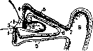
Figure 1. Lacrimal puncta, lacrimal canals, and their entry into the lacrimal sac: 1-plica semilunaris (semilunar fold); 2-lacrimal caruncle and lacrimal lake; 3 and 3'-lacrimal puncta; 4 and 4'-vertical part of the lacrimal canals; 5 and 5'-horizontal part of the lacrimal canals; 6-connection of both canals; 7 - entry of the canals into the lacrimal sac (8).
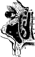
lacrimal fossa" (fossa lacrimalis), and together with the nasolacrimal duct, into which it directly passes, forms the vertical bend of the common lacrimal drainage pathway. The lacrimal fossa, containing the lacrimal sac, is formed in its anterior part by the frontal process of the maxilla, and in its posterior part by the lacrimal bone (os lacrimale), and it is bounded in front by the bony ridge of the first (crista lacrimalis anterior), and behind by a similar ridge of the second (crista lacrimalis posterior); downward these two ridges converge arcuately, forming the place where the lacrimal fossa transitions into the nasolacrimal duct (fig. 2). The lacrimal sac itself is a membranous tube, 10-12 mm long and 2-3 mm wide, the upper end of which is blind, while the lower end, narrowing, passes into the nasolacrimal duct, and the walls of the sac are closely applied to the bony bed only posteriorly and on the nasal side, whereas anteriorly and on the temporal side it is protected by a strong sheet of fascia, separating it from the orbital cavity and stretched between crista lacrimalis anterior and crista lacrimalis posterior; additionally, it is surrounded on both sides by the legs of lig. canthi internus, of which the anterior one, more substantial, crosses it somewhat below the apex anteriorly and attaches to crista lacr. ant., while the posterior one, weaker, goes around the sac posteriorly and attaches to crista lacrimalis post. together with that part of m. orbicularis which is known under the name of Horner's muscle. Lig. canthi internum can be easily palpated under the skin when the eyelids are stretched in the direction of the temple. The inner surface of the lacrimal sac is covered with a mucous membrane lined with stratified cylindrical epithelium, with a small number of mucous glands.
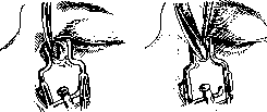
The nasolacrimal duct (ductus nasolacrimalis) is a direct continuation of the lacrimal sac, with its lower end opening into the nose, beneath the inferior nasal concha. The length of the duct averages 15 mm, its width 4 mm, and its general direction is from top to bottom, from front to back, and from outside to inside. The walls of the duct are rather closely fused with the periosteum of the bones, and between them there is a densely developed venous network, which is a continuation of the cavernous tissue of the inferior concha. The lacrimal fluid, while moistening the anterior surface of the eye, partially evaporates from it, and its excess collects in the lacrimal lake; from here, through the lacrimal puncta and along the lacrimal canals, it enters the cavity of the sac, and from there it flows down the nasolacrimal duct into the nose. The mechanism of lacrimal drainage is closely connected with the blinking movements of the eyelids. The main role in this process, based on recent research by some authors (Frieberg), is attributed to the pumping action of the lacrimal canals, the lumen of which apparently widens under the influence of the tone of their longitudinal muscular layer when the eyelids open, and their cavity fills with lacrimal fluid from the lacrimal lake. When the eyelids close, the lacrimal canals are compressed, and the lacrimal fluid is squeezed into the lacrimal sac. A certain auxiliary role in this process is obviously played by the suction action of the sac itself, which also expands during blinking due to the pulling of its anterior wall forward by the action of lig. canthi interni, as well as its upward traction by means of Horner's muscle. The further movement of the lacrimal fluid along the nasolacrimal duct occurs partly due to its weight, and partly as a result of the collapse of the sac walls as a consequence of the relaxation of the tone of the circular muscle. All diseases of the lacrimal pathways, starting from the lacrimal puncta and ending with the inferior nasal opening, lead to a disruption of the normal function of lacrimal drainage, which manifests as a delay of lacrimal fluid in the eye and subsequent epiphora (epiphora, illacrimatio). The latter, especially if it is persistent, represents a tormenting suffering. Under the influence of systematic moistening by tears, the skin of the eyelids and face is subjected to irritation and various inflammatory processes, which often leads to eversion of the lower eyelid and lacrimal punctum. Furthermore, the constant delay of tears disrupts the process of normal self-cleansing of the conjunctiva from bacteria and leads to chronic inflammation of the latter, which in turn is usually accompanied by increased secretion of tears, thus creating a kind of vicious circle in the end. Finally, and most importantly, epiphora disrupts vision: the tears 'blur the eyes' and interfere with seeing, thereby causing a decrease in work capacity. The immediate causes of epiphora basically come down to three fundamentally different factors, namely: either it is a matter of increased secretion of tears (see Lacrimal secretion) with normal outflow, or a weakening to complete absence of the expelling force, which is the blinking movements of the eyelids, which in turn can be caused by paralysis of the facial nerve or extensive scar changes that interfere with the free movement of the eyelids, or finally, it can be a matter of mechanical obstacles to the movement of tears in any part of the lacrimal pathways. Measures against epiphora should consist in eliminating one of the factors listed above, which underlie this suffering. The pathology of the lacrimal puncta is expressed by a violation of their correct position, a decrease in their lumen, and clogging with foreign bodies. A change in the correct position of the lacrimal puncta can occur in two directions: either the lacrimal puncta are turned inward too much (inversio puncti lacrimalis) or, conversely, they are everted outward (eversio puncti lacrimalis). The cause of such deviations from the norm is either corresponding changes in the normal position of the eyelids themselves or various kinds of scar changes in the skin or conjunctiva near the lacrimal puncta. Their eversion is sometimes caused by an increase in the papillae on the tops of which they are located, which is especially observed in old age. The openings of the lacrimal puncta can be reduced to their complete closure as a result of chronic inflammatory processes of the mucous membrane or the edges of the eyelids; sometimes the absence of lacrimal puncta (atresia) is observed as a congenital phenomenon. Foreign bodies in the lacrimal puncta are usually eyelashes, which are carried by the current of tears into their openings. Diseases of the lacrimal puncta lead to a violation of the capillary absorption of lacrimal fluid from the lacrimal lake and to subsequent epiphora. The treatment of these conditions in the case of eversion or narrowing of the lacrimal puncta consists in splitting the lacrimal canals with Weber's pug knife towards the eye, and in the case of inversion - in corresponding operations on the eyelid (fig. 3).
The pathology of the lacrimal canals consists of their narrowing or complete absence (atresia) or, conversely, splitting, and all these anomalies can be of a congenital nature. Narrowing of the lacrimal canals as a result of inflammatory processes of the conjunctiva and eyelids is a rather common phenomenon. Isolated inflammation of the lacrimal canals is rarely observed. The lacrimal canals sometimes are the site of formation of concretions or dacryoliths from the accumulation of fungi of various species of Streptothrix and even, as an exception, actinomycetes. This is manifested by swelling of the affected lacrimal canal, dilation of its opening at the lacrimal punctum, and the presence of hardening upon palpation. The treatment of these conditions consists in splitting the canal and removing the concretions. The pathology of the lacrimal sac is limited almost exclusively to its inflammations in various forms (see Dacryocystitis). Rarely, tumors of a benign nature in the form of cysts, polyps, fibromas, and of a malignant nature in the form of carcinomas and sarcomas can develop in the lacrimal sac. As a consequence of dacryocystitis (see), dropsy of the lacrimal sac (hydrodrops, s. hernia sacci lacr., s. dacryocystocele) may occur. In tumors of the lacrimal sac and its dropsy, the only correct measure is the complete removal of the lacrimal sac (extirpatio sacci lacrimalis). The latter is performed as follows: after subcutaneous anesthesia of the sac area, an arcuate incision is made
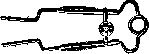
Fig. 5. Retractor for wound in the operation of removal of the lacrimal sac. (After Mi Peg.)
of the skin about 1/2 cm long through the ligamentum canthi interni (fig. 4), beginning 1/2 cm above it and directed outward; the lig. canthi int. is cut at its point of attachment to the crista lacrimalis anter.; next, an incision is made in the fascia covering the sac, and its anterior wall of grayish-blue color is revealed; with the tip of blunt scissors inserted between the fascia and the sac, the latter is separated from all sides of its bed, during which the lacrimal canals are cut with one stroke of the scissors; the head of the sac is grasped with forceps and pulled out of the wound, the lower end is carefully separated from all sides and cut off as deeply as possible at the entrance to the nasolacrimal duct, which is thoroughly curetted with a sharp spoon; three sutures on the skin wound. During the operation, the edges of the wound are usually dilated with the help of a Mueller mirror (fig. 5) or an Axenfeld dilator (see Ophthalmological Instruments). Removal of the lacrimal sac does not free the patient from tearing, which, on the contrary, remains forever due to complete disruption of tear drainage. Nevertheless, numerous observations show that over time this tearing ceases to bother patients, since with the removal of the sac, tear secretion is obviously reflexively reduced. In cases where it still bothers patients, sequential removal of the palpebral lacrimal gland is recommended. Injuries to the lacrimal sac are rare; they may be limited to only its soft parts or may also involve the bone. The pathology of the nasolacrimal canal is exclusively due to narrowing of its lumen (strictura canalis naso-lacrimalis), which develop on the basis of inflammatory processes from the side of the nasal mucosa of atrophic or hypertrophic nature, extending also to the mucous membrane of the canal. These narrowings may be temporary if they are caused only by swelling of the submucosal venous plexus and are anatomically expressed only by the adhesion of the canal walls, or permanent when ulcers form on its mucous membrane followed by the development of scars. Most often, narrowings of the nasolacrimal canal develop at its beginning and end (diagnosis and treatment of narrowings see Dacryocystitis). In addition to the spread of inflammatory processes to the mucous membrane of the lacrimal canal and the formation of scars, tear drainage can also suffer from mechanical compression of the nasal opening of the canal by a hypertrophied inferior turbinate, polyps, a deviated nasal septum, etc., especially if the membranous canal, after emerging from the bony part, continues for some distance under the nasal mucous membrane.
Related articles
Mentioned in
Cite this page
“Slavyansky.” Soviet Medical Encyclopedia. English translation of Bolshaya Meditsinskaya Entsiklopediya, 1st ed. (Moscow, 1928–1936), ed. N. A. Semashko. https://sovietmedicalencyclopedia.pages.dev/article/slavyansky/