Ovulation
Historical document, translated for reference. It reflects medical knowledge of the 1920s–30s and is not medical advice.
Summary
Ovulation is the rupture of a mature follicle with the release of an egg cell from the ovary. The process involves follicle maturation, egg maturation, follicle rupture, and the formation of the corpus luteum.
Encyclopedia article (1928–1936)
OVULATION (from Latin ovulum-egg), the rupture of a mature follicle with the exit of the egg cell from the ovary. Essentially, O. includes the following moments: 1) maturation of the follicle, 2) maturation of the egg, 3) rupture of the follicle, and 4) the formation of the corpus luteum directly associated with it. Development of the follicle. In the early period of embryonic development, the covering germinal epithelium of the ovary begins to grow into its stroma in the form of solid cellular (epithelial) strands-the so-called Pflüger's tubes (fig. 1), or Waldeyer's spheres. In their center, the egg cell differentiates, surrounded
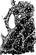
Figure 1. Pflüger's tubes (section of ovary of a five-month embryo).
along the periphery by a layer of epithelial cells, forming in this way the primary or primordial follicle: They were already found at the 6th month of intrauterine life (Strassmann), and at the 7th and 8th months, signs of simple and reverse development are noticeable. At the time of birth, the number of primordial follicles reaches 36,000 (Henle)-200,000 (Sapey) in one ovary. Multiplying intensively-by mitosis, the epithelial cells form the so-called follicular epithelium; simultaneously in the center of the follicle a small cavity filled with serous fluid (liquor folliculi) forms. Accumulating, it pushes the rows of cells and the egg surrounded by them to the periphery (see separate table, figure 6). Of the total number of primary follicles, no more than 500 reach full maturity (counting 13 per year for 30-35 years of a woman's sexual function). A significant part of the primordial follicles undergoes so-called physiological atrophy (growth of ovarian connective tissue together with phagocytosis), which should be distinguished from the process that Slavyansky called physiological dormancy (atresia). During atresia, fatty degeneration of cells sometimes occurs with subsequent resorption, more rarely-colloid degeneration, especially with pathological changes in the ovary itself (Slavyansky), or sclerosis of the surrounding connective tissue with the formation of a shiny membrane (Membrana) around the dying follicle. The follicle collapses, its contents are resorbed. Death begins with phenomena of chromatolysis of nuclear substance and cessation of karyokinesis. Seitz distinguishes two forms of atretic follicles-cystic and obliterating. Walthard believes that atresia of follicles is only obliterating, while the cystic form represents only a preliminary stage of obliterating. Seitz's view is more correct. In each ovary, atretic follicles can be found in various stages of regressive changes. The mature follicle, or Graafian follicle (fig. 2), was first described in 1672 by de Graaf, has from 15 mm to 26 mm (Leopold) in length, 15-17 mm in height, and protrudes on the surface of the ovary. The follicular epithelium is arranged in several layers along the inner
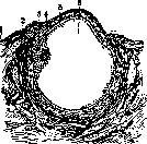
Figure 2. Graafian follicle: 1-germinal epithelium; 2-theca externa; 3-theca interna; 4-cumulus oophorus; 5-membrana granulosa; 6-liquor folliculi.
Figures 1-5. Maturation of the egg. Figure 1. Beginning of spindle formation. Fig. 2. Spindle. Fig. 3. Movement of the spindle to the pole. Figure 4. Separation of the first polar cell. Figure 5. Complete separation of the first polar cell. Fig. 6. Development of the follicle: 1-primordial follicle; 2-maturing follicles; 3-follicular epithelium; 4-egg; 5-theca folliculi; 6-follicle cavity. Fig. 7. Egg cell: 1-corona radiata; 2-zona pellucida; 3-deutoplasm; 4-protoplasm; 5-germinal spot; 6-germinal vesicle. Figure 8. Flowering corpus luteum: 1-blood center; 2-protrusions of the fibrous network; 3-theca interna; 4-lutein cells. To st. Ovulation, surface, forming the granular layer (membrana granulosa), in one of whose areas a tubercle with the egg cell in the center-discus oophorus, s. cumulus oophorus, s. cum. ovigerus, s. cum. proligerus protrudes. The stroma of the ovary forms around the follicle a
layer. This layer is penetrated by vessels, often dilated, reaching up to membr. granulosa, but never penetrating into it (Schickele) (figure 3). The egg cell (see separate table, figure 7). A fully developed human egg (ovulum) has a diameter of 0.2 mm (first described by Baer in 1827). Its main mass is the yolk (vitellus), in which large-grained nutrient yolk-deutoplasm and the more transparent cortical layer of formative yolk-protoplasm are distinguished. Inside the latter lies the nucleus or germinal vesicle (vesicula germinativa), and in it the germinal spot (macula germinativa). The egg has an amorphous glassy membrane (zona pellucida). In some animal species, it has radial striations. Between the yolk and the zona pellucida there is a slit-like perivitelline space filled with fluid. The entire outer surface of the egg cell is covered with radially arranged rows of cells (the so-called corona radiata). In this form, Waldeyer calls the egg 'ready', but to become capable of fertilization, it must go through a period of maturation, consisting of the following (see separate table, figures 1-5): the nucleus approaches the surface, and the chromosomes double by splitting, forming a spindle-shaped figure. Part of the chromosomes is extruded onto the surface of the egg cell in the form of the first polar cell or directing body of the first order. In the remaining part of the nucleus, a second mitotic division occurs with the extrusion of the second polar cell or directing body of the second order. Their fate is unknown. The remaining part of the spindle after the second division forms the nucleus of the mature egg-pronucleus femininus. It is smaller than the primary germinal vesicle and differs from it in the absence of a large germinal spot and a nuclear membrane. In mammals, the process of egg maturation occurs in the last days before follicle rupture, partly shortly before fertilization. According to R. Meyer, it is the development of the egg cell that has a stimulating effect on the growth of the follicle; Zondek and Aschheim, however, believe that the pituitary hormone causes the production of ovarian hormone and simultaneously promotes the maturation of the egg; the egg and ovarian hormone stand side by side, are coordinated, do not depend on each other, both being subject to the hormone of the anterior pituitary. Rupture of the follicle (properly O.). During follicle rupture, the release of the egg occurs in two moments: 1) detachment from the wall of the follicle due to loosening of the cells at the base of the cumulus oophorus-internal release and 2) exit from the ovary through the rupture of the follicle-external exit. The rupture of the follicle usually occurs at its outer pole at the point of greatest thinning of the wall and tunica albuginea, where there are almost no vessels. The immediate mechanical cause of the rupture is the accumulation of fluid, an increase in intrafollicular pressure, and stretching of the wall. A number of factors can accelerate follicle rupture: thermal and chemical irritations, so-called aphrodisiacs (cantharidin, yohimbine, muiracitin, alcohol), biogenic amines, psychic irritation, especially of an erotic nature, and coitus. Hormonal irritants have the same effect: the hormone of the anterior pituitary and folliculin. Less is known about substances that delay O.: boron-choline (Wintz), corpus luteum hormone (Haberlandt, Bokharev; according to Frenkel it delays only the rupture of the follicle, not the development), parenteral and paravaginal introduction of sperm, vitamin-poor nutrition. -The site of rupture (stigma folliculi) represents a slit-like or uneven opening several mm in diameter
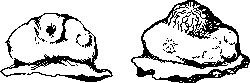
Figure 4.
Figure 5.
Figure 4. Ovary with ruptured follicle. Figure 5. Corpus luteum from the surface. (fig. 4). Bleeding from the site of rupture is slight, mainly into the cavity of the follicle itself. The opening is first closed by a fibrin clot, which then organizes, forming a delicate scar. According to observations by Honore in guinea pigs, the opening remains open even after 30 hours and only closes completely after 6 days. At the moment of rupture, the mature, fertilizable egg cell is expelled from the follicle along with the fluid, and this is where O. properly ends. This ovarian cycle occurs periodically, at more or less definite intervals, characteristic for each woman, most often-4-weekly. O. usually occur in both ovaries, but there is no complete uniformity in their alternation. When one is removed or diseased, the other functions with the same regularity. In exceptional cases, O. occurs simultaneously in both ovaries or two follicles rupture in one. O. stands in deep physiological connection with two processes: 1) the development of the corpus luteum and 2) menstruation. Anatomy and histology of the corpus luteum. The follicle freed from the egg undergoes a series of changes, which come down to three processes. 1. Hemorrhage. It It is observed in most cases, but not always, and occupies the central part of the former follicle. The corpus luteum in this stage protrudes sharply on the surface of the ovary (fig. 5), it is dark red in color, easily bleeds 1 2
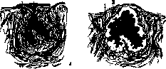
Figure 6.
Figure 7.
Figure 6. A freshly ruptured follicle: 1-theca int.; 2-hemorrhage. Figure 7. Development of the luteal layer: 1-theca int.; 2-membrana granulosa. The corpus luteum is 1.5-2 cm in diameter. Severe hemorrhages from the corpus luteum have been described, requiring surgery, more often immediately before or during menstruation. 2. The second process, which constitutes the essence of corpus luteum development (figs. 6-10), is the formation of lutein cells. The cells of the membrana granulosa, freed from the pressure of the follicular fluid, begin to proliferate and become filled with highly light-refracting grains of lutein lipid, from which they macroscopically acquire a yellowish tint and receive the name of lutein cells. As a result of enhanced proliferation, they form a wide wavy layer, the festoons of which penetrate into the blood center [see separate table (pp. 87-88), fig. 8]. Numerous capillaries grow from the theca interna side into the thickness of the layer between individual cells. At this stage, the corpus luteum stands out sharply on the ovarian section, representing a peculiar glandular organ. It was discovered by Volcher Coiter and named corpus luteum. The genesis of lutein cells remained controversial for a long time: older authors attributed them to the theca interna (Rokitansky, His, Kolliker, Hegar, Slavyansky), while later studies established their origin from the cells of the membrana granulosa (Sobotta, C. Ruge II, Cohn, Nowak, Schickele, Okintchits, Timofeev). 3. In parallel, the third process proceeds - the growth of connective tissue from the side of the theca interna, first - in the form of individual spindle-shaped cells, which then form strands, and then also connective tissue layers. The latter, in the form of a wedge, penetrate from the periphery into the layer of lutein cells, divide them into groups and, gradually thickening themselves, compress and lead to atrophy. This process is extremely constant, begins early in young corpus luteum and progresses steadily. The connective tissue formations in the place
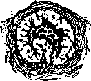
of the former corpus luteum are called corpora albicantia (fig. 10). Determination of the age of the corpus luteum. R. Meyer and Ruge II distinguish 4 stages of corpus luteum development. 1. Proliferation - transformation of membrana granulosa cells into lutein cells; numerous mitoses; characterized by the simultaneous presence of membrana granulosa cells and lutein cells. 2. Vascularization - penetration of endothelial cells from the vessels of the theca interna and development of capillaries in the thickness of the lutein layer proceeds very quickly, the beginning is difficult to notice. 3. Bloom - strong development of the lutein layer, polygonal shape of cells, richness in lipoids. 4. Regression of the corpus luteum - development of connective tissue, cell death. Ruge could not determine the duration of each period, he attributed follicular rupture to the middle of the intermenstrual period, while regression begins with the appearance of menstruation. The works of Leopold and his student Ravano give the following scheme of age


Figure 9.
Figure 10. Figure 9. Regression of the corpus luteum. Figure 10. Corpus albicans. Changes in the corpus luteum: 1st week-blood clot, by the end-a clear outline of the lutein layer at the periphery, blood in the center-red. 2nd week: yellowish border of the lutein layer up to 2 mm, by the end-individual connective tissue cells between lutein cells. 3rd week: the border of lutein cells is distinctly yellow, winding, blood in small amounts, strands of connective tissue from periphery to center. 4th week: the blood core is smaller, lutein cells penetrate the center, processes of connective tissue penetrate deeply. 5th week: reduction of the blood core, growth of connective tissue at the expense of lutein cells. 6th week: clear wrinkling of the border, still yellowish, the blood core has a brown color. 7th week: the blood core in the form of a point; center and periphery take the same color. According to Schickele, the duration of evolution of the corpus luteum is 8-9 days, the duration of the stage of bloom is unknown. He divides the regressive stage into 4 phases. 1st: a wide lutein band is divided by layers of connective tissue (according to the scheme of Leopold and Ravano corresponds to the 3rd week), 2nd: thick layers, small groups of cells in the stage of degeneration (4th and 5th weeks according to Leopold and Ravano), 3rd: changes in lutein cells, the cavity is flattened (sixth week according to Leopold and Ravano), 4th: remnants of lutein cells among connective tissue. The corpus luteum in case of the onset of menstruation undergoes regression much faster and is called corpus luteum spurium (false), s. corpus lutei menstruationis. In the case of pregnancy it reaches especially lush bloom and is called corpus luteum verum (true), s. corpus lutei gravidi-tatis. The difference between them is essentially only quantitative. Chemistry of the corpus luteum. The substance giving the corpus luteum its yellow color was obtained by Piccolo and Lieben (Piccolo, Lieben) and named lutein. At first it was considered identical with the yolk of a chicken egg, but this was later refuted. Lutein, or lipochrome, is a coloring substance found in blood serum and fat. Its formula is C40H56O2, crystals and concentrated solutions are orange-red in color. According to Mikulicz-Radecki, in the cells of membranae granulosae and thecae int. of the maturing follicle there are only insignificant traces of cerebrosides and phosphatides, cholesterol and neutral fats. Fresh corpus luteum in the period of transformation of cells of membranae granulosae into lutein cells gives no reaction to fat. In the stage of vascularization and proliferation, according to Mikulicz-Radecki's research, it contains only cerebrosides and phosphatides, in the period of full bloom cholesterol and traces of neutral fats appear. With the onset of involution, fatty degeneration begins in the corpus luteum. According to literary data cited by Bill, in the hemorrhagic corpus luteum cholesterol content is 1.99%, in the stage of maturation-5.84%, during regression-10.92%. Timofeyev considers the fat inclusions in lutein cells to be lecithin. Fatty infiltration represents neutral fat in combination with cholesterol or in mixture with it. Vignes considers that from the corpus luteum one can isolate up to 10 different bodies with the help of water, alcohol, ether, chloroform, acetone, etc. The menstrual corpus luteum contains more lipoids than the corpus luteum of pregnancy; the latter is richer in them in early stages, in later stages colloid and lime appear in it (especially with the beginning of the puerperium). Mikulicz-Radecki is inclined to consider the lipoids of the corpus luteum as ovarian secretions, but Preis-secker thinks that simple inclusions of fat substances cannot be considered as having hormonal properties and that the true carriers of hormonal properties are lipoids connected with the protein of protoplasm. They can be detected by microchemical reactions only by breaking this connection. Zondek also thinks that fats, lipoids and hormone are not identical; the hormone is only connected with them. It must first be freed from fats to transfer it to aqueous solutions. Seitz, Vince and Fingerhut isolated two lipoids from the corpus luteum: 1) Lipamin (lipo-proteid, Lecithalbumin), which causes menstruation in amenorrhea and promotes the growth of the sexual apparatus (according to Adler identical with the secretion of the follicle), and 2) Luteolipoid-from more mature corpus lutea; it stops bleeding, reduces the duration of menstruation. Thus, the question of the chemical composition of the corpus luteum at the present time cannot be considered finally settled. Physiol. role of the corpus luteum. Podvysotsky first in 1896 expressed the hypothesis that the corpus luteum is a gland of internal secretion. Simultaneously Prenant recognized it as a gland of internal secretion on the basis of research by Sobotta on the histological structure of the corpus luteum. At present this position is generally accepted. In 1901 Born first expressed the hypothesis that the corpus luteum has as its task to promote the attachment of the fertilized egg in the uterus. His students Frenkel, Magnus and Conn continued the study of the physiology of the corpus luteum and attributed to it a whole series of functions (up to 25). Frenkel's theory at first received wide recognition, then began to be tested, and at present his positions are considered refuted by many. A tremendous shift in this question was brought about by the work of Allen and Doisy, Zondek and Aschheim with the hormone isolated from the follicular apparatus and the hormone of the anterior pituitary gland. Frenkel formulated his main position as follows: the general protective influence of the ovary on the genitals is connected with the corpus luteum. Its main functions are the following: 1) the corpus luteum indicates trophic influence on the sexual apparatus, especially on the increase of the uterus, 2) it causes cyclic (premenstrual) changes in the mucous membrane of the uterus, 3) it promotes the implantation of the fertilized egg and 4) it exerts a protective influence on the egg in early stages. The influence of the corpus luteum on the growth and development of the uterus. The first position was refuted already by the work of Okinich (1913), in whose experiments injection of a follicular preparation into castrated rabbits delayed the atrophy of the uterus, while injection of a corpus luteum preparation did not. Later work by Courrier and Potvin established that under the influence of follicular fluid, the uterine horns of castrated rabbits reached normal size. According to research by Allen and Doisy (1923), follicular fluid in castrated mice causes estrus phenomena, and in sexually immature animals accelerates maturation by 20-40 days. This has been confirmed by the work of Pavlenko and Pchelina with the Russian preparation ovarikrin. Thus, the stimulating effect of the follicular hormone on the growth and condition of the sexual apparatus must be recognized as one of its essential properties. Research by Zondek and Aschheim established a close connection and specific influence of the hormone of the anterior pituitary gland-prolan-on the ovary and the entire sexual apparatus. Zondek on the basis of thorough research came to the following conclusions: 1) the production of follicular hormone is connected with the follicular apparatus, 2) it is acyclic, 3) the concentration of the hormone in different stages is different, 4) the ovarian function cannot be replaced by the function of another endocrine gland, but the dormant function of the ovary can be called into activity, but only by the pituitary gland (neither the thyroid, nor the thyroid, nor testes). The ovarian hormone acts on the structure of the uterine mucosa and on the entire complex of secondary characteristics. Relationship between Ovulation and the corpus luteum. As early as 1831 it was expressed that O. and menstruation are in causal and temporal connection (Negrier). The causal connection is recognized by all, and the position that without O. there is no menstruation, is indisputable. Bleeding appearing during menopause does not represent menstruation, but is a sign of a pathological process (sclerosis of uterine vessels, neoplasms). O. without menstruation can take place, as evidenced by the onset of pregnancy before the appearance of the first menstruation or during lactational amenorrhea. According to the modern view, menstruation itself-the appearance of bleeding-is only the final destructive act, ending the complex process in the uterine mucosa. Therefore, the essence of disagreements comes down to the question: do the cyclic changes in the uterine mucosa depend on the follicular apparatus, in particular on the growing or mature follicle, or on the corpus luteum. The data of Okinich, Bourcour, Vasic, Allen and Doisy and the work of Schickele speak that the changes in the uterine mucosa cannot be explained by the influence of the corpus luteum, but depend on the growing follicle. Pratt and Allen injected monkeys for 9-20 days 64-190 rat units and observed characteristic premenstrual changes. Cessation of the injection caused typical menstruation. Clinical observations on the application of ovarian preparations confirm the experimental data: extracts of the follicular apparatus are used in underdevelopment of the genitals, oligomenorrhea, opsomenorrhea, amenorrhea, because they promote the appearance and intensification of menstruation, and consequently the development of the endometrial cycle.
Zondek determined that the hormone is contained precisely in the maturing and mature follicle, and the closer it is to rupture, the more hormone there is in the follicular fluid and the follicle wall. The corpus luteum is credited with the property of delaying O. Galban and Keler removed the corpus luteum during laparotomy, and after 2-4 days bleeding occurred regardless of the time since the last menstruation and the age of the corpus luteum. The next menstruation occurred after 4 weeks. The authors see in this a refutation of Frenkel's theory and confirmation that the primary cause of menstruation lies in the contents of the rupturing follicle and the simultaneous influence of other glands. Frenkel himself does not deny the delaying influence. Loeb calls the corpus luteum a gland of sexual rest. The temporal relationship between O. and menstruation. There are 3 views: 1) O. and menstruation each have their own cycle, but there is no temporal relationship between them. 2) There is a temporal dependence, but no exact temporal coincidence. 3) O. and menstruation coincide. In resolving this question, various methods were used (the corpus luteum was studied at autopsy, it was examined in situ during abdominal operations, histological research was performed on removed ovaries, etc.). Most studied the corpus luteum histologically, comparing it with changes in the uterine mucosa. R. Schroeder presented a scheme of the temporal relationships between O. and menstruation. According to Schroeder, O. occurs between the 14th and 16th day from the beginning of the last menstruation; the peak of the corpus luteum coincides with premenstrual changes in the mucous membrane; with the onset of menstruation, the reverse development of the corpus luteum begins. Schroeder's scheme is very simple, but a number of facts and later observations do not confirm either the principle or the simplicity of this scheme. These facts are as follows: 1) The finding of a freshly ruptured follicle during menstruation in the absence of a corpus luteum. 2) The absence of a corpus luteum in the premenstrual week and during menstruation. Such cases were presented by Frenkel himself: during operations during menstruation in 4 out of 11 cases, he found no corpus luteum. Similar data are provided by Schickele, Terebinskaya-Popova, and others. 3) Complete discrepancy between the age of the corpus luteum and menstruation. Thus, the question of the connection between cyclic changes in the uterus (i.e., menstruation) and O. is resolved in recent years not in favor of the corpus luteum. But not the fact of follicle rupture itself - O. in the narrow sense - has significance in the appearance of menstruation, but the presence of a developed, mature specific formation of the ovary - the follicle. The time of O. can vary (before menstruation, during menstruation, immediately after menstruation) - more often closer to menstruation (Figure 11). The influence of other glands of internal secretion cannot be excluded, first of all the pituitary gland. The physiological role of the corpus luteum cannot yet be considered clarified and fully proven. Apparently the corpus luteum has some relation to future pregnancy, just as the pregravida changes of the uterine mucosa itself. The existence of the menstrual corpus luteum is extremely short-lived - at least in the stage of functional ability, and indeed, its stable existence is achieved only during pregnancy. Apparently this is where its physiological role must be sought.
Related articles
Mentioned in
Cite this page
“Ovulation.” Soviet Medical Encyclopedia. English translation of Bolshaya Meditsinskaya Entsiklopediya, 1st ed. (Moscow, 1928–1936), ed. N. A. Semashko. https://sovietmedicalencyclopedia.pages.dev/article/ovulation/