Corpus Luteum
Historical document, translated for reference. It reflects medical knowledge of the 1920s–30s and is not medical advice.
Summary
This article from the 1928–1936 Soviet Medical Encyclopedia details the anatomy, development, and endocrine function of the corpus luteum in women and animals. It describes the four developmental stages—proliferation, vascularization, full development, and degeneration—and discusses its role in preparing the uterine mucosa for pregnancy.
Encyclopedia article (1928–1936)
CORPUS LUTEUM (Malpighi), a formation arising in the ovary at the site of a ruptured follicle (Graafian vesicle) and now recognized as an endocrine gland. At the height of its development, the corpus luteum has a dense consistency,
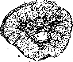
Figure 1. Section through a corpus luteum from a woman's ovary at the 5th month of pregnancy: 1 - surface of the ovary; 2 - theca externa folliculi; 3 - layer of lutein cells; 4 - connective tissue core; 5 - remnant of the cavity; 6 - remnants of blood extravasate and hyaline mass. (After Koelliker.)
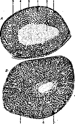
round or elongated in shape; dimensions: in humans 15–25 mm, in dogs 7–8 mm, in rabbits about 1 mm. It is covered by the outer shell of the follicle (theca externa) and consists of a connective tissue stroma with a large number of blood vessels and the bulk of epithelial-like cells containing fat inclusions and yellow pigment (lutein cells); inside the corpus luteum there is a cavity, partly filled with connective tissue (Fig. 1). Various views have been expressed regarding the origin of lutein cells: some (v. Baer, Koelliker, His) derived them from the cells of the inner membrane of the follicle (theca interna), others (Bischoff, Pflüger) from the membrana granulosa, and still others (Waldeyer, van der Stricht, H. Rabl) from both sources together. Currently, following precise observations by Sobotta on mice, most scientists recognize the origin of lutein cells from the membrana granulosa; for women, this origin of lutein cells is proven by Villemin and R. Meyer. Four periods can be distinguished in the developmental cycle of the corpus luteum. 1. Proliferation. After the rupture of the follicle and the escape of fluid with the egg and part of the cells of the membrana granulosa, its walls collapse, forming folds, and the cavity is filled with blood (in animals this is far from always the case) (Fig. 2). The remaining cells of the membrana granulosa begin to multiply and hypertrophy, transforming into lutein cells; it is possible that in some cases they are replenished at the expense of the cells of the inner membrane; as a result, a thick-walled closed sac is formed, in the middle of which a blood clot and fluid remain (Fig. 3). These phenomena occur in the first hours after rupture. 2. Period of vascularization. Plates of connective tissue with blood vessels begin to grow inward from the follicle wall in a radial direction, cutting the cell layer into lobules. When the connective tissue reaches the cavity, it spreads along the inner wall of the lutein layer, also giving plate-like outgrowths from there into its interior; simultaneously, the organization of the blood clot begins.
3. The corpus luteum reaches full development, according to some data (Schroeder) on the 3rd–4th day, according to others (Villemin) on the 10th day (in the mouse on the 3rd, in the rabbit on the 8th day). This period lasts a varying length of time: if fertilization has followed, the corpus luteum continues to exist approximately until the 5th month of pregnancy (it is then called the true corpus luteum—corpus luteum verum, s. graviditatis); otherwise, until the onset of the next menstruation (the so-called false corpus luteum—corp. lut. spurium, s. menstruationis). There is no difference in structure between these two types of the corpus luteum. 4. Period of degeneration, or involution. Lutein cells begin to atrophy or undergo fatty degeneration, being replaced by connective tissue (corpus luteum fibrosum), as a result of which the corpus luteum decreases in size. This process is slow during pregnancy, and the corpus luteum at the time of childbirth has a size of several millimeters; the false corpus luteum degenerates after the second menstruation. The result is a white scar (corpus luteum albicans), consisting of a convoluted fibrous bundle undergoing hyaline degeneration; sometimes pigment of blood origin is deposited in its middle, giving the scar a dark color (corpus luteum nigrum). With atresia of the follicle, accompanied by the death of the egg and degeneration of the membrana granulosa, a small corpus luteum (corpus luteum atreticum) also develops in the follicle, upon the degeneration of which a convoluted "vitreous strip," the remnant of the inner membrane, remains. Lutein cells have a polygonal shape, dimensions of 25–45 µ; in the middle is a nucleus, around which protoplasm with small granules is located, the rest of the cell being filled with fat droplets
Figure 3. Corpus luteum of a mouse (A — 5–7 hours after follicle rupture; B — 50 hours): 1 — connective tissue processes of the theca interna; 2 — coagulated liquor; 3 — theca externa folliculi; 4 — theca interna; 5 — follicular epithelium; 6 — lutein cells; 7 — central connective tissue core.
of various sizes, blackened by osmium acid and containing dissolved yellow pigment lutein. These cells are very similar to the interstitial cells of the testis and ovary. Malpighi already considered the corpus luteum to be a gland supplying nutrients to the embryo; subsequently, it began to be considered granulation tissue, and only recently (Prenant, 1897; Born) has it begun to be recognized as an endocrine gland that influences the proliferation of the uterine mucosa, preparing it for pregnancy. This was experimentally proven by Fraenkel (1901), who showed that the removal of corpora lutea in the first half of pregnancy entails the interruption of pregnancy; surgical removal of corpora lutea in women causes the premature onset of menstruation; on the other hand, delay in the involution of the corpus luteum causes amenorrhea (in cows, it delays estrus).

V. Karpov. Pathology. The Yellow Body plays a prominent role in the pathology of the ovaries. In the Yellow Body, ovarian abscesses may develop, and Yellow Bodies can serve as foci of tuberculosis; some authors have described true malignant tumors—cancers and sarcomas—that have arisen from Yellow Bodies or their cysts. The greatest clinical significance belongs to cysts of the Yellow Body. Attention was first drawn to them by Rokitansky, who described them only macroscopically. Cysts of the Yellow Body may be single or, more often, multiple. Their size usually varies from that of a hazelnut to a mandarin, but as an exception, cysts can reach the size of an adult human head. The cysts are always located at the periphery of the ovary and contain either serous or bloody fluid. Even macroscopically on a section, a very characteristic, mottled white membrane can be distinguished in the wall of the cysts, and on the inner pole, a crescent-shaped segment of yellow color, which upon microscopic examination consists of lutein cells. Usually, the cysts lack an epithelial lining, but sometimes it is present and represents remnants of the membranae granulosae. The structure of the wall can vary even within the same cyst; most often it consists of an outer connective tissue, a lutein, and an inner connective tissue layer (the latter is the most variable). Frenkel distinguishes 3 types of cysts: the 1st type has an outer connective tissue and an inner lutein layer, the 2nd type has an outer lutein and an inner connective tissue layer, and finally the 3rd type has an outer lutein, then connective tissue, and an inner epithelial layer. The presence of Yellow Body cysts in the ovary apparently has a delaying effect on menstrual bleeding. The clinical symptoms of Yellow Body cysts are in general similar to the symptoms of ordinary cysts and cystadenomas of the ovaries. Sometimes a rupture of the cyst into the abdominal cavity occurs with the formation of a hematoma of the broad ligament, which can simulate an ectopic pregnancy. In general, bleeding into the abdominal cavity from the Yellow Body and its cysts has been described by many authors. The bleeding can be so extensive as to require surgical intervention; fatal cases have also been described. The causes of these bleedings are various and not fully clarified; they can be divided into 3 groups: 1) external mechanical factors (strenuous work, gymnastic exercises, direct trauma to the ovaries during surgery or during gynecological massage, etc.); 2) general infections or intoxications that predispose to bleeding; 3) physiological fluctuations in the blood supply of the internal genital organs during ovulation and menstruation. Aschoff, Marcotti, Robert Meyer, and others have shown that during the premenstrual period, which is always accompanied by hyperemia of the ovaries, secondary hemorrhages often occur in the Yellow Body, which usually do not have great significance. In some cases, due to a particular richness in blood vessels, such a predisposing hyperemia can be accompanied by extensive hemorrhage with a rupture into the abdominal cavity. This view is fully consistent with the fact that in the vast majority of cases, bleeding from the Yellow Body occurs precisely in the premenstrual interval. In rare cases, the Yellow Body or its cysts can completely prolapse into the abdominal cavity (prolapse of the Yellow Body—prolapsus corporis lutei). In such a prolapse, separation occurs in the theca externa layer, and the prolapsed Yellow Body can sometimes implant, i.e., adhere to neighboring organs and have the possibility of further growth. Mechanical factors (e.g., surgery, gynecological massage) or inflammatory processes around the ovary and in the ovary itself can contribute to the prolapse of the Yellow Body. In hydatidiform mole, multiple cystic degeneration of the ovaries is sometimes observed, in which the cysts are lined with a layer of lutein cells (so-called lutein cysts) [see separate table (article: Congested Papilla), Fig. 1]; in structure and content, such cysts are completely similar to cysts of the Yellow Body. Some believe that lutein cysts form around atretic follicles, while others identify them with cysts of the Yellow Body. Grudzev believes that the formation of lutein cysts and in general excessive formation of lutein tissue in the ovary is not specific to hydatidiform mole and that such degeneratio polycystica luteinalis can occur as a disease sui generis.
Related articles
Mentioned in
Cite this page
“Corpus Luteum.” Soviet Medical Encyclopedia. English translation of Bolshaya Meditsinskaya Entsiklopediya, 1st ed. (Moscow, 1928–1936), ed. N. A. Semashko. https://sovietmedicalencyclopedia.pages.dev/article/yellow-body/