Pelveoperitonitis
Historical document, translated for reference. It reflects medical knowledge of the 1920s–30s and is not medical advice.
Summary
Pelveoperitonitis is a localized inflammation of the pelvic peritoneum, typically secondary to infection from adjacent organs. The article describes its anatomical features, causes, pathological changes, and clinical manifestations.
Encyclopedia article (1928–1936)
PELVEOPERITONITIS (pelveoperitonitis, s. peritonitis pelvis circumscripta), a local, limited inflammation of the peritoneum of the pelvis. Free peritoneal space in the pelvis in vivo practically does not exist—there are only slit-like (capillary) spaces between adjacent organs, containing a small amount of serous fluid, which allows these organs to move without friction. The anatomical-physiological features of the pelvic peritoneum, which play a role in its inflammation, are as follows: 1) the peritoneum here is abundantly supplied with blood and lymphatic vessels, especially in the roots of the mesenteries (mesosalpinx, mesovarium, mesodesma of the round ligament). 2) The pelvic peritoneum has great absorptive capacity (although significantly less compared to the upper parts of the abdominal cavity), and absorption here occurs in two ways—blood vessels (water-soluble substances) and lymphatic vessels (colloidal suspensions and microbes). 3) The parietal pelvic peritoneum is extremely sensitive to painful irritations, in contrast to the visceral leaf, which in a healthy state is little or almost insensitive to pain; the mesenteries of pelvic organs are very sensitive to painful irritations, but the floor of the posterior Douglas pouch has particularly great sensitivity. 4) The pelvic peritoneum in women, unlike men, communicates through the vagina, uterus, and tubes with the external world—a circumstance playing a very important role in the spread of infection within the pelvis. 5) The peritoneum of the pelvis, like other parts of the abdominal cavity, has great plasticity—with any kind of inflammation, it quickly glues to neighboring organs due to rapidly developing processes of exudate organization, and this gluing limits and localizes the infection. 6) The omentum (see) plays a major role in pathological processes in the pelvis. 7) In women, unlike men, a relatively large amount of serous fluid accumulates in the pelvis; such a 'physiological ascites' Novak (Novak) associates with the development of the follicle and the formation of the corpus luteum and attributes to it an important role in the transition of the egg from the ruptured follicle into the tube canal. 8) The pelvic peritoneum has great resistance to infection and inflammatory processes coming from the outside; extra- and retroperitoneal foci of pus rarely rupture into the pelvic cavity, and only in those places where the peritoneum is firmly fused with the underlying organs or tissues [e.g. through the wall of the Fallopian tube in pyosalpinx (see)]. Limited peritonitis (pelveoperitonitis) in most cases is a secondary disease, i.e. infection enters here from some neighboring organ. In women, the most frequent cause of P. is infection coming from the tubes (P. of tubal origin). With acutely proceeding salpingitis, when the abdominal opening of the tube has not yet had time to close, microbes can directly pass from the tube canal onto the peritoneum and infect it, while with obliteration of the abdominal opening, microbes can be carried to the serous membrane of pelvic organs along lymphatic vessels. P. can also occur as a result of rupture of pyosalpinx, after opening of an ovarian abscess or parametric abscess. Finally, it can be caused by an infected parametrial blood tumor. In addition to the causes mentioned, the wall of the uterus itself can serve as the starting point for P.—either directly (in uterine cancers, in rupture of degenerating myomas), or through the lymphatic system (especially in postpartum diseases, infected miscarriages), or finally due to trauma and injuries to the uterus (and sometimes vaginal vaults) when attempting to induce abortion (unclean instruments, introduction of infected or irritating fluids). Sometimes an exudative P. occurs in the area of the stump after surgery. The third cause of P. are diseases of the intestine (especially purulent perityphlitis and sigmoiditis).—Most often the causative agent of P. is the gonococcus, while in severe forms of P., leading to the formation of a parametrial abscess (abscess of Douglas), its causative agent is more often the streptococcus (first of all - Streptococcus putrificus). Heynemann in 33 bacteriologically examined abscesses in the posterior Douglas pouch found sterile pus (probably of gonorrheal origin) in 14, anaerobic streptococcus in 14, intestinal bacillus together with other microbes in 6, and intestinal bacillus in pure culture in 2. Jaegerroos in purulent douglitis found predominantly strepto- and staphylococci in 65%, para-streptococci in 10%, gonococci in 15%, and hemolytic streptococcus in 10%. Pathological-anatomical changes in P. are very diverse, and they often do not correspond to the accompanying clinical phenomena. In general, here one can first distinguish the initial stage of inflammatory irritation of the pelvic peritoneum, characterized by active and passive hyperemia, serous impregnation of tissues, inflammatory edema, then further stages, when with the intensification of inflammatory phenomena, secretion becomes more abundant and exudate forms. Serous, serofibrinous, purulent, hemorrhagic, ichorous P. are distinguished. At the same time, different microbes can give an exudate of different character: gonococci lead to the formation of an exudate rich in fibrin, which contributes to the rapid elimination of the process in terms of its limitation to the pelvic cavity, streptococci give a more or less liquid effusion with a large number of flakes, in staphylococcal P. the exudate usually has a purely purulent character, the intestinal bacillus and anaerobic microbes give the exudate a foul odor, pneumococci cause the formation of fibrinous deposits of yellow and even green color and a thick, cream-like consistency effusion, without any smell. In practical terms, the mentioned features do not play a significant role. In the vast majority of cases, the exudative stage in P. is at the same time the stage of localization of the inflammatory process, the stage of its delimitation. In the area of the affected peritoneum, adhesions, fusions with the omentum and other organs form depending on anatomical relations; thus in P. it is delimited from the rest of the peritoneum either the entire peritoneum of the pelvis or one or another of its parts. If there is a lot of fibrin in the exudate and secretion is not too abundant, then adhesions and effusion form in the immediate vicinity of the organ that served as the source of the disease—perimetritis, perisalpingitis, perioophoritis, s. periadnexitis are obtained. Thus perimetritis, periadnexitis and P. in this sense are coinciding concepts (Heynemann). If adhesions and gluing do not prevent the spread of exudate, then the latter accumulates in the deepest place of the peritoneum, on the floor of the posterior Douglas pouch, forming a parametrial abscess (see. Pyocele retrouierina) or abscess of Douglas space. The intestines, especially the sigmoid, and the omentum form the roof for the accumulated inflammatory effusion and thereby block the path for the infection to spread further. If the infection has captured the vesicouterine pouch, then the urinary bladder also participates in the delimitation of the effusion and in creating the roof. When the inflammatory process in the pelvic peritoneum has ended, and its irritation was insignificant and few adhesions formed, e.g. in aseptic P., then the previous normal relations are restored (outcome in restitutio ad integrum). Under other conditions, after the exudate is resorbed, large adhesions may remain. Often such adhesions also resorb to such an extent that the pelvic organs retain their mobility, but in most cases the consequences of P. in the form of displacements of the uterus and appendages remain for life. Not so rarely the exudate in the final stage of P. becomes purulent; such an outcome occurs especially often in parametrial abscesses. Symptomatology, clinical course and diagnosis of P. In the clinical picture of P., local signs predominate; this is precisely the peculiarity of P., which distinguishes it from general peritonitis. Among these symptoms are first of all pains in the lower abdomen and in the pelvic area, not the same in intensity and character, in some cases they are not particularly sharply expressed, in others, on the contrary, they are very strong. The pains are especially strong at the beginning of the process, in the stage of active hyperemia; later, in the stage of effusion and edema formation, their intensity significantly dulls. At the same time, gonorrheal processes always proceed very painfully, because in them active hyperemia lasts much longer than in septic P. In addition to independent pains in P., pain and sensitivity on palpation are observed, a clearly expressed Blumberg's symptom, pain during defecation and urination. Increase in temperature is usually moderate (up to 38°), sometimes rather significant (up to 40°), but without chills and sweats.
Depending on the stage of the disease, the type of temperature curve is also different: at the beginning of the disease, the temperature has the type of constant fever (only with small remissions); but after the process, becoming delimited, becomes local (encapsulation of the exudate), the temperature (especially in puerperal P.) takes on a clearly remittent character (large drops to normal). Such a temperature is also present when the exudate begins to melt. Attempts to determine the type of temperature curve characteristic of one or another causative agent of P. have not been successful. The pulse in P. is somewhat more frequent than corresponds to the temperature (above 100). Among other symptoms of P., nausea, sometimes vomiting, hiccups, meteorism, and a dry coated tongue should be noted. The general condition of the patient in pelveoperitonitis is as a rule significantly disturbed, although all phenomena of this order usually pass quickly. On bimanual examination at the beginning of the disease, there is resistance and tenderness in the region of the posterior Douglas, resp. one or the other posterior fornix, while in the stage of exudate-the presence of exudate, located either behind the uterus or behind and to the side, not merging with the walls of the pelvis, with clear contours from below (the vaginal mucosa is not infiltrated, movable) and indistinct, receding upward boundaries. This exudate can be palpated indistinctly also on external palpation, and on percussion its dullness borders do not coincide with the palpation data (the adherent intestine masks the picture). Usually the stage of acute phenomena in P. passes quickly, in 3-5 days, after which the process passes into a subacute state, which lasts for several (3-5) weeks. As the final form of P., chronic P. is distinguished, in which there are so-called residual phenomena-connective tissue adhesions and thickenings of the peritoneum, displacements of the uterus, appendages, etc. However, chronic P. can also arise immediately as such, without a preceding acute stage, for example in tuberculosis. Recovery and restoration of working capacity after P. occurs on average in 6-8 weeks; with complications (thrombophlebitis, opening of purulent exudate into the bladder, rectum, etc.) recovery is delayed (up to 3-4 months, even up to 6 months and more). When establishing the time for restoration of working capacity after P., one must always take into account the tendency of the process to frequent relapses under the influence of increased movements, rough medical manipulations (forced treatment), colds, excessive sexual intercourse, etc. The prognosis quoad vitam in P. should in general be considered favorable, although one must never forget that P. can pass into general peritonitis (cases of perforating peritonitis after rupture of pyosalpinges, abscesses of Douglas' space, etc.). The presence of secondary infection worsens the prognosis in P., especially in gonorrheal P. (addition of bacillus coli, anaerobic streptococcus, etc.), as well as the opening of purulent exudate into the intestine and bladder (intoxication, cachexia, pyelitis, etc.). As for the prognosis of P. in the sense of restitutio ad integrum of the sexual sphere in anatomical and functional relations, it should be considered, on the contrary, unfavorable-P. leaves behind a large number of rather serious changes in the female sexual apparatus, and moreover, on this basis, very serious subsequent diseases can develop even after a long time, such as internal strangulation of the intestines, etc. In recognizing P., one must first find the starting point of the disease. A carefully collected and properly interpreted history, as well as comprehensive objective examination, usually make it possible to establish the diagnosis. The occurrence of the disease at the end of menstruation, the presence in the history of leucorrhea, gonorrhea, pregnancy with suspicion on an improperly done abortion, phenomena from the bladder and rectum-all these moments must be taken into account in recognizing P. In differential diagnosis from a practical point of view, the greatest interest is the timely exclusion of inflammation of the vermiform appendix (different therapy: in appendicitis-immediate operation, in P.-expectant treatment). As recognition points, in addition to the above historical moments, it is necessary in case of suspicion of appendicitis to keep in mind the following: earlier and more sharply expressed defense musculaire in appendicitis, more frequent vomiting in it, the characteristic for appendicitis primary localization of independent pains and tenderness on palpation at the beginning of the disease (symptoms of Mc Burney, Lanz, Kümmel, Rovsing, Obraztsov, Sitkovsky, etc., see Appendicitis), pain and sensitivity in the region of the great psoas muscle with active and passive movements of the right leg, reflected pains in the region of the navel and stomach, absence of SR or its slight acceleration in the first 30 hours of the disease appendicitis, etc. The distinctive recognition of P. from a uterine blood tumor should be based on clinical data characteristic of an abscess of Douglas' space (more severe tenderness on pressure, swelling of the mucosa of the posterior fornix, absence of anemia, absence of convulsive and colicky pains, presence of leukocytosis and clearly expressed SR); as decisive diagnostic procedures, puncture of the posterior fornix or posterior colpotomy can serve. From parametrial exudate, the exudate in P. differs by the following signs: the exudate in P. more often occupies the posterior Douglas' space, does not extend to the bones of the pelvis, the vaginal mucosa is movable in it, the Henter symptom (dulling of the percussion sound on percussion of sp. ilii ant. sup.) is absent, the uterosacral ligaments are palpated distinctly and firmly. For the practical physician, it is extremely important to differentiate P. from general diffuse peritonitis, which is important both in the sense of prognosis and in relation to therapy. In general diffuse inflammation of the peritoneum, the entire clinical picture has a much more severe character than in P. (see Peritonitis). On objective examination in cases of general peritonitis, attention should be paid to tenderness, increased sensitivity and sharp tension in the upper part of the abdominal cavity, which we usually do not have in P. Prevention of P. in general coincides with those measures which are recommended in obstetrics in relation to postpartum diseases, and in gynecology-in relation to gonorrhea (see Gonorrhea and Postpartum period). As a special preventive measure applicable to P., one should persistently recommend rest in local diseases which are the starting point for P. (puerperal ulcers and endometritis, acute gonorrhea of the lower part of the genital tract). Treatment. In the acute stage of P., the overwhelming majority of gynecologists strictly adhere to conservative therapy. Early operation here has not become widespread, operative intervention is indicated in P. only in the presence of life-threatening phenomena (see below). Conservative therapy in acute P.: strict rest (bed rest, abstinence from sexual intercourse), ice on the abdomen, analgesics ( pantopon, morphine, opium, papaverine), care of the intestine (enemas), with high temperature-pyramidon (some recommend quinine), starvation diet (tea without sugar for the first days, subsequently-liquid diet) and elimination of all kinds of trauma (vaginal examination, etc.). In the subacute stage of P., the following are applied: rest (sexual and physical), instead of narcotic agents-light hypnotics (adaline), instead of ice, after the temperature drops, careful resorption treatment (vaginal douches and tampons with ichthyol), protein therapy, autogenous blood therapy, injections of turpentine according to Klingmüller (the injections must be carried out carefully, under control of SR and blood examination-leukocytosis; in any case they should not be prescribed earlier than 8-10 days after the cessation of the acute stage). Protein therapy should be applied especially carefully during menstruation (relapses of the inflammatory process). In the chronic stage of P., the entire arsenal of anti-inflammatory heat treatment is applied (hot douches and showers, hot sitz baths, light procedures, electric hot-water bottles, hot sand, linen poultices, ionization, diathermy, muds). For poorly resorbing exudates, some recommend treatment by weight (Belastungstherapie), others-x-ray therapy. Surgical treatment of P. is indicated for complications (suppuration of exudate, opening into the abdominal cavity), in the transition of pelveoperitonitis into general diffuse peritonitis and in the added secondary infection.
M. Malinovsky. PELVIOTOMIA (pelviotomy), a collective name for operations aimed at expanding the pelvis in cases of its narrowing (introduced Aitken in 1786, but in a narrower sense; see Obstetric operations). Pelvis-expanding operations are divided into two groups—one is used exclusively during childbirth (symphysiotomy, pubiotomy, ischiopubiotomy); the other is performed beforehand with the aim of expanding and preparing the bony ring for future pregnancy and the act of childbirth (so-called prophylactic operations—pelvi-, osteoplasty, symphysectomy, etc.). Symphysiotomy (pubiotomy, synchondrotomia, operatio sigaultiana, s. camperiana), an operation of incising the pubic symphysis to increase the size of the pelvis in cases of its narrowing; it belongs to one of the first modifications of this type of surgical intervention. At present, there are two methods of symphysiotomy, the so-called open and subcutaneous. The first method is almost never used, and the second has also almost completely been replaced by subcutaneous pubiotomy, but in recent years it has again attracted the attention of certain schools, especially the Argentinean one, where it is performed in the latest modification by Zarate (1925); the school of Hernandez (Cuba) also willingly resorts to it (where Ortiz-Pereyra has proposed a new type of symphysiotome-tenotome). Open symphysiotomy.—After the usual preparation, the parturient is placed in the position for lithotomy or the Walcher position (see Walcher's hanging position), an incision is made in the skin to the pubic symphysis (or longitudinal—Galbiati—along the midline slightly above the pubic symphysis, see 1st fig., and reaching the base of the clitoris, or transverse—Carbonai—at the same distance from the pubic symphysis). To avoid injury to the vessels, the clitoris is displaced to the side and downward. After incising the lig. suspensorii (the lig. arcuatum is visible), the tissues lying behind the pubic symphysis are bluntly separated, and the cartilage of the lig. arcuatum is incised from front to back (a hollow probe is previously inserted to protect the clitoris, bladder, and plexus pubo-vesicalis from being cut by the scalpel). The characteristic crack that appears from the divergence of the pubic bones is the moment indicating the need to stop the incision. The incision of the pubic symphysis is usually made with a simple scalpel; special instruments have also been proposed, but they have not become widespread (falcetta Galbiati resembling a crescent knife, symphysiotome Spinel, etc.). The wound is sutured (with the legs closed) or by applying a bone suture (wire) followed by a suture of the fascia and skin, or without a bone suture (the latter was only used in the first cases, but now most believe that it is sufficient to suture the periosteum and fascia with catgut). Some also drained spatii praevesicalis with gauze. If it is possible to wait until after delivery, a gauze drain is inserted into the prepubic space, and the wound is sutured after delivery. Subcutaneous symphysiotomy is performed either by making an incision in the skin or
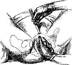
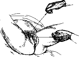
Figure 1.
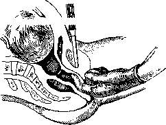
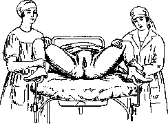
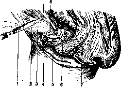
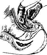
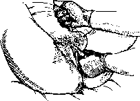
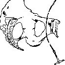
by a simple puncture with a scalpel. In the latter method (developed by Frank), there is no blunt separation of soft tissues, the scalpel is directly inserted in front of the pubic symphysis, and it is incised forward and downward through the small opening thus formed. The operation takes less time and is performed under the mandatory and constant control of two fingers inserted into the vagina (fig. 1 and 2) (the urethra and bladder are displaced to the side). In the first method, after an incision in the skin and fascia (transverse
Figure 2.
or longitudinal) to the bone, the soft tissues on the posterior surface of the pubic symphysis are separated with the index finger, and the bladder is displaced to the side. The pubic symphysis is incised in a downward direction, the lig. arcuatum is also incised, diverging with a
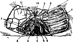
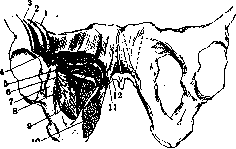
Figure 3. Venous plexus of the external genitalia: 1-rami v. obturatoriae; 2-m. constrictor cunni; 3-v. labii minoris; 4-introitus vaginae; 5-urethra; 6-bulbus vestibuli; 7-muscles; 8-v. pudenda dorsalis; 9-crus clitoridis; 10-v. dorsalis; 11-v. labii majoris; 12-adipose tissue.
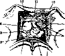
characteristic crack. At the beginning of the incision, the patient's thighs are spread apart by assistants. The main danger lies in possible bleeding due to injury to the clitoris and in general to the venous plexuses (fig. 3 and 4) (in most cases, it stops by applying a tampon while simultaneously bringing the legs together). This method of incision was mainly developed by Zweifel, Kehrer and others; to it a number of technical modifications have been proposed, of which some especially recommend the preliminary insertion of a catheter and its holding by an assistant.—Method Zarate. (Chondrotomia symphysaria, symphysiotomia partialis) differs in that with it the lig. pubicum superius is not incised (see Pubic symphysis). The incision begins below the edge of the symphysis by 1/3 cm; the operation (legs in abductio maxima, fig. 5 and 6) is also performed with the help of two fingers inserted into the vagina; the urethra is displaced, the upper edge of the pubic symphysis is protected, the scalpel is inserted perpendicular to the skin. The elasticity of the lig. pub. super. allows achieving an expansion of 4 cm, which is sufficient.
Figure 5. Position abductio maxima. (According to Zarate.)
Pubiotomy (lateral section of the pubic bone—Gilli, hebostotomy, hebotomy—Van de Velde). The term was first introduced by Galbiati (1832). The operation was proposed as early as 1821 by Champion de Barle Due; a more detailed description was given by Stolz (1841), but it began to be spread and studied only from 1894 thanks to the works of Gilli and the special needle he invented [first successfully performed on a living person by Bonardi (1897), in Russia—Kitner (1904)].
Figure 6. Section of ossis pubis: 1,2, 3-position of the legs during turns; 4-gl. clitoridis; 5-v. dorsalis clitoridis; 6-urethra; 7-vagina; 8-cartilage; 9-cavum Retzii. (According to Zarate.)
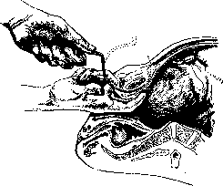
There is no unanimity regarding the name itself (some stand for the old name pubiotomy, while others adhere to the name hebotomy). All methods of pubiotomy can be divided into open (with bone exposure—Schnittmethode) and subcutaneous (puncture and incision without bone exposure—Stichmethode).—Open pubiotomy is now rarely used, and the opinion of most gynecologists on it can be expressed in the words of Bumm, that when cutting and exposing the bone, both the trauma and other unfavorable conditions are the same as in symphysiotomy. Gilli based his proposal on two principles: that the place of incision should not be in the joint itself but in the bone, and that the incision should be made outside the location of the lig. pubo-vesicalia. For this purpose, the place of incision by him was chosen as the line from the tuberculum subpubicum upward to the middle. The bone is grasped with the index and thumb of the left hand (along this line), the skin is pulled down as far as possible; then an incision is made in the skin (from the upper edge of the pubic symphysis somewhat obliquely to the lower edge of the pubic bones), the tissues are bluntly prepared, and a guide is inserted (in case of bleeding it is sufficient to apply a cotton tampon). The guide easily passes behind the bone (under the control of two fingers inserted into the vagina). At the same time, a silk thread is introduced, to which Gilli's needle is attached, with reverse movements of the guide the needle is withdrawn, and the bone is sawn through. After delivery, the wound is either drained from above or after suturing an immovable bandage is applied. The modification by Van de Velde consists in that the skin incision goes in the direction from tuberc. pub. to tuberc. subpubic, and that before passing the needle into the upper end of the wound, a finger is inserted to prevent damage to the soft parts and the main part of the bladder.—Some peculiarities is the method of Tandler, the distinguishing features of which are 1) finding and ligating before sawing the corpora cavernosa (to avoid bleeding), 2) passing the needle under the periosteum and 3) performing the sawing in a vertical direction. Despite the fact that the method, according to the author himself and those who resorted to it, is based on anatomical data, is safer and more surgical, better prevents bleeding, hematoma formation, thrombosis than the subcutaneous methods, better protects against possible injuries to the bladder, it has not found supporters. Subcutaneous pubiotomy (method Doderlein's). When introducing subcutaneous pubiotomy, Doderlein aimed to avoid
Figure 7. Anatomical conditions during pubiotomy: 1-v. femoralis; 2-a. femoralis; 3-p. femoralis; 4-m. pectineus; 5-m. adductor brevis; 6-a. obturatoria; 7-v. obturatoria; 8-p. obturatorius; 9-m. obturator ext.; 10-m. adductor magnus; 11-m. adductor longus; 12-m. gracilis.
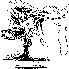
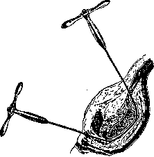
of a large incision and traumatization of the bone when it is significantly exposed (compared to symphysiotomy, it has the advantages that the bone is sawed not along the midline, there is less chance of injuring the corpora cavernosa, and there is no significant divergence of the pelvis thanks to the mm. gracilis and adductor longus) (fig. 7). A transverse incision (through the fascia of the rectus abdominis muscle) 2-3 cm long is made above the tubercle of the pubis closer to the midline; after the fascia is incised, the operation proceeds bluntly with the introduction of the index finger—the finger is directed toward the posterior wall of the pubic bone and reaches its lower edge, where after introducing a needle a small incision is made on the large lip in order to pass the needle. Dederlein's needle is directed between the bone and the index finger (figs. 8 and 9); when it is brought out in the lower incision, a Gigli saw is passed and with two handles the bone is sawed without sharp pressure (it is recommended prophylactically that when introducing the finger, the bladder be pushed toward the pubic symphysis, avoiding traumatization of the crus clitoris). The upper wound, like the lower incision, is tightly sutured with catgut (previously Dederlein introduced drainage into the prevesical space). Bumm's method. After grasping the pubic symphysis with the thumb and index finger directly under the bone, a needle is injected at the border of the large lip (between it and the small lip); the needle is carried upward along the inner surface of the pubic bone, upon reaching its upper edge the skin is pierced under some pressure (the passage of the needle is constantly controlled by the index finger in the vagina); the needle should be brought out closer to the midline (to the pubic symphysis) (figs. 10-14). The sawing takes 1-2 minutes, the wire must be well tightened. After delivery, a tampon is introduced into the vagina, closely applied to the bone wound. Besides these methods, there are a number of modifications of subcutaneous pubiotomy (Walcher's, Kanner's and others); these modifications differ either in the method of needle insertion, in the starting point of needle passage (from top to bottom or vice versa), in the location of the sawing itself, in passing the needle between the periosteum (with its detachment) and the bone (Henkel), etc. Ischio-pubiotomy (Farabeuf's ischio-pubiotomy)—a completely special modification of pubic bone sawing, proposed by Farabeuf before the spread of pubiotomy as a replacement for symphysiotomy, mainly for incorrect (coxalgic) forms of the pelvis. Ischio-pubiotomy consists in sawing not only the pubic but also the ischial bone, and was justified by the fact that with symphysiotomy there is no sufficient increase in pelvic capacity and that for the passage of the head it is also necessary for additional divergence of the sacroiliac joints—the latter being impossible with the indicated forms of the pelvis. Ischio-pubiotomy now has only historical interest, and even earlier this operation was applied only in isolated cases (for the first time successfully by Pinard in 1892).
Figure 8. Pubiotomy according to Dederlein.
of the posterior wall of the pubic bone and reaches its lower edge, where after introducing a needle a small incision is made on the large lip in order to pass the needle. Dederlein's needle is directed between the bone and the index finger (figs. 8 and 9); when it is brought out in the lower incision, a Gigli saw is passed and with two handles the bone is sawed without sharp pressure (it is recommended prophylactically that when introducing the finger, the bladder be pushed toward the pubic symphysis, avoiding traumatization of the crus clitoris). The upper wound, like the lower incision, is tightly sutured with catgut (previously Dederlein introduced drainage into the prevesical space). Bumm's method. After grasping the pubic symphysis with the thumb and index finger directly under the bone, a needle is injected at the border of the large lip (between it and the small lip); the needle is carried upward along the inner surface of the pubic bone, upon reaching its upper edge the skin is pierced under some pressure (the passage of the needle is constantly controlled by the index finger in the vagina); the needle should be brought out closer to the midline (to the pubic symphysis) (figs. 10-14). The sawing takes 1-2 minutes, the wire must be well tightened. After delivery, a tampon is introduced into the vagina, closely applied to the bone wound. Besides these methods, there are a number of modifications of subcutaneous pubiotomy (Walcher's, Kanner's and others); these modifications differ either in the method of needle insertion, in the starting point of needle passage (from top to bottom or vice versa), in the location of the sawing itself, in passing the needle between the periosteum (with its detachment) and the bone (Henkel), etc. Ischio-pubiotomy (Farabeuf's ischio-pubiotomy)—a completely special modification of pubic bone sawing, proposed by Farabeuf before the spread of pubiotomy as a replacement for symphysiotomy, mainly for incorrect (coxalgic) forms of the pelvis. Ischio-pubiotomy consists in sawing not only the pubic but also the ischial bone, and was justified by the fact that with symphysiotomy there is no sufficient increase in pelvic capacity and that for the passage of the head it is also necessary for additional divergence of the sacroiliac joints—the latter being impossible with the indicated forms of the pelvis. Ischio-pubiotomy now has only historical interest, and even earlier this operation was applied only in isolated cases (for the first time successfully by Pinard in 1892).
Figure 9. Pubiotomy according to Dederlein.
not only the pubic, but also the ischial bone, and was justified by the fact that with symphysiotomy there is no sufficient increase in pelvic capacity and that for the passage of the head it is also necessary for additional divergence of the sacroiliac joints—the latter being impossible with the indicated forms of the pelvis. Ischio-pubiotomy now has only historical interest, and even earlier this operation was applied only in isolated cases (for the first time successfully by Pinard in 1892). Indications, contraindications, time and conditions for performing P. The main indication is a narrow pelvis, but not in all cases, only with moderate narrowing—since with both symphysiotomy and pubiotomy the expansion of the pelvis from incision of the pubic symphysis and sawing of the pubic bone reaches only a certain degree. Thus, Morisani indicated that with this the true conjugata increases from 7 to 9 cm; subsequently Pinard, based on his research, also came to the conclusion that this expansion occurs with the true conjugata from 6.0 to 8.4, from 8.0 to 9.8, from 10.8 to 12.4, and that on average it equals 2 cm. In view of this, Pinard considered the lower limit for symphysiotomy to be a true conjugata of 6.5 cm for a flat pelvis and 7.0 cm for a generally narrowed one. Dimant's research on cadavers also showed that the maximum expansion of the anteroposterior diameter of the pelvic inlet equals 1.5 cm. The same figures were given by precise measurements on 17
Figure 10. Pubiotomy according to Bumm.
cadavers (van Canvenberghe, 1905) with pubiotomy; they showed that sawing of the pubic bone increases the true conjugata by 1.5 cm and not more than 2 cm. Furthermore, he came to the conclusion that primiparas with pubiotomy are less subject to traumatization than
Figure 11. Pubiotomy according to Bumm.
with symphysiotomy. Valuable anatomical research was also carried out by Rosenfeld (Kosenfeld; 1905); from it it is evident that the capacity for expansion exists in every pelvis (true conjugata up to 6 mm) and that
^ this expansion must be
^ added to the results obtained after pubiotomy. There is no particular difference in the expansion of the pelvis with symphysiotomy and with pubiotomy, since the distance of the incision site differs only by 172 cm. In recent times (1928) Vaudescal, Horst studied on cadavers a new operation by Zarate; they speak of its expediency and advantages and also consider the resulting expansion sufficient while preserving the superior pubic ligament.
Figure 12. Pubiotomy according to Bumm.
Figure 13. Resawing of the bone. (According to Bumm.)
Figure 14. Position of the Gigli needle. (According to Bumm.)
Based on these as well as clinical data, most consider that the lower boundary is the conjugata vera at 7 cm, and the upper boundary is 8 cm (some establish these dimensions at 6.75-9 cm); individual authors also attribute importance to the shape of the pelvis; thus, they believe that in flat pelves the lower boundary should be less than in generally narrowed ones (6.75 or 7.5 for flat, 7.5 or 8.0 for generally narrowed). Henkel, however, rightly notes that one cannot place the degree of narrowing in the first place, since one must also take into account the size of the fetus, the thickness of the bones, and the shape of the pubic arch. Other authors attribute importance to the relative size of the fetus as the main factor; there are even proposals to perform the operation even with less narrowing of the pelvis in a large fetus, with incorrect insertion of the head (brow presentation, positio occipitopubica alta), or even with an additional promontory. The opinion of Couvelaire, that indications for pubiotomy begin where indications for cesarean section end, can only be accepted for certain degrees of narrowing. Kolosov, considering the operation "technically easier than high forceps and no more difficult than cesarean section," says that it is difficult to establish indications even for a specialist with extensive obstetric experience. Contraindications to P., besides severe degrees of narrowing, are large varicose nodes of the external genital organs, a narrow vagina, foul-smelling waters, corrosive leucorrhea, while fever is mainly a contraindication for open methods and for symphysiotomy. There are indications that symphysiotomy should not be performed in prolonged labors, threatening rupture of the uterus, in eclampsia, and that generally it gives the best results in cephalic presentations. The overwhelming majority also believes that symphysiotomy and pubiotomy are inadvisable in primiparae (Seeligmann, Brindeau and many others) or can only be performed in extreme cases (Frank).-The question of the timing of the operation and the so-called "early" pubiotomy was discussed. The proposal to apply the operation prematurely, i.e., before the onset of labor, is shared by very few, is considered irrational, and even Pinar said that although this idea is very interesting, in practice it may happen that the operation will be performed where the head could have passed. Almost everyone agrees that pubiotomy should be applied at the end of the act of labor with sufficient and even complete opening of the uterus. Regarding the permissibility of preliminary application of trial forceps, opinions are also divided: some categorically object to any attempts at delivery with forceps, version, etc. (Krivsky, Lichkus, Döderlein and others), while others allowed it (Kitner and others) depending on the case, and some even made it a rule (Ols-hausen). There is no unanimity regarding the choice of side for sawing the pubic bone (right or left). Some believe that the side where the occiput of the fetus will pass should be cut (Ilyin, Fenomenov), while others indicate that the saw should always be made on the side opposite the location of the occiput. Many either do not attribute importance to this factor or consider it more convenient to saw the left bone. Undoubtedly, among the conditions must be the presence of a living fetus (some also indicate the need for the consent of the parturient). The situation itself also plays a role-pubiotomy should only be performed with sufficient assistants and in a well-equipped institution. Delivery in cases of P. One of the shadowy aspects of P. is that it is only a preparatory operation, not a delivery operation, and that with it one often has to resort additionally to extraction of the fetus. This has special significance, since observations show that a number of further complications are caused precisely by the subsequent delivery operation (which in the opinion of some represents the main danger). Naturally, for these reasons quite a number of authors (generally those who resort to P.) believe that labor after pubiotomy and symphysiotomy should be left to the forces of nature and that only in extreme cases should forceps or version be resorted to (this is in the interests of both mother and fetus), while others believe that the delivery operation with prepared birth canals is fundamentally necessary, since P. itself is an indication for ending labor: it is cruel, inhumane in their opinion to subject a woman to new torments, it is impractical and a secondary application of anesthesia. Fenomenov also adhered to this view; he advocated always performing artificial delivery (forceps), since it better preserves the life of the fetus and shortens the mother's suffering.As for the nature of the delivery operation itself, most prefer the application of forceps and speak not only against the wide application of version recommended by some, but also generally against its performance after P. In recent times, some strongly recommend to avoid subsequent operative assistance to inject pituitrin. Complications of symphysiotomy. The main ones are injuries to the urethra and bladder, bleeding, ruptures (often penetrating) of the soft birth canals. The postpartum morbidity is also great, and the percentage of maternal and child mortality is significant. In the postpartum period-and not as an exception-hematomas with subsequent suppuration, thromboses, embolisms, disorders of urination, etc., are observed. If statistics of the most recent time are more favorable compared to statistical data of the 19th century and the first decade of the 20th century (when more than half of the patients had fever and mortality reached 10% and higher), they still leave much to be desired both in immediate and in remote results. As an example, one can point to Puppel (1929), who in analyzing 73 cases over the last 10 years noted frequent injuries, a high percentage of mortality (mothers-4.3%, children-9.6%), difficulty in establishing indications, and says that one should not approach all this optimistically. The results of Bauch (1929) over 20 years of performing subcutaneous symphysiotomy according to Frank are also indicative. Thus, in 293 cases, 1.3% maternal mortality, 6.8% child mortality were noted, in 62.8% it was necessary to resort to operative intervention, in 2% injuries of the bladder were observed, in 7% hematomas, in 5% thromboses, in 1.3% severe bleeding, in 1.7% disorders of gait. Pfanmüller (1930) points out that despite waiting only in 1/3 of cases labor ended spontaneously, he notes complications in almost 50% (often severe, e.g., in 6.8% vesicovaginal fistulas, in 10% disorders of gait) and explains this by the fact that the operation is not surgical and the indications for it are still unclear. Bumm considers the average mortality to be 10%, but says that in individual statistics of some authors there is not a single fatal case. There are indeed individual statistics with better outcomes, but even in them these complications were noted by no means as an exception, and mortality is also great in them, especially child mortality (thus, in the collective German statistics of Menge on 316 cases the percentage of maternal mortality is 2.48%, child mortality-10%; Kehrer on 111 cases had 4.5% maternal mortality). Fewer complications are observed with the new method of Zarate, but even with it the author himself on 60 cases had a mortality rate of up to 5% (child-2%). Complications of pubiotomy are not so rare and in general are similar to those of symphysiotomy. Among them are also injuries to the bladder, bleeding with subsequent formation of hematomas, injuries and ruptures of the soft parts. All these complications were demonstrably presented in the statistics of Schlafli (700 cases, 1909) and Romer (300 cases, 1911). Although in the latter they are noted less frequently due to refinement of technique and greater experience, the percentage still seems quite large (for example, severe bleeding was noted in 15.4%, mortality from them 0.33%, hematomas in 14%, injuries to the bladder in 5.2%, penetrating ruptures of the vagina in 7%, thrombophlebitis in 3.5%, maternal mortality 2.7%, child mortality 6.7%). Yurasovsky at one time collected cases with bladder injury, which was observed by 25 authors in 106 cases in 13.2%. Cases were also observed of such severe bleeding that it was necessary to resort to symphysiotomy or open pubiotomy to stop it. Cases (though single) of death from acute anemia were described. For some, the breaking of the saw was also a reason for switching to symphysiotomy. And in the latest data this percentage of complications is great. Thus, in one of the best world clinics of Döderlein (Brandes; 1924-85 cases) the percentage of maternal mortality is 3.5%, child mortality-9.4%, ruptures of the vagina were noted in 23.8%; new data from the same clinic (Gross, 1928-25 cases) also indicate the relative frequency and high percentage of mortality (4% maternal and the same percentage child). Remote consequences are also not uncommon; thus, in 110 cases in 4 there was a disorder of gait and in 1 case of incontinence of urine. Menge in his cases had no maternal deaths (child-6.73%), while in the collective German statistics the percentage of mortality was calculated for mothers at 3.7% and children at 8.5%.
Postoperative period. The question of the patient's position attracted the most attention from the very beginning. Previously, a 'hanging' bed was recommended for this purpose, and special devices (Krasovsky, Ott) for immobilizing the pelvis were also proposed, but these were soon abandoned. At present, the majority consider it necessary to have the patient in a quiet position on their back (or with the upper half of the body elevated). Usually, a bandage made of adhesive plaster is applied around the pelvis (which is removed on the 8th-10th day), and in addition, the pelvis is held from the sides with sandbags. The previously used catheter a demeure is now inserted much less frequently. Tying the legs is not recommended to avoid lochiometra (Fenomenov advised giving the patient a lateral position in the following days for this purpose). On average, it is considered that the operated patient should remain in bed for at least 2-3 weeks; attempts to get up earlier should be avoided. Some have noted that recovery proceeds more quickly after pubiotomy. The long-term results of P. are of considerable interest. The main questions in this direction are as follows: how does healing occur after symphysiotomy and pubiotomy, how often and for how long are walking disturbances observed, does persistent widening of the pelvis remain, and how do subsequent births proceed? The bone wound after P. usually heals quickly, and a strong union is already formed in 3-4 weeks. However, cases have been described of greater mobility of the sawn bones, later healing of the bone wound, and finally even the absence of a bone scar at the time of subsequent births. The formation of pseudoarthroses has also been mentioned. Healing of the wound can be of 4 types: 1) connective tissue with a callus on the periosteum, 2 and 3) periosteal and endosteal calli, and 4) complete bony union with endosteal callus. In symphysiotomy, connective tissue callus forms, which some supporters cite as proof of its advantage over pubiotomy. However, non-union of the symphysis after incision leads to much worse consequences than non-union of the bone. Fehling says that in pubiotomy, in such cases, the pelvic girdle remains stronger than with poor healing of the pubic joint (formation of a loose joint). Bumm also believes that after symphysiotomy, walking remains disturbed for life. Opinions on the possibility of persistent widening of the pelvis, both in general after P. and in particular after various methods, differ. Thus, some believe that the widening is less with P., while others think there is no significant difference and that the widening depends on the shape of the pelvis. Reifferscheid found the following increase in pelvic dimensions after P.: conjugata externa 17 instead of 16.5, and conjugata vera 7.5 instead of 7 before the operation. However, there are not a few authors who believe that neither with symphysiotomy nor with pubiotomy does persistent widening of the pelvis occur, and as proof of this, they cite cases of repeated P. This is also the basis for the proposal to subsequently induce premature labor in women who have undergone P., or even to perform a cesarean section. On the other hand, a number of cases have been described where spontaneous births or births ending only with forceps or version were observed after symphysiotomy and pubiotomy. liotomy 21 & Particularly interesting is the case of Beghin (1926), who observed normal births in the 3rd pregnancy, with cesarean section in the first, and symphysiotomy in the second. He points out that persistent widening of the pelvis after symphysiotomy is confirmed by radiography and the subsequently observed normal births.-Among the most severe and unpleasant consequences of pelvic expansion operations are walking disturbances, urination disorders, and fistulas. The two cases of walking disturbances in peasant women during heavy rural work described by Gross should make one take into account to a large extent the profession when performing the operation and generally observe all precautions.
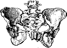
Fig. 15. Abundant callus formation after pubiotomy. (After Winter.)
In addition, the literature contains not a few cases of changes in the position of the bladder, cystocele, prolapse and prolapse of genital organs (Niedermeyer and others). Cases of exostosis formation (Fig. 15) and even a case of paralysis of the calf muscles with severe sensory disturbances have been described. Plastic operations: pelvi-osteoplasty, symphysectomy, auto-osteoplasty. Due to insufficient pelvic expansion after symphysiotomy, Fenomenov and Kochetkov (1894), after studying the issue on cadavers and animals, proposed during symphysectomy to saw out a piece of the pubic joint in the form of a triangle or quadrangle (Fig. 16) and to perform autoplasty to compensate for the resulting
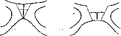
Fig. 16. Fenomenov-Kochetkov operation.
bone defect. Crede with the same purpose proposed after pubiotomy to saw out a piece of bone about 2 cm long from the horizontal branch of the pubic bone and insert it into the place of the sawn pubic bones (in large constrictions, a similar piece is also inserted on the other side). This operation (pubioplasty) was successfully performed by Crede on a living person (long-term results were also favorable-normal walking, persistent pelvic widening; the effect on births was not traced, as pregnancy did not occur), but it found no imitators. This type of operation is closely related to Costa's (Costa; 1921) latest proposal for partial symphysectomy (symphysectomia partialis). It can be definitely said that of all plastic operations of the pelvis, it deserves more attention and has received relative dissemination. Costa performed it in 100 cases and, based on radiography and clinical observations, concludes that it provides persistent pelvic widening. Thus, of 100 cases, births per vias naturales were subsequently observed in 11 (10 spontaneous, 1 with forceps, in 2 cases repeated normal births). Partial symphysectomy was performed by him primarily (before birth) or secondarily during birth concurrently with cesarean section (of 11 cases, Costa had primary symphysectomy in 9 and secondary in 2). In 3 cases, Costa after cesarean section and partial symphysectomy also performed the Rotter-Schmid operation (in this combination, the operation was successfully performed by Fossati in 2 cases-persistent pelvic widening of 2.2 cm and 2.3 cm was obtained). The limit for performing the operation is conjugata vera not lower than 8.3-8.5 cm. Symphysectomy was also successfully tried by others and studied on cadavers. Thus, Scotti (1922) performed it on cadavers and studied the relationship of the head and pelvis before and after symphysectomy using a special rubber balloon and found that it provides certain benefit (especially in a flat pelvis with moderate constriction). Scotti, like Costa, considers the operation technically easy, safe, and not violating the strength of the pelvis and the statics of the trunk (so

Fig. 17. Costa's teeth.
it should not be performed locally with other pelvic expansion operations). The technique of partial symphysiotomy according to Costa—an incision (6 cm) along Pfannenstiel or vertical 1 cm above the upper edge of the pubic symphysis; to protect the peritoneum and bladder, a compress is introduced into cavum Retzii, along the line of both tubercula pubica the periosteum is cut, separated from the upper edge of the pubic symphysis (posteriorly) approximately to its middle, and from top to bottom a bone-cartilage piece is cut out to the site of the prepared part of the posterior surface of the pubic symphysis. The periosteum is not sutured. Wound closure is usual. Removal of bone is performed either with a scalpel or Gigli's saw or Costa's special forceps resembling a parrot's beak (Fig. 17). Resection of the sacral promontory (operation of Rotter-Schmidt) has been used in isolated cases; it consists in sawing off the sacral promontory to increase the direct diameter of the pelvic inlet. The size of the resected bone piece depends on the shape of the sacrum, a double promontory is also excised. The resection should reach S1, but sometimes without harm it can reach even to half of S2. The periosteum is carefully separated before resection. The operation is done either primarily or simultaneously with cesarean section. In 1923 Schmid reported on 15 cases, of which in 12 cases it was performed during cesarean section, once at 4 months of pregnancy and once simultaneously with radical hernia operation. Schmid subsequently observed natural deliveries. In the literature Schmid has collected 18 cases with 3 fatal outcomes (which also points to postoperative complications—cystitis, parametrial abscess, etc.). Regarding the methods presented (with the exception of ischiopubicotomy), there is no unanimity among supporters of pelvic expansion operations; however, the majority still adheres to subcutaneous pubiotomy, a significantly smaller part to the open method; a small group of authors until very recently preferred symphysiotomy, especially according to the latest method of Zarate. Thus, many supporters of pubiotomy point out that it is a more humane operation and that a smaller percentage of complications is observed with it; it is also noted that subsequently the normal support of the bladder is better preserved with it, that the tension of the muscles prevents excessive divergence of the bones, and finally that the bone heals better at the joint. Among the methods, Dederlein's method is preferred. Thus, Fehling, Küttner, and others indicate that the data apparently speak in favor of Dederlein's method rather than Bumm-Walcher's method; Pobedinsky, however, wrote that Bumm's method is not quite safe and not quite suitable for sufficient expansion of the pelvis. Prozorovsky specially studied on corpses (for the purpose of evaluating the technique) and came to the conclusion that the best method is Dederlein's, the passage of the guide is safe and it is more aseptic than open methods. Symphysiotomy (whose obstetric significance in the opinion of Gruzdiev should be evaluated not highly) continues to be performed by some schools and now almost exclusively according to Frank's method. The method of Zarate, which caused considerable revival of the question of symphysiotomy, requires further observations; it is premature to draw conclusions; it is hardly correct to fully compare it with cesarean section and the opinion that supposedly obstetricians, applying it, will better know the mechanism of labor and the principles of the physiology of the act of labor (Zarate). But a definite percentage of mortality in the hands of the author himself and the already described disturbances of gait allow one to fully join Correa (Correa), who advises not to accept with enthusiasm this new method. Some caution in the application of other pelvic expansion operations is also observed by authors who performed them. Thus, regarding pubiotomy, there are not isolated indications that this operation is not as easy, innocent, and safe as was thought earlier. Yurasovsky already wrote about the seriousness of the operation, and apparently along with frequent complications this gave the right to some (Krivsky; 1912) to speak of the fact that the enthusiasm for the operation is beginning to cool down. In recent times (1929) it is also indicated (Pal'mov), that the prestige of the operation has declined and that for its adherents it has taken a secondary role in the therapy of a narrow pelvis. A fairly large group of gynecologists, considering all the conditions that accompany the performance of the operation itself, the complications associated with it, and especially the remote results, always regarded it with more than restraint. Many schools did not apply and do not apply it at all. It was more often applied in Germany and France, in England its application is very limited. But apparently the number of its supporters in Germany is also small, which is explained by the fact that supporters are constantly improving the methods, since there is no method that would be quite satisfactory. In the USSR there are also few supporters of P.; only a few dozen operations have been performed, and in recent years it has almost not been applied. It did not enjoy and does not enjoy success with a number of individual schools. Undoubtedly, one of the main reasons for such limited application of it is the quite justified fear of possible complications, the possibility of reducing a woman's working capacity, and in some cases partial disability. And one must think that with a deeper preventive orientation in obstetrics, with observation of the woman during pregnancy, with timely and early placement in an institution, and consequently with the reduction of doubtful cases, the obstetrician-preventive specialist will often prefer pelvictomy to cesarean section even in lesser degrees of pelvic narrowing. It is difficult to agree with those authors (Shpolyansky; 1926) who consider that refusal of pubiotomy means deterioration of the prognosis for mothers and pointless increase in cases of perforation of living children. The most correct point of view appears to be that of Niedermeyer, who considers that pubiotomy can compete only with perforation, and by no means with cesarean section, and can be performed where cesarean section is not feasible.
Related articles
Mentioned in
Cite this page
“Pelveoperitonitis.” Soviet Medical Encyclopedia. English translation of Bolshaya Meditsinskaya Entsiklopediya, 1st ed. (Moscow, 1928–1936), ed. N. A. Semashko. https://sovietmedicalencyclopedia.pages.dev/article/pelveoperitonitis/