Omentum
Historical document, translated for reference. It reflects medical knowledge of the 1920s–30s and is not medical advice.
Summary
The omentum (omentum, epiploon) refers to the duplicatures of peritoneum connecting abdominal organs. This article describes the anatomy, embryological development, and clinical significance of both the greater and lesser omenta in mammals.
Encyclopedia article (1928–1936)
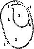
OMENTUM (omentum, epiploon), large duplicatures of peritoneum extending from one abdominal organ to another and consisting of peritoneal sheets, the greater and lesser peritoneal sacs (fig. 1). Usually, the omentum, i.e., the peritoneal sheets, encompasses the vascular pedicle extending from one organ to another. By location, they are distinguished as the greater omentum (omentum majus) and the lesser omentum (omentum minus). Testut distinguishes even four omenta: besides the greater and lesser, also omentum gastro-lienale and omentum pancreatico-lienale, but these are parts of the same greater and lesser omenta. A characteristic feature of all omenta is their connection with the stomach. Phylogenetically, the omentum is a young organ. It is found only in mammals and is particularly well developed in carnivorous animals. Ontogenetically, the lesser omentum should be considered as the primary duplication of peritoneum formed in the posterior part of mesenterium ventrale (remnants of it), and the greater omentum-as the primary duplication of mesenterii dorsalis-mesogastrium. By the 4th week of embryonic life, both omenta have a vertical orientation and are located strictly along the median line: the lesser in front of the stomach (between the stomach and liver), the greater behind (between the stomach and posterior abdominal wall). By the 6th week, the stomach has largely completed its rotations around vertical and horizontal axes, and both omenta assume a frontal position, extending from left to right. The right edge of the lesser omentum, formerly the lower end of the vertical mesentery, remains free and is called lig. hepato-duodenale. The remaining part of the lesser omentum, which extends from the inferior surface of the liver to the lesser curvature of the stomach, is called lig. hepato-gastricum. To the right and behind lig. hepato-duodenale there is an opening-foramen epiploicum, s. Winslowi, which leads into bursa omentalis. The latter formed as a result of stomach rotations around two axes and represents a slit bounded in front by the lesser omentum, the posterior wall of the stomach, and during the embryonic period and sometimes in childhood by two peritoneal sheets descending from the greater curvature of the stomach, i.e., the anterior lamina of the greater omentum. Behind and below, bursa omentalis is bounded by peritoneum covering the posterior wall of the abdominal cavity and the organs beneath it-the pancreas, abdominal aorta, inferior vena cava, and the diaphragmatic pillars with accompanying vessels. Above, it is mainly bounded by the posterior part of the inferior surface of the liver (Spigelian lobe). The upper part of bursae omentalis is called the vestibule of the omental cavity. It is bounded from the proper omental cavity by the opening-foramen pancreato-gastricum, bounded in front by the posterior wall of the stomach, behind by the peritoneum covering the anterior surface of the pancreas. In the adult, under normal conditions, the omental cavity is absent. In cases where embryonic development has not completed and where the fusion of the omental sheets has not occurred, we find the omental cavity as a slit between four peritoneal sheets. The anterior wall of this cavity is formed by the stomach and the anterior lamina of the omentum descending from it. Its posterior wall, rising from below upward, is the second lamina (posterior lamina of the omentum), covering the anterior surface of the transverse colon and fusing above it with mesocolon, so that in its upper part the omentum consists as if of 6 sheets (fig. 2).
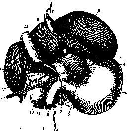
The lesser omentum is especially visible when the liver is lifted upward. It has the shape of an almost quadrangular sheet, in which two surfaces-anterior and posterior-and four edges can be distinguished: superior, inferior, right, and left (fig. 3). Both surfaces are even, smooth, and represent a continuation of both peritoneal sheets of the stomach. The superior edge is connected to the liver, mainly to the quadrate lobe, and extends to the left to the right edge of the esophagus. The inferior edge is connected to the initial part of the duodenum, the lesser curvature of the stomach, the pylorus, and the cardia. At the inferior edge, both sheets of the omentum separate from each other, leaving space for arteries, veins, nerves, and lymphatic vessels of the lesser curvature. This space, in case of gastric ulcer perforation, may fill with gases or fluid, which thus infiltrate the wall of the lesser omentum. The left edge of the lesser omentum approaches the diaphragm for a short distance, forming a fold known as lig. phrenico-oesophageum, marking the posterior boundary of Spigelian lobe. The right edge of the lesser omentum is free. It bounds in front foramen Winslowi, which above is bounded by lobus caudatus, below by the superior part of the duodenum, behind by the peritoneum covering the inferior vena cava, and lig. hepato-renale. In the sheets of the lesser omentum are laid the elements of the 'hepatic pedicle': the rightmost-ductus choledochus, the leftmost-hepatic artery, and between them behind-the portal vein. Both sheets of the lesser omentum are separated from each other by a layer of fatty connective tissue, which in some cases is particularly pronounced-'fatty omentum'. Overall, the lesser omentum is a fragile structure. Its strength is associated with the vessels contained within it. During traction, which is performed during operations on the stomach and liver, it is not the omentum that is subjected to it, but the vessels and nerves (branches of the vagus nerve) contained within it, which has great practical importance for the surgeon, as it can cause shock during stomach or liver surgery.
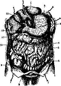
The greater omentum in an adult hangs freely from the greater curvature of the stomach into the abdominal cavity like an apron, lying between the posterior surface of the abdominal wall and the anterior surface of the intestinal loops (fig. 4). The length of the greater omentum varies from 7.5 to 70 cm. In the latter case, it extends into the pelvis. Its shape varies depending on length; short-it is square-shaped with scalloped edges, long-semicircular. In adults, the omentum is dense, thick, and in full people contains a large amount of yellow fat, which collects in numerous lobules making it opaque. In children-it is thin, transparent with numerous visible vessels, in the loops of which white spots are visible in places. In the greater omentum, we distinguish four edges and two surfaces. Only the superior edge is not free, but is connected to the stomach along the entire greater curvature, with the pylorus, with the duodenum up to a. gastro-duodenale, and to the left its anterior lamina approaches the hilum of the spleen, and some authors consider it as a separate omentum-epiploon gastro-lienale (Testut). The posterior lamina is connected to the transverse colon at the boundary of its first and middle thirds and passes completely onto the mesocolon transversum. At this place, the posterior wall can be separated from mesocolon. The omentum is supplied with vessels extremely abundantly, and there are many more than its nutritional needs require. The arteries in it form two arches. It should be noted that each lamina of the omentum has its own individual blood supply (fig. 5). One arch consists of both aa. gastro-epiploicae and extends along the greater curvature of the stomach; the right-from a. hepatica, the left-from a.lienalis, with the right supplying mainly the anterior lamina, the left-the posterior. The other arch consists of vessels proper to the omentum and is located under the transverse colon.
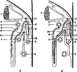
Figure 2. Development of the omental bursa, transverse colon, and primary mesentery. A-before adhesion: 1-liver; 2-first part of duodenum; 3-body of pancreas; 3'-head of pancreas; 4-transverse colon; 5-third part of duodenum; 6-ascending colon; 7-omental bursa; 8-lesser omentum; 9-anterior lamina of greater omentum with a. gastro-epiploica dext.; 10-posterior lamina of greater omentum with a. gastro-epiploica sin.; 11-a. hepatica; 12-mesocolon transversum; 13-lig. pancreatoduodenale with a. parapancreaticoduodenale sup. B-after adhesion: 1-liver; 2-first part of duodenum; 3-body of pancreas; 3'-head of pancreas; 4-transverse colon; 5-third part of duodenum; 6-ascending colon; 7-omental bursa; 8-lesser omentum; 9-anterior lamina of greater omentum with a. gastro-epiploica dext.; 10-posterior lamina of greater omentum with a. gastro-epiploica sin.; 11-a. hepatica; 12-surface of adhesion between posterior lamina of greater omentum and superior surface of primary mesocolon transversus; 13-surface of adhesion of posterior lig. pancreatoduodenale, forming ligament of Treitz; 14-surface of adhesion of mesentery of small intestines and duodenum.
Figure 3. Peritoneum, view from the inferior surface of the liver: 1-right lobe; 2-left lobe; 3-quadrate lobe; 4-Spigelian lobe; 6-stomach; 7-duodenum; 8-part of lesser omentum containing 'hepatic pedicle'; 9-completely transparent part of omentum, containing neither fat nor vessels; 10-lig. hepato-renale; 11-right kidney; 12-right adrenal capsule; 13-gallbladder; 14-grooved probe passing through foramen Winslowi into omental bursa. (According to Testut.)
The greater omentum in an adult hangs freely from the greater curvature of the stomach into the abdominal cavity like an apron, lying between the posterior surface of the abdominal wall and the anterior surface of the intestinal loops (fig. 4). The length of the greater omentum varies from 7.5 to 70 cm. In the latter case, it extends into the pelvis. Its shape varies depending on length; short-it is square-shaped with scalloped edges, long-semicircular. In adults, the omentum is dense, thick, and in full people contains a large amount of yellow fat, which collects in numerous lobules making it opaque. In children-it is thin, transparent with numerous visible vessels, in the loops of which white spots are visible in places. In the greater omentum, we distinguish four edges and two surfaces. Only the superior edge is not free, but is connected to the stomach along the entire greater curvature, with the pylorus, with the duodenum up to a. gastro-duodenale, and to the left its anterior lamina approaches the hilum of the spleen, and some authors consider it as a separate omentum-epiploon gastro-lienale (Testut). The posterior lamina is connected to the transverse colon at the boundary of its first and middle thirds and passes completely onto the mesocolon transversum. At this place, the posterior wall can be separated from mesocolon. The omentum is supplied with vessels extremely abundantly, and there are many more than its nutritional needs require. The arteries in it form two arches. It should be noted that each lamina of the omentum has its own individual blood supply (fig. 5). One arch consists of both aa. gastro-epiploicae and extends along the greater curvature of the stomach; the right-from a. hepatica, the left-from a.lienalis, with the right supplying mainly the anterior lamina, the left-the posterior. The other arch consists of vessels proper to the omentum and is located under the transverse colon.
Figure 4. Position of the omentum during opening of the abdominal cavity: 1-left lobe of liver; 2-stomach; 3-spleen; 4-greater omentum; 5-descending colon; 6 and 8-sigmoid colon; 7-peritoneum; 9-cecum; 10-ascending colon; 11-transverse colon; 12-duodenum; 13-gallbladder. (According to Testut.)
The greater omentum in an adult hangs freely from the greater curvature of the stomach into the abdominal cavity like an apron, lying between the posterior surface of the abdominal wall and the anterior surface of the intestinal loops (fig. 4). The length of the greater omentum varies from 7.5 to 70 cm. In the latter case, it extends into the pelvis. Its shape varies depending on length; short-it is square-shaped with scalloped edges, long-semicircular. In adults, the omentum is dense, thick, and in full people contains a large amount of yellow fat, which collects in numerous lobules making it opaque. In children-it is thin, transparent with numerous visible vessels, in the loops of which white spots are visible in places. In the greater omentum, we distinguish four edges and two surfaces. Only the superior edge is not free, but is connected to the stomach along the entire greater curvature, with the pylorus, with the duodenum up to a. gastro-duodenale, and to the left its anterior lamina approaches the hilum of the spleen, and some authors consider it as a separate omentum-epiploon gastro-lienale (Testut). The posterior lamina is connected to the transverse colon at the boundary of its first and middle thirds and passes completely onto the mesocolon transversum. At this place, the posterior wall can be separated from mesocolon. The omentum is supplied with vessels extremely abundantly, and there are many more than its nutritional needs require. The arteries in it form two arches. It should be noted that each lamina of the omentum has its own individual blood supply (fig. 5). One arch consists of both aa. gastro-epiploicae and extends along the greater curvature of the stomach; the right-from a. hepatica, the left-from a.lienalis, with the right supplying mainly the anterior lamina, the left-the posterior. The other arch consists of vessels proper to the omentum and is located under the transverse colon.
intestine.-The veins are more numerous than the arteries, follow the course of the arteries, have valves, and empty into the portal vein system. The venous network is very powerful, which surgeons utilize by attaching the omentum to the abdominal wall to form anastomoses with the inferior vena cava system in cases of liver cirrhosis-Talm's operation (ascites).-The omentum has its own lymphatic vessels; they collect into trunks that go behind the pylorus together with the a. gastro-epipl. dext., and empty into the inferior gastric lymph nodes, partly (on the left side) into the splenic and celiac lymph nodes. There are no anastomoses between the lymphatic vessels of the greater omentum and the transverse colon mesentery.-The innervation of the omentum is the same as that of the peritoneum. Histologically, the omentum is an organ consisting of a dense network of delicate connective tissue fibers, with a large number of elastic fibers and numerous bundles of collagen fibers. On a thin basal membrane there is a layer of flat simple epithelium--endothelium (tunica serosa). In the embryo, the greater omentum is a delicate membrane with a properly arranged network of vessels. Only after birth do the first small openings appear between the connective tissue beams and along the vessels. Their number gradually increases with age. (Seifert). For the omentum, the distribution of capillaries in the so-called "vascular tufts" is characteristic. In the area of these tufts in newborns, delicate white spots, the so-called "milk spots" (Ranvier), are visible. These are accumulations of cellular elements that play a very important biological role-wandering cells (plasma cells, histiocytes, adventitial cells, etc.). In these milk cells, individual fat cells form, which gradually increase in number and later completely transform into fat nodules. Attention should be paid to the fact that fat cells can disappear under certain conditions and be replaced again by wandering cells (secondary "milk nodules" of Seifert). Marshan considers the adventitia of the capillaries of the fat nodules as the site of formation of milk cells. A direct transition of fat cells into wandering cells has been proven. The latter have phagocytic properties, which they exhibit when bacteria invade. They also have amoeboid movements. These cells are directed to dangerous places in the abdominal cavity, in order to later attach themselves again in clusters to the omentum (Seifert). Subsequently, the omentum becomes stronger and denser through an increase in connective tissue and fat inclusions. The structure of the omentum can be used to judge the age of its owner. From its complex anatomical structure, it is evident that the omentum is a biologically very important organ: due to
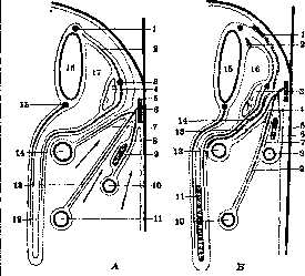
Figure 5. Development of the omental bursa, transverse mesocolon, and mesentery of the small intestine in a sagittal section along the median line. A-before adhesion: 1- coronary artery of the stomach; 2- artery of the greater curvature of the stomach; 3- lienal artery; 4- body of the pancreas; 5- left gastroepiploic artery; 6- aorta; 7- transverse mesocolon; 8- mesentery of the duodenum with the inferior pancreaticoduodenal artery; 9- head of the pancreas; 10- third part of the duodenum; 11- small intestine; 12- greater omentum; 13- mesentery of the small intestine; 14- transverse colon; 15- right gastroepiploic artery; 16- stomach; 17- omental bursa. B-after adhesion: 1- adhesion of the parietal layer of the gastric mesentery; 2- additional site of adhesion behind the stomach; 3- body of the pancreas; 4- additional site of adhesion behind the mesentery of the small intestine; 5- head of the pancreas; 6- site of adhesion forming the ligament of Treitz; 7- site of adhesion of the peritoneum in front of the pancreas; 8- third part of the duodenum; 9- mesentery of the small intestine; 10- small intestine; 11- greater omentum; 12- transverse colon; 13- transverse mesocolon; 14- site of adhesion between the transverse mesocolon and the posterior layer of the greater omentum; 15- stomach; 16- omental bursa. Due to its rich blood supply, it can serve as a regulator of blood circulation (Blutregulator-Gundermann) and as an organ protecting the abdominal cavity (Schutzorgan). Clinically and experimentally it has been proven that in the absence of the omentum, infection of the abdominal cavity proceeds more severely: guinea pigs, when staphylococci were injected into the opened abdominal cavity, died in 2-3 days if the omentum had been removed. The controls with the same dose survived. In the fight against infections, an important role falls to the omentum-the resorption of bacteria and toxins. Absorption occurs1 directly through the blood and lymph vessels, but more often it is not direct, but through phagocytosis from the milk and fat nodules that produce wandering cells (Seifert, Koch). The omentum is able to absorb entire organs or their parts, for example, parts of a destroyed spleen, injured kidney tissue, etc. An exceptionally important feature of the omentum for surgeons is its plasticity: it is laid over damaged or infected areas, glues them together and thereby isolates, delimits the pathological focus from the rest of the abdominal cavity, for example, in injuries to the gastrointestinal tract. In injuries to the bladder, it lies in the defect of the bladder and protects the abdominal cavity from infection, shielding it from communication with the bladder, the inflamed appendix is wrapped by it, etc. The exceptional mobility of the omentum and its plastic ability, combined with the ability of resorption, rightly give reason to call it the 'protective organ of the abdominal cavity' (in Germany even 'Polizeiorgan'). When the omentum delimits the affected area, it can be assumed that here a change in the colloidal state of the visceral peritoneum and the omentum plays a role. The rich blood supply gives the omentum the ability to quickly and widely form vascular anastomoses and thus help nourish the organ which the omentum surrounds. For example, tumors that had become separated from their organ on a pedicle remained alive if they were wrapped in the omentum. Many authors observed in extensive resections of the stomach for cancer with ligation of the mesocolon significant nutritional disorders of the corresponding section of the intestine, which threatened with gangrene; nevertheless, the patients survived if the intestine was wrapped in the omentum and gangrene thus did not occur. However, the author does not recommend relying on this property of the omentum and when ligating the middle colic artery to resect the corresponding sections of the intestine. The great elasticity in combination with the plasticity of the omentum also plays a major role in surgery, as it is used to close wounds of abdominal organs, to protect unreliable sutures of the gastrointestinal tract, for live tamponade in injuries to the liver and spleen. According to Koch, the omentum is a protective organ not only for the abdominal cavity but also for the inner surface of the gastrointestinal tract: Koch introduced tubercle bacilli into the intestine of a rabbit through laparotomy. At relaparotomy after several weeks he found on the omentum numerous tubercles, while the intestinal mucosa was intact. Based on the anatomical position of the omentum, attempts were made to construct a theory of its physiology. Thus, Franzen assumed that the omentum provides the 'roundness' of the small intestines and thereby promotes their peristalsis. Fabricius thought that the omentum is a reserve fold which the stomach fills when it is overloaded with food. Even earlier, Aristotle, Galen and others considered that the omentum is rich in fat to protect the organs it covers from cold. Bauhin, Glisson considered the omentum as a reservoir for fat. The latter view was not confirmed, as it is known that the fat content in the omentum in a corpse is parallel to the fat content in the entire body and that often a fat corpse has a lean omentum, but the reverse phenomenon has not been observed. Franzen considered the function of the omentum exclusively as mechanical and called it the 'filling of the abdominal cavity' on the grounds that it is especially often found in the hernial sac in hernia. The latter however only speaks of the great mobility of the omentum. Broman, based on his work, considers the omentum an organ of lymphatic vessels (Lymphgefassorgan). This is also confirmed by the work of Koch. What role the omentum plays in the formation of antibodies has not yet been clarified. From all that has been said, it is clear that the omentum is a parenchymal organ, with a specific structure and specific cells, and in its function can be compared with the thyroid gland and bone marrow.
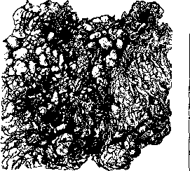
Figure 6. Spindle cell sarcoma of the greater omentum.
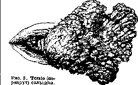
Twisting (torsion of the omentum). 90% of all cases occur in hernias (epiplocele). Twisting can be abdominal, purely hernial, and combined. The latter is most common (fig. 8). The pathogenesis is unclear. It is assumed that hypertrophy of the omentum plays a role. Being in the hernial sac for a long time, the omentum becomes chronically inflamed, thickens, gathers into a ball, more often at the end of a thin pedicle. Enhanced
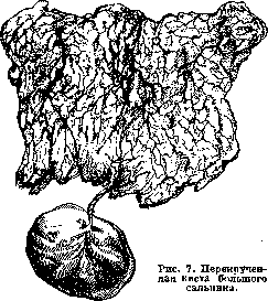
peristalsis of the intestines, sharp turning of the body can be the cause of twisting in a spiral around the corresponding artery-Payer's hemodynamic theory. In the twisted area, cyanosis, edema, partial or complete necrosis occur, in the abdominal cavity-serous or hemorrhagic effusion. Further-peritonitis, intestinal bleeding due to
to press into the surrounding parts of the remaining omentum. Under no circumstances is it sufficient to simply untwist the omentum: in this case, we eliminate only the symptom, not the cause, and therefore may get recurrences. 7) Abnormal obesity: cases have been described where a very large, hypertrophied and fatty omentum caused symptoms from the stomach, as in ulcers, even with bleeding from the gastrointestinal tract. The latter was explained by the appearance of erosions in a retrograde embolic way from chronically damaged and thrombosed vessels of the abnormal omentum. Many patients were cured by resection of the changed part of the omentum. 8) Hernias of the omentum (epiplocele, hernia omentalis). After the intestine, the omentum is most often the content of a hernia. A necessary condition for this is a certain length of the omentum. Since in the first years of life the omentum is only a short appendage of the stomach, it does not occur as the content of a hernia in early childhood. In the hernia sac, the omentum does not lie in the form of an apron, but rolled into a lump, twisted, often adherent, almost always in front of the intestinal loops if the hernia contains intestine. In an umbilical hernia sac, the omentum is extremely rarely absent. It must be remembered that a very long omentum may have part of its surface adhere to the bottom of the hernia sac and from there return, turning back its cone through the hernia opening, freely indenting into the abdominal cavity. This can have fatal consequences: if, as is usually done, such an omentum is ligated below the hernia opening and the stump is returned into the abdominal cavity, then part of the omentum, deprived of nutrition from all sides and doomed to necrosis, will be found in the abdominal cavity. This can be easily avoided if, before ligation, the omentum is pulled to determine if there is a free part of the omentum in the abdominal cavity. If the omentum has not been returned to the abdominal cavity for a long time, changes occur in the form of fibrous and lipomatous thickenings due to chronic inflammation, which sharply increases the volume of the prolapsed part of the omentum and makes the hernia irreducible. 9) Prolapse of the omentum is observed in wounds of the abdominal wall. Any prolapse should not end only with its elimination. It absolutely requires laparotomy, since prolapse of the omentum without injury to the abdominal viscera is possible only in exceptional cases. The prolapsed part of the omentum must be resected. It is a mistake to return it through an infected wound. The prolapsed part should be resected first, and the stump returned after preliminary expansion of the abdominal wound, in order to verify the absence or presence of other injuries to abdominal organs. When ligating the omentum, one should not approach too close to the large intestine, as its nutrition may be impaired. 10) Thrombosis and embolism of the large vessels of the omentum lead to gangrene of the corresponding area, to peritonitis, etc. After resection of the omentum, embolism of the veins of the stomach and intestines is observed, leading to postoperative bleeding; Eiselberg and Recklinghausen in such cases found fresh small ulcers of the gastric and duodenal mucosa and explained their appearance by the formation of retrograde emboli.
apron, but rolled into a lump, twisted, often adherent, almost always in front of the intestinal loops if the hernia contains intestine. In an umbilical hernia sac, the omentum is extremely rarely absent. It must be remembered that a very long omentum may have part of its surface adhere to the bottom of the hernia sac and from there return, turning back its cone through the hernia opening, freely indenting into the abdominal cavity. This can have fatal consequences: if, as is usually done, such an omentum is ligated below the hernia opening and the stump is returned into the abdominal cavity, then part of the omentum, deprived of nutrition from all sides and doomed to necrosis, will be found in the abdominal cavity. This can be easily avoided if, before ligation, the omentum is pulled to determine if there is a free part of the omentum in the abdominal cavity. If the omentum has not been returned to the abdominal cavity for a long time, changes occur in the form of fibrous and lipomatous thickenings due to chronic inflammation, which sharply increases the volume of the prolapsed part of the omentum and makes the hernia irreducible. 9) Prolapse of the omentum is observed in wounds of the abdominal wall. Any prolapse should not end only with its elimination. It absolutely requires laparotomy, since prolapse of the omentum without injury to the abdominal viscera is possible only in exceptional cases. The prolapsed part of the omentum must be resected. It is a mistake to return it through an infected wound. The prolapsed part should be resected first, and the stump returned after preliminary expansion of the abdominal wound, in order to verify the absence or presence of other injuries to abdominal organs. When ligating the omentum, one should not approach too close to the large intestine, as its nutrition may be impaired. 10) Thrombosis and embolism of the large vessels of the omentum lead to gangrene of the corresponding area, to peritonitis, etc. After resection of the omentum, embolism of the veins of the stomach and intestines is observed, leading to postoperative bleeding; Eiselberg and Recklinghausen in such cases found fresh small ulcers of the gastric and duodenal mucosa and explained their appearance by the formation of retrograde emboli.
injuries to abdominal organs. When ligating the omentum, one should not approach too close to the large intestine, as its nutrition may be impaired. 10) Thrombosis and embolism of the large vessels of the omentum lead to gangrene of the corresponding area, to peritonitis, etc. After resection of the omentum, embolism of the veins of the stomach and intestines is observed, leading to postoperative bleeding; Eiselberg and Recklinghausen in such cases found fresh small ulcers of the gastric and duodenal mucosa and explained their appearance by the formation of retrograde emboli.
Related articles
Mentioned in
Cite this page
“Omentum.” Soviet Medical Encyclopedia. English translation of Bolshaya Meditsinskaya Entsiklopediya, 1st ed. (Moscow, 1928–1936), ed. N. A. Semashko. https://sovietmedicalencyclopedia.pages.dev/article/omentum/