MESENTERIC ARTERIES (VENAE)
Historical document, translated for reference. It reflects medical knowledge of the 1920s–30s and is not medical advice.
Summary
This article describes the anatomy of the superior and inferior mesenteric arteries and veins, their branching patterns, and associated pathologies including aneurysms and thrombosis. It also compares the vascular architecture of human and animal mesenteric systems.
Encyclopedia article (1928–1936)
MESENTERIC ARTERIES, VENAE. The right colic artery (a. colica dextra) sometimes departs independently from the superior mesenteric artery. In these cases, it anastomoses with the ileocolic artery and the middle colic artery. 4. The middle colic artery (a. colica media) (fig. 2); departs from the right side of the superior mesenteric artery, first lying in front of the pancreas, then goes to the right and forward in the thickness of the transverse colon mesentery to the walls of the transverse colon. It anastomoses with the ileocolic artery (or the right colic artery) and with the left colic artery (from the inferior mesenteric artery system).-According to the nature of branching, two types of superior mesenteric artery are distinguished: main and diffuse. Pathology. On the significance of the superior mesenteric arteries in the occurrence of obstruction in the lower part of the duodenum-see Ileus. Aneurysms of the superior mesenteric artery occur very rarely. They give the same symptoms as aneurysms of the abdominal aorta: pains in the stomach and intestines, pains in the back, increasing with the slightest change in body position, a feeling of compression in the abdomen, meteorism, vomiting and painful belching, a pulsating tumor in the epigastric region, giving a noise on auscultation. If the aneurysm is of large size, displacement of neighboring organs (e.g., the liver) and compression of individual sections of the intestine, obstruction (e.g., of the duodenum) and rupture with subsequent fatal bleeding may occur. The inferior mesenteric artery, a. mesenterica inferior (fig. 1), is unpaired, significantly smaller in size than its superior counterpart, is the sixth branch of the abdominal aorta and departs from the anterior surface of the latter at the level of the interval between LIII and LIV. It lies behind the parietal peritoneum and goes from top to bottom to the left iliac fossa, forming an arc convex downward and to the left. It has the following branches: 1) a. colica sin.; in front of the left kidney it goes to the left and upward (lying retroperitoneally) to the descending colon and connects on the right and above with the middle colic artery, and below with the artery of the sigmoid colon. At some distance from the sigmoid colon, it divides into 2 (sometimes into 3) branches, which, connecting further with each other, form arches, from which the proper intestinal branches depart. 2) A. sigmoidea; is sometimes double, lies retroperitoneally and goes to the left and downward to the medial edge of the sigmoid colon. Anastomoses with a. colica sin. and with a. haemorrhoidalis sup. Connecting with them, it forms arches, from which branches go to the intestine. 3) A. haemorrhoidalis sup.; is the terminal branch of a. mesent. infer.; descends downward in front of the common iliac arteries and the left v. iliaca comm.; lies in the thickness of the mesorecti. A. haem. sup. usually gives 2 branches to the walls of the rectum, which descend downward on the sides of the latter. Above they anastomose with the arteries of the sigmoid colon, below-with a. haemorrhoidalis media (a branch from a. hypogastrica).-Just like its superior counterpart, the inferior mesenteric artery may show features of both main and diffuse types of division. Mesenteric Veins. The superior and inferior mesenteric veins are distinguished. The superior mesenteric vein, v. mesenterica sup., about 1 cm thick, accompanies its同名 artery, lying somewhat to the right and in front of it; merging with the splenic vein (and sometimes also with the inferior mesenteric), it forms the portal vein (see). V. mesenterica sup. collects blood from part of the stomach, pancreas, small intestines, cecum and vermiform appendix, ascending and transverse colon, and part of the parietal peritoneum. In the embryo, the v. omphalo-mesenterica flows into v. mesent. sup. V. mesent. sup. is composed of branches that generally repeat the branching of a. mesent. sup. (see Portal vein). The v. mesenterica sup. system has valves in places, forming two pockets. There are no valves on the peritoneal veins and in the large collecting veins of the intestine; valves are first encountered only in veins 1-2 mm thick (Köer). V. gastro-epiploica dextra in the embryo and newborn has well-expressed valves, which become insufficient in adults (Wilkie). The inferior mesenteric vein, v. mesenterica inf., corresponds to its同名 artery and is composed of veins of the descending and sigmoid colon, as well as veins of the upper parts of the rectum (see vol. V, fig. 1, separate table, p. 616-617). It begins in the pelvic region as the superior hemorrhoidal vein and goes upward, forming an arc convex to the left. In the upper parts, v. mesent. inf. lies behind the pancreas. Along its path, it receives vv. sigmoideae and v. colica media and small veins of the preperitoneal cellular tissue. V. mesent. inf. flows either into the splenic vein (most often), or into the superior mesenteric, or into the angle formed by the confluence of v. mesentericae sup. and v. lienalis. A. and v. mesenterica inf. usually shine through the parietal peritoneum of the abdominal wall. When comparing the nature of branching of intestinal veins in humans and in some animals, one feature inevitably catches the eye: the vessels going in the mesentery to the loops of intestines in animals form a series of arches approaching very edge of the intestine; in humans, however, the arches are relatively less developed and do not reach the intestinal loop, but give off at some distance from it a series of branches (Walker) (fig. 3 and 4). Pathology. Thrombosis of the superior and inferior mesenteric veins occurs relatively rarely. It proceeds with phenomena resembling symptoms of obstruction, intestinal bleeding, and irritation of the peritoneum (due to necrosis of the intestines). Of the roots of v. mesent. sup., v. ileo-colica is thrombosed more often than others. The cause of this is usually the spread of the process from the small vessels of the vermiform appendix in suppurative appendicitis. In thrombosis of v. ileo-colicae of appendicular origin, to prevent further spread of the process, Wilms proposed to ligate the named vein. Braune developed the technique of this operation as follows: the patient is in the Trendelenburg position, the incision is made as in appendectomy. Abscesses and the vermiform appendix are isolated from the abdominal cavity. After separating the edges of the wound, the cecum, ascending colon, and small intestines, as well as the appendix, are pulled aside. The anterior leaf of the mesentery is incised, v. ileo-colica is prepared to its confluence with the superior mesenteric vein and ligated. Ligation cannot be performed close to the intestine, as severe circulatory disorders may occur.-In the mesenteric veins, mainly in the superior ones, retrograde embolisms are sometimes observed, leading to necrobiotic changes in the walls of the intestine. Such cases may occur, for example, in thrombophlebitis in the area of the vermiform appendix.
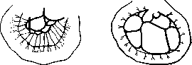
Figure 3. Intestinal veins of man.
Figure 4. Intestinal veins of animal.
of man.
of animal.
Related articles
Mentioned in
Cite this page
“MESENTERIC ARTERIES (VENAE).” Soviet Medical Encyclopedia. English translation of Bolshaya Meditsinskaya Entsiklopediya, 1st ed. (Moscow, 1928–1936), ed. N. A. Semashko. https://sovietmedicalencyclopedia.pages.dev/article/mesenteric-arteries/