HERNIAS
Historical document, translated for reference. It reflects medical knowledge of the 1920s–30s and is not medical advice.
Summary
This article from the 1928-1936 Soviet Medical Encyclopedia provides a comprehensive overview of hernias, including their etiology, prevention, diagnosis, various types, complications, and treatment. It details the anatomical components of hernias and discusses their prevalence and hereditary factors.
Encyclopedia article (1928–1936)
HERNIAS. Contents: Etiology....................237 Prevention..................239 Diagnosis...................240 Various types of H...............241 Inguinal H....................241 Femoral H......................246 Umbilical H...................247 Hernia of the white line of the abdomen.........248 Lateral abdominal hernia............250 Lumbar H......................252 Obturator H....................252 Ischiatic H....................253 Perineal H.....................254 Diaphragmatic H..................255 Complications...................256 Treatment of H..................259 Hernia of the brain...................275 In the strict sense of the word, hernia, hernia (from the Latin hira-intestine), should refer only to the protrusion of abdominal cavity organs beyond its limits (which is also indicated by the Latin term). The exact etymological meaning of the terms for H. among Western peoples (der Bruch-German, rompure-French, rupture-English) indicates the pathogenesis of this disease-the presence of a forcible violation of the integrity of the cavity wall. When one speaks of "hernia of the brain," "hernia of the lung," "muscular hernia," one is rather guided by analogy with abdominal hernia, thereby neglecting the scientific interpretation.-Abdominal hernia should be defined as a formation which consists of the following three parts: 1) hernial gates, i.e., either expanded natural openings in the abdominal wall (inguinal, femoral, umbilical channels, etc.) or artificial openings formed at the site of a wound or injury; 2) hernial sac, i.e., that part of the parietal peritoneum which exits through the defect (hernial gates) in the abdominal wall; 3) one or another abdominal organ protruding into the hernial sac. Sometimes the hernial sac may be absent from these three components (for example, when organs located behind the peritoneum or only partially covered by it, such as the ureter, bladder, large intestine, etc., exit through the hernial gates). Also, if during an injury there is a rupture of the peritoneum and muscles and the organ protrudes through this rupture, then clinically there is a hernia, while patho-anatomically there is a prolapse (prolapsus) of the organ.-Hernial sacs vary in shape and size, most often single, rarely double; there are multichambered sacs and sacs with diverticula. In a fully developed hernial sac, one distinguishes the orifice, neck, body, and fundus. The orifice is the place where the sac communicates with the abdominal cavity; the shape, size, and direction of the orifice vary depending on the type and size of the hernia. The neck connects the orifice of the sac with its body and is always a narrowed and less yielding part of the hernial sac; therefore, strangulation of the organ most often occurs in the region of the neck.-Microscopically, the hernial sac has the structure of the peritoneum, usually with sclerotic or scar changes; sometimes infiltrates are noted. The contents of the hernia are most often the small intestine, which is a more mobile organ, one or with the omentum, but all abdominal organs located both inside and behind the abdominal cavity have been found in hernias. The pancreas seems to be the only organ that has not been found in a hernia (except for diaphragmatic). If, on the one hand, only the vermiform appendix of the cecum, Meckel's diverticulum, a portion of the intestinal wall, fatty appendages of the large intestine can protrude into the hernial sac, then, on the other hand, almost the entire intestine and sometimes other organs (spleen, female genital organs) have been found in the hernial sac; this condition is called eventration. Etiology. Abdominal hernias occur in approximately 3-4% of cases (Malgaigne, Wer-nher, Macready, and others). The greater or lesser development of industry affects the percentage of hernia diseases; in countries with developed industry, the percentage of hernia diseases is higher. According to Tikhon's data, in industrial provinces, the percentage of hernia diseases is almost 2 times greater than in agricultural provinces. Heredity has enormous significance. The works of Berger (Be^ger) and others showed that heredity in hernias is observed in approximately 25% of cases. Regarding age, it can be noted that the maximum incidence of hernia falls on small children and middle age. Men get sick more often than women, which should be connected with heavier physical labor. Tailors ....
2.3% Textile workers ....
1.7% Light industries
1.5% by works. Therefore, a profession associated with physical labor exerts its influence. Golyanitsky gave such a table of the percentage of hernias in various industries: Porters .... 18.8% Metalworkers . . . 14.2% Rubber production 8.2% Tramway..... 7.0% Printing, prod. 2.9% The causes of hernia formation are conventionally divided into 2 categories-general and local. The latter consist in the special anatomical structure of the area where the hernia appears. The general causes are divided into predisposing and producing. Predisposing causes include: nationality, heredity, age, sex, physique, musculature, weight loss of the body, changes in the internal organs of the abdomen. Producing causes can be divided into 2 groups. The first includes: 1) disorders of defecation (constipation), 2) cough, 3) screaming, 4) difficulties in urination (strictures of the urethra, phimosis), 5) tight tightening of the abdomen, 6) singing and playing wind instruments, 7) difficult childbirth, 8) heavy physical labor (lifting weights, carrying loads, work in a bent position, etc.), i.e., all moments that repeatedly and long-term increase intra-abdominal pressure. The combination of several harmful conditions enhances the harmful effect. The second group includes: 1) pregnancy, especially repeated, which stretches and thins the abdominal wall; 2) old age and general diseases causing weakening of the body's musculature; 3) all kinds of traumatic injuries to the abdominal wall-all factors that lower the tone of the abdominal wall. If intra-abdominal pressure is balanced by the resistance of the abdominal wall, then a hernia does not form. If the abdominal wall cannot withstand it, then a hernia appears in the weakest places and places of greatest pressure (inguinal in men, femoral ring in women). Under the name of congenital hernias are meant not only those hernias with which people are born, but generally hernias which can form at any age of a person, but for its occurrence requires congenital developmental anomalies of the area where this hernia appears. This congenital anomaly consists not only in the structure of the abdominal wall of this area, in the presence of a hernial sac as a diverticulum of the peritoneum, but also often in various kinds of anatomical peculiarities of the organ which protrudes into the hernia-Hernia of "weakness" in some cases is very difficult to distinguish from a congenital hernia. In hernia of "weakness" we are dealing with a combination of pathological conditions of the organs of the abdominal cavity and abdominal wall of an exclusively acquired nature, which can for example be observed in weakening of the body on the basis of prolonged hunger or after various kinds of diseases.-Traumatic hernias are divided into 4 categories: 1) purely traumatic hernias, 2) forced hernias, 3) scar hernias, 4) artificial hernias. Purely traumatic hernias form at the site of a blow to the abdomen with any blunt object. Due to the blow, a subcutaneous rupture of the underlying muscles and fasciae occurs, while the peritoneum remains untouched and under the influence of intra-abdominal pressure protrudes into the rupture along with the internal organs, forming a hernia. True traumatic hernias, studied during surgery, are few, a much larger number were observed only clinically. Of the 28 traumatic hernias described by various authors and counted by Krymov, 24 are in men and 4 in women. By location these 28 cases are divided as follows: 13 - in the inguinal area, 6-in the lumbar, 4-along the white line of the abdomen, 2-in the femoral area, 2-in the right iliac and 1-in the middle part of the abdomen, at the edge of the rectus muscle. The question of forced hernias is still little clarified. French authors hold the opinion that forced hernias can be called those which suddenly form in healthy people with a strong abdominal wall, under the influence of various kinds of efforts. German authors believe that to recognize such a hernia, only the exact establishment of the fact that the hernia formed under the influence of one or another effort and that the patient did not previously have a hernia is required, while the physical condition of the victim is not important. According to Russian law of 2/VI 1903, "A hernia is subject to compensation only when it is a consequence of an accident that suddenly caused all the objective signs of hernia with signs of its incarceration, requiring medical help." At present, hernia is classified as a disability, and under a disability received in connection with work by hire, should be understood as a sudden injury to health caused by an external event (such is the decision of the NKT of the Ukrainian SSR of 19/VI 1924).-Scar hernias are divided into 2 categories: the first category includes those cases when a hernia develops at the site of any accidental injury to the abdominal wall, and the second-postoperative hernias.-Artificial hernias are usually found in persons called upon to perform military service and wishing to be exempt from it. They occur only in the groin and more often on the left. Krymov divides artificial hernias into 4 categories: 1) hernias which are produced through a rupture of the subcutaneous opening of the inguinal canal; 2) hernias in which the rupture of the abdominal wall occurs above the subcutaneous opening of the inguinal canal, so that the latter remains intact; 3) artificial hernias produced through a rupture of only the transverse fascia, with the integrity of the subcutaneous opening of the inguinal canal; 4) artificial hernias produced by gradual stretching of the tissues of the inguinal canal. In the latter case, they differ in no way from the natural direct inguinal hernia, while in the other cases scars always form at the site of injury. Prevention of hernia is of great importance. It has already been noted above that hernia in most cases appears in small children and people of middle age. When describing the oblique inguinal hernia, as the most common, it is found that the hernial sac is the remnant of the peritoneal appendix. The study of its obliteration shows that the disappearance of the appendix is delayed up to 1 year and that the cause of its closure is compression by the adjacent muscles. There is no doubt that this involutionary process should not encounter obstacles from the side of the child's intra-abdominal pressure. Therefore, everything that leads to an increase in intra-abdominal pressure should be eliminated from the physical care of the infant: tight swaddling (Baginski), early vertical position of the child, tossing and throwing upward of newborns, which is sometimes done by mothers and nannies. It is self-evident that good and proper nutrition of the child, beneficially affecting the whole body, also strengthens the muscular system. The fact that hernias more often appear in weak and atrophic children and as a rule in premature infants is well and long known. Owen drew attention to the fact that inguinal hernias are more common in children growing on artificial feeding. What significance should be attached in the matter of the appearance of umbilical hernias to the care of the navel, is rather difficult to say, but the fact of the more frequent appearance of umbilical hernias in children who had this or that irregularity in the process of healing of the navel has been noted by many pediatricians. It is clear that all moments contributing to an increase in intra-abdominal pressure should be prevented in children as much as possible. In this respect, disorders of the gastrointestinal tract, cough, strong screaming and disorders of urination (phimosis) are particularly important. The role of the latter in the appearance of hernias has long been established. Therefore, the treatment of phimosis in children becomes necessary. In adolescence, physical education as a way of strengthening muscles can have significance in the prevention of hernias. Good influence on the strengthening of the inguinal areas is exerted by properly applied cycling, skating and skiing. Bandaging the abdomen, wearing a corset during pregnancy and abdominal massage after childbirth have already become common practice. With obesity, which is a predisposing cause of hernia formation, one should also fight. Heavy physical labor, especially with a weak body, often causes the development of hernia; this forces one to strive for the correct selection of workers for various enterprises.-Prevention of postoperative hernias should be in the hands of surgeons. In this respect, attention deserves: 1) the method of suturing the abdominal wound, 2) the direction and place of the incision, 3) the difference in material for the suture, 4) the quality of the material, 5) the postoperative course (cough, vomiting) and care (dressing, early rising, etc.), 6) the nature of healing. Diagnosis. The characteristic sign of a reducible hernia is its ability to appear outside the abdomen spontaneously or with tension and to disappear easily-both spontaneously in certain positions of the body and with pressure on it. If the hernial tumor appears in the usual places for hernias (inguinal, femoral, umbilical areas), then the hernia is easily recognized. The appearance of such a tumor on the perineum, in the gluteal region or in the region of the obturator opening first makes one think of the possibility of a hernia here also. The second characteristic sign of a hernia is the "cough impulse." If you put your hand on the tumor and make the patient cough, the hand clearly feels the impulse. Tapping, palpation of the hernial tumor, as well as digital examination of the hernial gates establish the diagnosis. Recognition of the internal organs contained in the hernia is facilitated by the use of X-rays.-Irreducible hernias. Hernias that have lost the ability to be reduced into the abdominal cavity are called irreducible.
Irreducibility can be temporary and permanent, congenital and acquired, complete and partial. In the latter case, part of the hernia is reducible, while the other remains in the hernial sac. The causes of irreducibility of hernias are divided into predisposing and precipitating factors. The former include: age (old age), sex (more common in women), occupation (heavy physical labor), type of hernia (more often femoral), and size of hernia (large hernias). The causes that precipitate irreducibility can be divided into 3 groups: 1) causes lying outside the hernial sac, 2) causes lying within the hernial sac itself, 3) causes lying inside the hernial sac. In most cases, the cause of irreducibility is adhesions of the abdominal organs to each other and to the hernial sac. Irreducible hernias, however, can still increase in size when the hernial gates are free and sometimes reach very large dimensions. Irreducible hernias are more dangerous than reducible ones, as they are more prone to various complications. Various types of hernias According to their mode of formation, hernias are divided into congenital and acquired. Depending on the condition of the protruding organs, hernias are distinguished as: 1) reducible, 2) irreducible, 3) with symptoms of coprostasis, 4) with symptoms of inflammation, 5) incarcerated. According to the site of protrusion, hernias are distinguished as: 1) inguinal hernias, 2) femoral, 3) umbilical, 4) hernias of the white line and tendinous intersections of the rectus abdominis muscles, 5) lumbar, 6) obturator, 7) sciatic, 8) perineal, 9) diaphragmatic. The latter is called an internal abdominal hernia, while all others are external. Inguinal hernia (h. inguinalis). The formation of the inguinal canal occurs in boys during the descent of the testicles, and in girls during the formation of the round ligament of the uterus. In normal adults, there is essentially no canal, but only a slit where the spermatic cord is located in men and the round ligament of the uterus in women. The canal exists only when the peritoneal diverticulum, which forms in embryos and closes in newborns, remains open, or when a hernia forms. In boys under 1 year old (according to Krymov's research), the peritoneal diverticulum remains open entirely or partially (only from the abdominal cavity) in 54%, of which 32% are open bilaterally, 14% on the right, and 8% on the left; in girls under 1 year old, the peritoneal diverticulum is open in 42%, of which 23% are open bilaterally, 11% on the right, and 7% on the left. In adults, an open peritoneal diverticulum, entirely or partially (only from the abdominal cavity), is observed in 4-5%. The inguinal canal has 4 walls and 2 openings-external (subcutaneous) and internal (abdominal). The anterior wall of the inguinal canal is formed by the aponeurosis of the external oblique muscle, the posterior wall by the transverse fascia, the upper wall by the free edge of the internal oblique and transverse muscles, and the lower wall by the Poupart's ligament. The length of the inguinal canal in men is 4-4.5 cm, and in women it is somewhat longer. If one looks at the anterior abdominal wall from the abdominal cavity side, 5 folds covered by peritoneum are visible in its lower half. One of these folds is located along the midline of the abdomen, and the other 4 are on the sides, 2 on each side. The middle of these folds is formed by the elevation of the peritoneum caused by the obliterated urachus, which extends from the umbilicus to the apex of the bladder. On either side of this fold, symmetrically, runs a second fold, which also begins at the umbilicus but, going downward, diverges from the middle fold and extends to the lateral surface of the bladder; this fold is formed by the obliterated umbilical artery, which in embryos extends from the hypogastric artery to the umbilicus. Even more laterally, also symmetrically on each side, runs a second fold formed by the inferior epigastric artery passing under the peritoneum. Between these folds, 3 depressions are formed on each side, called the inguinal fossae. In the etiology of inguinal hernias, only two of these depressions are important and significant, because the depression located between the obliterated urachus and the obliterated umbilical artery lies behind the rectus abdominis muscle and, when this muscle is intact, cannot serve as a site for hernial protrusion. Inguinal hernias protrude from the abdominal cavity only through two fossae: one lying between the obliterated umbilical artery and the inferior epigastric artery, and the other located lateral to the inferior epigastric artery. The internal opening of the inguinal canal is located in the latter. A hernia protruding through this fossa is called an indirect inguinal hernia, while one protruding through the first fossa is called a direct hernia. Several degrees of development of indirect inguinal hernia are distinguished: 1) initial degree, when the tumor is barely visible to the eye, but when the patient strains the abdominal muscles, the examining finger introduced into the inguinal canal feels a tense tumor (protrusion of peritoneum) at the internal opening of the canal, which disappears when the patient stops straining (hernia inguinalis obliqua incipiens); 2) canal form of hernia, when the fundus of the hernial sac does not protrude beyond the external opening of the canal (hernia obliqua canalis inguinalis); 3) when the hernia, having exited the external opening of the inguinal canal, distributes at various levels along the spermatic cord (hernia inguinalis obliqua funicularis); 4) the hernia descends into the scrotum (hernia obliqua inguino-scrotalis), in women into the labium majus (see figures 1 and 2). If the entire peritoneal diverticulum remains open and the protruding organ lies together with the testis, the hernia is called testicular indirect inguinal hernia (hernia inguinalis obliqua testicularis). The indirect inguinal hernia always lies within the spermatic cord. The direct inguinal hernia lies medial to the spermatic cord, only adjacent to it. Direct inguinal hernia is more common in elderly individuals and in men much more often than in women. The indirect inguinal hernia has several varieties that the surgeon must know in order to properly orient himself during the operation. Of all inguinal hernias, the interstitial (h. interstitialis) is most frequently observed, characterized by the hernial sac being located in a greatly dilated inguinal canal, sometimes extending between the muscles. The hernial sac can be in the form of only a cavity, but it can have a continuation of varying lengths and shapes, which can extend toward the umbilicus, the anterior superior iliac spine, or into the scrotum. The testis is located in the inguinal canal in the vast majority of cases. - The preperitoneal inguinal hernia (h. properitonealis) is characterized by the fact that the hernial sac is in the vast majority of cases bicameral, with one chamber lying in the usual location of an indirect inguinal hernia and the other in the form of a diverticulum in the preperitoneal tissue. In rare cases, only the latter may be present. The preperitoneal sac can have different directions, and depending on this, 1) h. properitonealis antevesicalis, 2) h. properit. iliaca, 3) h. properit. obturatoria are distinguished. The testis was in a state of ectopia in 2/3 of all cases. - The inguino-superficial hernia (h. inguino-superficialis) is characterized by the fact that the hernial sac, having exited the inguinal canal through its external opening, does not extend, as is usual with indirect hernia, into the scrotum, but emerges into the subcutaneous tissue, where it spreads. The hernial sac is thus in the form of two halves-one lies in the inguinal canal, and the other in the subcutaneous tissue. The latter can extend toward the umbilicus, the anterior superior iliac spine, or descend downward onto the thigh, as well as onto the perineum, located behind the
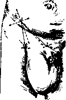
Figure 1. Right-sided inguino-scrotal hernia. Anesthesia according to Braun: 1, 2, 3, and 4-points of needle insertion, arrows indicate the direction of injection of anesthetic substance. (After Bier-Braun-Kummell.)
stok and the protruding organ lies together with the testis, then the hernia is called testicular indirect inguinal hernia (hernia inguinalis obliqua testicularis). The indirect inguinal hernia always lies within the spermatic cord. The direct inguinal hernia lies medial to the spermatic cord, only adjacent to it. Direct inguinal hernia is more common in elderly individuals and in men much more often than in women. The indirect inguinal hernia has several varieties that the surgeon must know in order to properly orient himself during the operation. Of all inguinal hernias, the interstitial (h. interstitialis) is most frequently observed, characterized by the hernial sac being located in a greatly dilated inguinal canal, sometimes extending between the muscles. The hernial sac can be in the form of only a cavity, but it can have a continuation of varying lengths and shapes, which can extend toward the umbilicus, the anterior superior iliac spine, or into the scrotum. The testis is located in the inguinal canal in the vast majority of cases. - The preperitoneal inguinal hernia (h. properitonealis) is characterized by the fact that the hernial sac is in the vast majority of cases bicameral, with one chamber lying in the usual location of an indirect inguinal hernia and the other in the form of a diverticulum in the preperitoneal tissue. In rare cases, only the latter may be present. The preperitoneal sac can have different directions, and depending on this, 1) h. properitonealis antevesicalis, 2) h. properit. iliaca, 3) h. properit. obturatoria are distinguished. The testis was in a state of ectopia in 2/3 of all cases. - The inguino-superficial hernia (h. inguino-superficialis) is characterized by the fact that the hernial sac, having exited the inguinal canal through its external opening, does not extend, as is usual with indirect hernia, into the scrotum, but emerges into the subcutaneous tissue, where it spreads. The hernial sac is thus in the form of two halves-one lies in the inguinal canal, and the other in the subcutaneous tissue. The latter can extend toward the umbilicus, the anterior superior iliac spine, or descend downward onto the thigh, as well as onto the perineum, located behind the
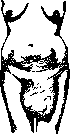
Figure 2. Hernia labii majoris.
scrotum. The testicle in most cases was found in a state of ectopia. Cooper's hernia, or hernia with a double sac, is characterized by the fact that one sac lies within another, and the fusion of these sacs can occur at various points of the inguinal canal, starting from its internal opening to the external, and sometimes even below it. The testicle can lie at the bottom of the external sac or be suspended from the bottom of the internal sac, or it can have its own cavity, separate from the sacs, which then lie above this cavity. The internal sac may communicate with the external sac through an opening, and a serous fluid accumulation may be observed between the sacs. Inguinal hernia is sometimes encountered in combination with hydrocele of the testicle, both simple and communicating with the abdominal cavity, as well as with a cyst of the spermatic cord, which can complicate both diagnosis and operation. The contents of an inguinal hernia can be almost all organs of the abdominal cavity, but most often one encounters the small intestine and omentum. Hernias of the cecum and S-Romani deserve attention. Several types are distinguished: 1) a completely free hernial sac in which the large intestine is freely accommodated; 2) a hernial sac fused with the mesentery of the large intestine at the level of its neck, and sometimes this fusion extends to the bottom of the sac; 3) fusion of the hernial sac with the large intestine may be observed over a greater or lesser extent in the upper part of the intestine, while in the lower part the hernial sac may be completely free; 4) the hernial sac is fused with the large intestine over a large area, and its free part represents either a diverticulum of the peritoneum or a rather voluminous sac in which usually lie loops of the small intestine or omentum; 5) the entire hernial sac is fused with the large intestine, so that a picture of the absence of a sac is obtained. The last 4 types represent so-called sliding hernias. The relationship of the urinary bladder to the hernial sac also deserves attention. The following classification of hernia of the urinary bladder is accepted: 1) hernia vesicae urin. intra-peritonealis, 2) hernia vesicae urin. extraperitonealis, 3) h. vesicae urin. paraperitonealis. In the first case, the urinary bladder lies within the hernial sac, in the second case, a part of the bladder not covered by peritoneum protrudes through the hernial ring, i.e., there is no hernial sac, and in the third case, the urinary bladder lies near the hernial sac. This last form must be recognized as the most common. Eight cases of the presence of a pregnant uterus in an inguinal hernia have been described. Etiology. Inguinal hernia occurs in men more often than in women. On the right side, inguinal hernia in men occurs almost twice as often as on the left, in women this difference is not so sharply expressed. Bilateral inguinal hernias are more common in adults than in children, and in women they are rarely observed. Bilateral hernias can be of the same type on both sides or different. In the formation of inguinal hernia, the anatomical structure of the inguinal area plays a major role, the presence of a peritoneal diverticulum in the form of an unobliterated peritoneal appendage as a ready-made hernial sac also has considerable importance. Direct hernia in children also more often forms when there is a peritoneal diverticulum here. It is known that in counteracting intra-abdominal pressure, the muscles of the abdominal wall play a primary role. Therefore, if the inguinal area is poorly protected by muscles, then it should be recognized as a locus minoris resistentiae for intra-abdominal pressure. This condition of the abdominal wall is further aggravated if the connective tissue parts of the wall do not possess elasticity and strength. The causes of the protrusion of abdominal contents into the hernial sac can be divided into 2 groups: 1) congenital and 2) acquired. Sometimes an organ fuses with the testicle already in the embryo and descends into the scrotum with the testicle. One of the main conditions for the protrusion of abdominal contents into a hernia is a certain degree of their mobility, which can be of either congenital or acquired nature. The second cause for the protrusion of abdominal contents into a hernia is the abnormal position of the organ, which can be of either congenital or acquired nature. The third cause must be recognized as the pulling of one organ by another or by an enlarging hernial sac as part of the parietal peritoneum (sliding hernias, see figure 12). Finally, the fourth cause can be various anatomical changes in organs.
Diagnosis. Distinguishing between indirect and direct inguinal hernia is relatively not difficult: at the beginning of their development, the first has an oblique direction, an oval shape, lies within the spermatic cord, while the second has a round shape, lies medial to the spermatic cord, and upon examination through the external opening of the inguinal canal, a depression can be palpated behind the pubic bone. The inferior epigastric artery in an indirect hernia lies medial to the hernial sac, while in a direct hernia it lies lateral. With certainty, other varieties of inguinal hernia can only be distinguished during operation. Simple hydrocele of the testicle differs from a hernia in that it has sharp boundaries both below and above, gives a dull sound on percussion, and transilluminates. Hydrocele of the testicle communicating with the abdominal cavity also gives a transillumination phenomenon. Varicose veins of the spermatic cord can also give a tumor with a cough impulse, but careful examination of the spermatic cord and visibly dilated veins resolve the diagnosis. New formations of the scrotum, spermatic cord, and inguinal canal can also cause confusion with an irreducible hernia, but the history and careful examination usually help to make the correct diagnosis. Femoral hernia (h. femoralis s. cruralis). A peritoneal diverticulum is sometimes found in the femoral canal in newborns. Krymov examined 500 children's corpses (200 boys and 300 girls), and a peritoneal diverticulum was found once (in a three-week-old boy) on the right side, in 31 cases an indentation of the peritoneum was found at the site of the internal opening of the femoral canal. The femoral canal, like the inguinal canal, exists only when there is a hernia or a peritoneal diverticulum lies within it. When describing the inguinal canal, it was noted what should be understood under this name. It is not so easy to do this with respect to the femoral canal, as the opinions of different authors on this question are far from being in agreement among themselves. Most recognize that the femoral canal is the space lying under the Poupart's ligament, medial to the vessels passing here, between the femoral vein and the edge of the Gimbernat's ligament, with its internal opening being considered this space, and its external opening the so-called oval fossa formed by the layers of the broad fascia of the thigh. The oval fossa is filled with fatty tissue, which communicates with the preperitoneal tissue. The space located between Poupart's ligament and the horizontal branch of the pubic bone is divided by the obliquely positioned lig. ilio-pectineum into 2 parts (lacunae): lateral-muscular and medial-vascular. In the first are contained muscles (iliacus, psoas and between them the femoral nerve), and in the second lie vessels (femoral artery and vein). The lateral part (muscular lacuna) is tightly filled with muscles, while in the medial part, medial to the femoral vein, a space remains-the above-mentioned femoral ring. The distance from the femoral vein to the Gimbernat's ligament in men on average is 1.2 cm, and in women 1.8 cm. Krymov developed a classification of femoral hernias in the following form. I. Vascular-lacunar femoral hernia: 1) the hernia occupies the entire width of the vascular lacuna (hernia cruralis vasculo-lacunaris totalis); 2) the hernia occupies only part of the vascular lacuna: a) median (h. cruralis vasculo-lacunaris medialis)-typical femoral hernia, b) middle, or proper vascular sheath (h. cruralis intravaginalis vasculo-lacunaris), c) lateral (h. cruralis vasculo-lacunaris lateralis); 3) hernias with multichambered hernial sacs or diverticula: a) hernia cruralis properito-RL i nealis, b) h. cruro-inguinalis; 4) h. cruralis pectinea (Gloquet). II. Femoral hernia of the Gimbernat's ligament. III. Muscular-lacunar femoral hernia. Recently Krymov studied during operation a hernia that lay only above the femoral vein and was named by Krymov hernia of the femoral sheath.
ja. n. of the vein. A typical femoral H. is the one that exits through the described femoral canal (see figure 3). Three types of femoral H. are distinguished: complete, incomplete or interstitial, and the initial degree of H. - A complete femoral H. exits through some opening in the superficial fascia into the subcutaneous tissue, while an incomplete H. is one that remains all the time near the vessels and does not extend beyond the superficial fascia. In the initial degree of H., it does not extend beyond the femoral ring. The most common should be recognized as the femoro-preperitoneal H.; it, like the inguinal, has a two-chamber hernial sac. H. of Cloquet begins to exit from the abdominal cavity as a typical femoral H., but then directly at the pubic bone it passes under the fascia pectinea and lies on the muscle of the same name. Hernia vasculo-lacunar-is (or praevascularis) most often, as shown by the research of Narath, is associated with congenital diseases of the hip joint. H. of the ligament of Gimbernat (Laugier's) usually passes through a congenital fissure in the ligament. H. of the muscular lacuna (Hesselbach's) exits onto the thigh through the muscular lacuna and is located on the thigh, between the anterior superior iliac spine and the femoral vessels. The hernial tumor has a flat shape, because it has to make its way under the dense fascia. - The contents of a femoral hernia are most often the small intestine and omentum. As an exception, Keller encountered the stomach, and Skey-the gallbladder. - Etiology. In women, femoral H. occurs 2 times more often than in men. On the right side, femoral H. occurs more often than on the left. In newborns and children up to 1 year, femoral H. is extremely rare, in men it begins to occur more often after 25 years, and in women-after 15. An anatomical predisposition to the formation of femoral H. should be considered the presence of a more or less pronounced diverticulum of the peritoneum and increased size and weakness of the structure of the femoral ring. - Recognition. By the shape and direction of the hernial tumor, 3 types of femoral H. can be distinguished: 1) when it is a round, spherical tumor, 2) when the tumor hangs down, and 3) when the tumor rises upward to Poupart's ligament and, covering the latter, is arranged along its length. When diagnosing femoral H., two questions arise: Is this a H., and is it femoral? Lipomas, enlarged lymph glands, abscesses (dropsies), nodular varicose veins are differentiated by careful clinical examination, although it should be noted that lipomas located in the femoral canal present difficulties in diagnosis. Femoral H. in obese people can be confused with inguinal, but if the external opening of the inguinal canal can be palpated, then diagnosis becomes easy; if not (in women), then one should be guided by the position of Poupart's ligament in relation to the hernial tumor: the femoral H. lies entirely or for the most part below the ligament. Umbilical H. (h. umbilicalis). Hernias of the navel are divided into 3 categories: 1) embryonic hernias, 2) hernias of childhood, 3) hernias of adults. Embryonic H. are also divided into 3 categories: a) proper embryonic hernias, b) hernias of embryos, c) mixed hernias. Proper embryonic H. are malformations, the beginning of the formation of which should be attributed to the very earliest period of embryonic life. The outer covering of the embryonic H. is a thin, translucent membrane, which however consists of 2 plates (outer and inner), separated by a thin layer of Warton's jelly. The inner membrane is a primitive membrane of Rathke, which has stopped in its development and has not passed into the peritoneum. Hernia of the embryo or H. of the umbilical cord is formed after three months of intrauterine life, when the abdominal wall has already been formed to a sufficient degree and when the peritoneum has formed, so that the hernia of the embryo has a true hernial sac. In cases of mixed H., 2 hernias are observed, one with a primitive membrane, and the other with a true hernial sac from the peritoneum. - Umbilical H. of childhood are most often formed before 6 months of age, when the formation of the umbilical ring has not yet been completed. They are formed under the influence of a sharp increase in intra-abdominal pressure. - Umbilical H. of adults are divided into 2 categories: direct H. and oblique H. (H. of the umbilical canal). The contents of H. of newborns Perren presented in the following table: y
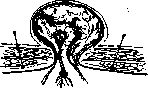
show the direction and depth of injection of anesthetic substance.
Small intestine ...
35 Small and large intestine
15 Liver and intestine ...
32 Meckel's diverticulum Stomach....... 2 Heart ........ 1 The contents of childhood H. and H. of adults is most often the small intestine, in adults together with omentum or only omentum (see figure 4). As an exception, various organs were found. - Etiology. Umbilical H. in women occur twice as often as in men. Regarding age, it can be said that the curve of the disease of umbilical H. reaches its highest point 2 times- the first time before 1 year (and this point is the highest), and the second time around 40 years. An anatomical predisposition is a diverticulum of the peritoneum in the umbilical ring and imperfection of the structure of the navel. - Recognition. Umbilical H. is easily recognized, but to determine H. of the umbilical canal with certainty can only be done during surgery. H. of the linea alba (h. lin. albae). The linea alba, located between the xiphoid process and the pubic symphysis and having the navel in the middle, represents a crossing of the aponeuroses of the broad abdominal muscles. This connective tissue strip does not have the same width everywhere. Its beginning and end look like a thin cord. A little below the xiphoid process, the cord begins to gradually expand, approximately to 2 cm, and then, descending 3-4 cm below the navel, suddenly narrows and continues so to the end. In women, the linea alba is on average wider than in men. Sometimes in the so-called divergence of the rectus muscles, the width of the linea alba reaches large dimensions. By its structure, the linea alba is divided into 2 types: 1) a solid plate, 2) perforated with openings of various sizes. - H. of the linea alba are divided into 3 categories: 1) suprapubic, 2) periumbilical, 3) subumbilical. The latter are very rare. H. of the linea alba rarely reach the size of a chicken egg, usually they are the size of a forest nut. The most common content is omentum, less often one or another part of the intestine is encountered. As an exception, the stomach, gallbladder, etc. were observed. - Etiology. Men get sick more often than women. In children, these H., except for suprapubic, are rare, most often they are observed at the age of 30-40 years. Trauma can play a major role in the origin of these H., heavy physical labor can also be included here, because the linea alba is easily torn. Golyanitsky in Astrakhan loaders found depending on the size of professional experience from 6.5% to 19.5% of patients with hernias of the linea alba, and Khesin in Moscow loaders-from 7.7% to 29.2%. An anatomical predisposition should be recognized as the existence of through openings and fissures in the linea alba, where sometimes diverticula of the peritoneum may even be located. - Recognition. From a clinical side, H. of the linea alba can be divided into 2 categories: 1) H. without subjective sensations and 2) H. with various painful phenomena. Patients usually complain of colicky pains in the stomach area. Often hernias are combined with various other diseases of abdominal organs (stomach ulcer, gallstone disease, etc.). Often instead of H., preperitoneal lipomas, which have passed through an opening in the linea alba into the subcutaneous tissue, are observed (see figure 5; in the figure is depicted a subperitoneal lipoma of the linea alba above the navel behind; the posterior surface of the anterior abdominal wall is visible; above the navel the peritoneum is partially separated and turned aside; thus the lipoma, which through a transverse fissure in the linea alba goes forward on a thin stalk, becomes visible). These lipomas are often difficult to distinguish clinically from H. containing omentum. Preperitoneal lipomas, like H., can be reduced, but they can also be irreducible, which is also often observed. In 50 cases of H. of the linea alba operated on by Krymov in recent years, a hernial sac was found only in 16 cases, which is 32%. Divergence of the rectus abdominis muscles is often observed together with the prolapse of the abdominal organs.
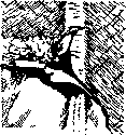
Figure 5.
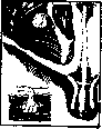
Figure 6. Interstitial hernia of Spiegel's line.
Lateral abdominal hernia (h. lateralis abdominalis). Three types of hernias are observed on the lateral abdominal wall: 1) hernia of the rectus sheath, 2) hernia of Spigelian line, 3) hernia in other locations of the lateral abdominal wall. According to origin, lateral abdominal wall hernias can be divided into 1) traumatic hernias, formed at the site of accidental trauma, or postoperative hernias, 2) hernias formed due to arrested development of the abdominal wall. Hernias of the rectus sheath usually develop due to rupture of the muscle under the influence of trauma, most often in the lower third of the muscle, where there is no posterior sheath. The Spigelian line is called the semilunar line, which is formed at the transition of the muscular part of the transverse muscle into the tendinous aponeurosis; in the upper 2/3 it lies behind the rectus muscle, and in the lower third - to the side of it, where the hernia is formed (see figure 6). Spigelian line hernias usually occur at the sites of slits that exist in the tendinous aponeurosis of the abdominal muscles and through which vessels pass. Sometimes these hernias occur after appendectomy, when the abdominal wall is opened according to the Lennander method. The Spigelian line hernia is observed more frequently than others. It is observed much more often in women than in men. Among etiological factors, obesity is noted. Lateral abdominal hernias are divided into 3 categories: 1) simple, subcutaneous hernia, 2) interstitial hernia, 3) preperitoneal hernia. If the hernia is located under the aponeurosis of the external oblique muscle, it is called interstitial, and in these cases the hernial orifice is formed by the tendinous aponeuroses of the transverse and internal oblique muscles, but if the hernia protrudes under the skin, then the hernial orifice passes through the tendons of all 3 muscles. The size of the hernia varies; the hernial orifice either barely admits a finger or has a diameter of 7-8 cm. Usually they occur on one side, but Berger observed a bilateral hernia in one patient. As contents of the hernia, the small intestine is more frequently observed, less often - the cecum or transverse colon. Small hernias, especially in obese women, can present great difficulties in diagnosis. Right-sided Spigelian line hernias may be confused with appendicitis. Lumbar hernia (h. lumbalis). Sites of exit for hernias in the lumbar region are considered: 1) Petit's triangle, 2) the Grynfeltt-Lesshaft space and 3) aponeurotic slits. Two fingerbreadths posterior to the midpoint of the distance between the anterior superior and posterior iliac spines, there is a small triangular space in the abdominal wall, not covered by either the broad muscle of the back or the external oblique muscle of the abdomen. This space, first described by Petit (Petit), has the appearance of a triangle, whose base is directed downward and lies on the iliac crest, while the vertex is directed upward. The posterior side of this triangle is formed by the lateral edge of the broad muscle of the back, and the anterior side by the posterior edge of the external oblique muscle. At this place, under the superficial fascia and a thin aponeurosis, lies the internal oblique muscle, under which is a thin layer of transverse muscle, and sometimes immediately, when there are no muscle fibers, it is covered by the deep lamina of the lumbo-dorsal fascia, behind which are the transverse fascia, preperitoneal cellular tissue, and peritoneum. According to Lesshaft and Baracz (Baracz), Petit's triangle is absent in 20-30%. Its dimensions vary: sometimes it appears as a slit, sometimes with a fairly broad base. - The Grynfeltt-Lesshaft space can have various shapes and is located between the internal oblique muscle of the abdomen anteriorly and inferiorly, the longitudinal muscles of the spine and quadratus lumborum muscle internally, the serratus inferior muscle and XII rib superiorly. The floor of this space is the aponeurotic part of the transverse muscle of the abdomen, and it is covered superiorly by the broad muscle of the back. Often this space has the appearance of a triangle, with its base lying either on the XII rib, or on the lower edge of the serratus inferior muscle, or directed toward the quadratus muscle, in which case the upper rib was formed by the edge of the serratus muscle, and the lower by the edge of the internal oblique muscle. Lesshaft noted that this space has larger dimensions and is more constant than the former. According to Baracz, the Grynfeltt space is absent in only 6.5%. - Aponeurotic slits are divided into 2 groups: 1) some serve as passages for vessels and nerves, 2) others are a developmental defect or a consequence of aponeurosis rupture. Hernias pass through the Grynfeltt-Lesshaft space more often than through Petit's triangle. Lumbar hernias, in relation to the hernial sac, are divided into 2 categories: 1) true hernias, with a hernial sac, 2) hernias without a hernial sac. The latter are less common than the former. The hernial tumor, emerging through the lower triangle or through aponeurotic slits, is usually located under the skin. When passing through the upper space, it is most often located under the broad muscle of the back and less often under the external oblique muscle. The contents of lumbar hernias are most often the small intestine and omentum; the ascending or descending colon is also frequently found. Jeannel (Jeannel) provides the following data regarding the frequency of internal organs found in lumbar hernias: 1) small intestine - 8, 2) large intestine - 2, 3) omentum - 4, 4) kidney - 1. - E T I O L O G Y. In men, lumbar hernia was observed more often than in women; thus, out of 43 cases of Jeannel's, 27 were men and 16 women. By age, 27 cases were distributed as follows: up to 10 years - 5, from 20 to 40 years - 8, from 40 to 60 years - 5, from 60 to 75 years - 9. On the left side, lumbar hernia occurred more often than on the right. Lumbar hernias are distinguished as congenital and acquired. The causes of acquired hernias are various types of trauma and pathological processes. Among other etiological factors, multiple births, atrophy of muscles in the lumbar region on the basis of nerve changes, cold abscesses, phlegmons are mentioned. - D I A G N O S I S. In typical cases of reducible lumbar hernia, its recognition does not present particular difficulties. Examination of the hernial orifice confirms the diagnosis. It is more difficult to distinguish irreducible hernias, but here the history clarifies the matter. The maximum size of a reducible lumbar hernia is obtained when the patient lies on the affected side in the middle position between lateral and dorsal. Obturator hernia (hernia obturatoria). The obturator canal is formed as follows: the obturator membrane (internal and external), closing the obturator
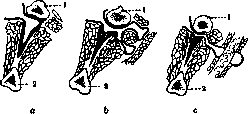
Figure 7. Varieties of obturator hernia: a-interstitial; b-posterior to the pectineus muscle; c-anterior to the pectineus muscle; 1-pubic bone; 2-ischial bone. The opening formed by the pubic and ischial bones does not reach the upper edge of the obturator foramen, and some space remains between them. The free edge of the membrane is located directly under that part of the pubic bone where it has a groove, known in anatomy as the obturator groove. The obturator canal has an oblique direction, the same as the inguinal canal. Its upper wall, and partly its lateral walls, is formed by the pubic bone, namely its sulcus obturatorius, while the lower wall is the fatty tissue, which lies between the two obturator membranes. This canal, according to Maydl, is about 3 cm in length, and according to Schmidt-1.5 cm. Its width is not a constant value, but on average equals 1 cm. Thus, the obturator canal appears to be bone-membranous, little yielding, and is a site for hernia incarceration. The peritoneum covering it often forms a small depression, and sometimes a clearly expressed diverticulum of the peritoneum has been found here. The anterior opening of the obturator canal does not have the appearance of a complete ring, but consists of two separate semicircles-the upper one formed by the pubic bone, and the lower one formed by the upper edge of the external obturator muscle. This opening is closed by the deep fascia of the thigh. In the obturator canal are the obturator nerve (n. obturatorius), obturator artery and vein (a. et v. obturatorius), surrounded by fatty tissue. From a practical standpoint, when providing surgical assistance, 3 forms of obturator hernia should be distinguished: 1) when the hernia remains in the obturator canal (h. obturatoria interstitialis, see figure 7, a), 2) when the hernia, having exited the obturator canal, remains lying under the pectineus muscle (h. obturatoria recto-pectinea, see figure 7, b), 3) when the hernia, passing between the pectineus and adductor longus muscles of the thigh, is located either under the deep fascia of the thigh or emerges into the subcutaneous tissue (h. obturatoria antepectinea, see figure 7, c). As content of obturator hernia, the small intestine was most often found, alone or with the omentum, and in incarceration, a portion of the intestinal wall was most commonly encountered.-Etiology. Obturator hernia occurs much more frequently in women and rarely in men. It is characteristic of old age. On the right side, obturator hernia is more common than on the left. It was often observed on both sides. The frequency of observation of obturator hernia in women is explained by 1) a more pronounced inclination of the pelvis, 2) a larger size of the pelvis, and 3) a larger size of the obturator foramen. Among the anatomical causes predisposing to obturator hernia, the existence of a peritoneal diverticulum should be recognized.-Diagnosis of obturator hernia was established far from often: out of 107 incarcerated hernias, Englisch counted only 25 correct diagnoses and 12 presumptive ones. As for non-incarcerated hernias, one should distinguish hidden, invisible, and obvious forms. The first give no symptoms, neither subjective nor objective, in the second there is no tumor. The characteristic sign of obturator hernia is considered the Howship-Romberg symptom, which consists in the appearance of pain along the course of the obturator nerve due to pressure from the hernia. This pain can be localized only in the upper part of the thigh on its median and anterior surface, but can also radiate upward, sometimes spreading throughout the abdomen, and downward, reaching the toes of the foot. Ischiatic hernia (h. ischiadica). The ilium and ischium, with the help of ligaments (ligamentum sacrotuberosum and ligamentum sacrospinosum), form two openings-the greater and lesser sciatic foramina. The greater sciatic foramen, through which the piriformis muscle passes, is divided into two parts, i.e., an opening lying above the muscle and an opening lying below. Through both of these openings pass vessels and nerves. Through all three of these openings, hernias can emerge. Therefore, 1) ischiatic hernia emerging above the piriformis muscle, 2) ischiatic hernia emerging below the piriformis muscle, 3) ischiatic hernia emerging through the lesser sciatic foramen are distinguished. The first variety of ischiatic hernia is most commonly encountered. Koppl gave such figures: the first form was encountered 10 times, the second 7 times, and the third once. The hernia sac is usually located between the superficial and deep layers of the muscles of the gluteal region (see figure 8). Ischiatic hernia was more often observed in women than in men,
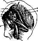
Figure 8. Location of ischiatic hernia: 1-posterior superior iliac spine; 2-gluteal artery; 3-greater sciatic notch; 4-exposed hernia sac; 5-piriformis muscle; 6-middle gluteal muscle; 7-superior gluteal nerve.
The explanation for this is found in the greater width of the female pelvis and the larger size of the openings through which hernias exit in women. Right-sided sciatic hernias were observed more frequently than left-sided ones. In general, this type of hernia was more commonly observed in middle-aged individuals, although some cases were observed in newborns and small children. Anatomical predisposition to hernia is manifested in the pre-existence of a hernial sac in the form of a diverticulum of the peritoneum and in abnormally widened openings. The difficulty in diagnosing sciatic hernias depends mainly on the size and nature of the hernia: if the hernia has a more or less significant size and is reducible, then its diagnosis is not difficult. The hernia usually descends down the thigh along the course of the sciatic nerve and the surrounding tissue. Colossal sciatic hernias have been described that descended to the middle of the thigh and even below the popliteal fossa. If the hernia is so insignificant that it cannot even be palpated, or in the case of irreducibility, its diagnosis can be very difficult. Sometimes patients experienced pain in the sciatic region, which was intensified under the influence of walking. Perineal hernia (h. perinealis). A line drawn from one ischial tuberosity to the other divides the perineum into 2 halves-anterior and posterior. The anterior part is called the urogenital area, and the posterior part is called the anal region, since the first passes the urethra in men and the urethra and vagina in women, while the second passes the anus. The urogenital septum in women is wider than in men, since the pelvis itself is wider; both aponeuroses are less pronounced. The peritoneum descends into the pelvis in the form of a sac, covering the walls of the pelvis and to a greater or lesser degree the organs located there. In men, the peritoneum, covering the anterior and lateral surfaces of the rectum, passes to the posterior surface of the bladder. Between these organs, the so-called rectovesical pouch is formed. In women, between the rectum and bladder there is the uterus, therefore the peritoneum, covering the rectum, passes to the uterus, and from there to the bladder; thus, 2 pouches are formed here-rectouterine and uterovesical. The anterior pouch always lies higher than the posterior one. The depth of the latter is subject to large fluctuations depending on both the individuality of the subject and age; sometimes the peritoneum lies so low that it forms a kind of diverticulum. Two categories of perineal hernias are distinguished: 1) anterior perineal hernias and 2) posterior ones. The boundary separating them is the linea terminalis. Due to the different structure of the perineum in men and women, hernias in them also differ. Anterior hernias in men occur only as traumatic: external violence is necessary to weaken the anterior septum; in women, however, whose urogenital septum is weaker, both anterior and posterior hernias are observed. Anterior perineal hernia in women originates from the vesicouterine pouch of the peritoneum, passes through slits between the muscles, and usually emerges in the labium majus, in its central part. Posterior perineal hernia originates in men from the rectovesical pouch of the peritoneum, in women from the rectouterine pouch, passes posterior to the interischial line through slits in the muscle that raises the anus, and emerges into the interstitial space of the ischiorectal fossa. The size of posterior hernias is much larger than anterior ones. Perineal hernias are also divided into complete and incomplete; the latter do not emerge into the subcutaneous tissue. The content of anterior hernias is most often the urinary bladder, and female genital organs were also frequently encountered. Perineal hernias are more commonly observed in women than in men. Up to 20 years of age, perineal hernia is rarely observed. Irreducible perineal hernias can present diagnostic difficulties: cases have been described where such tumors were mistaken for abscesses, an incision was made, and only opening revealed the true state of affairs. A case is described where a tumor on a stalk, located between the labia majora, was mistaken for a polyp and removed. The patient died, and on autopsy it was found that the omentum and large intestine had been subjected to resection. Prolapse of female genital organs and the rectum can also be closely associated with perineal hernia. Diaphragmatic hernia (h. diaphragmatica). Two periods are distinguished in the development of the diaphragm-the embryonic period and final formation. In the very earliest period of development of the embryo, there is only one so-called primary body cavity, which with further growth of the fetus divides into the pericardial cavity, thoracic and abdominal. The embryonic period of development of the diaphragm is divided into 2 parts: during the first, earlier part, the anterior part of the diaphragma appears, and during the second, later part, its posterior part is formed. The exact time of fusion of the two halves has not been clarified. Usov and Nau accept that it occurs on the 8th week of intrauterine life. The second, final period of development of the diaphragm usually begins on the 3-4th month of intrauterine life and continues until the end of pregnancy. Cases of complete absence of the diaphragm as well as cases of defects in both the muscular and tendinous parts of the diaphragm have been described. The diaphragma consists of 2 parts-a central tendinous and a peripheral muscular. It has a series of openings both for the passage of organs (esophagus, aorta, inferior vena cava) and free ones: in the sternal part-between muscle bundles, in the costal part-Morgagni's foramen, in the lumbar part-Bochdalek's fissure. Diaphragmatic hernias are divided into: 1) congenital, owing their origin to the arrest of development of the diaphragma; 2) acquired, penetrating through the aforementioned openings, and 3) traumatic, due to injury or rupture of the diaphragma. Diaphragmatic hernias are also divided into true ones, having a hernial sac, and false ones - without a hernial sac (see figure 9). The hernial sac may consist only of peritoneum or peritoneum fused with the underlying pleural leaflet. In some cases, the hernial sac may be the stretched and thinned part of the diaphragma, which is then called eventration of the diaphragma. Many more false than true diaphragmatic hernias have been described. Rochard, in 330 cases, gave the following table of the relative frequency of abdominal organs present in diaphragmatic hernia: stomach.......187 Large intestine ... 177 Small intestine ... 113
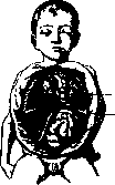
congenital hernia diaphragmatica vera syn.: 1-hernial gates in the thickness of the diaphragm; 2-hernial sac. Duodenum. Cecum . . . Stomach. Pancreas. Left kidney . . . right kidney . . . Omentum......107 Spleen...... 78 Liver........ 60 Congenital diaphragmatic hernia occurs more often on the left than on the right, while acquired hernia almost always occurs on the left. Congenital hernias occur equally often in boys and girls, while acquired hernias occur more often in men. According to the course, acute and chronic cases are distinguished; the former are very often accompanied by incarceration of the intestines. The clinical picture of diaphragmatic hernias is formed both from the subjective complaints of patients and from the data of objective examination. At present, diagnosis has been significantly facilitated by the use of X-ray examination, thanks to which a diaphragmatic hernia can be seen. Complications. Complications of hernias include coprostasis, inflammation, incarceration, contusion, and tumors. Coprostasis is observed in patients with a sluggish intestine who sometimes suffer from prolonged constipation. Often such patients, accustomed to irregularities of their intestine in general, do not pay any attention to the onset of the disease at first and miss the time for treatment. The cause of coprostasis, especially in children, can be coarse, indigestible food (often observed in famine-stricken areas). If there was sluggishness of the intestinal wall, coprostasis is more often observed in women with umbilical hernia of the large intestine, and in men-when the same intestine is located in the inguinal hernia. Coprostasis develops slowly, gradually, the hernial tumor is not particularly sensitive, the cough impulse is present (difference from incarceration), the general condition is little disturbed.-Inflammation of a hernia can occur from three sides: 1) from the side of the skin coverings (especially with umbilical hernia in obese women with untidiness of the navel), 2) from the hernial sac, 3) from the side of the organ that is in the hernia (e.g. hernial appendicitis). Sometimes tuberculous inflammation of the hernial sac is observed. Tuberculous bacilli can be carried into a hernia in three ways: 1) through blood and lymphatic vessels, 2) through the organ that is in the hernia and which primarily develops tuberculosis, 3) by the direct transition of tuberculosis from the peritoneum to the hernial sac. Tuberculosis of a hernia can be observed in three forms: 1) miliary form, 2) ulcerative, or fibro-caseous, 3) purely fibrous form. Clinically, the following forms are observed: 1) latent form (accidental finding during surgery), 2) form with painful phenomena, 3) form with acute or chronic inflammatory phenomena.- Incarceration, the most dangerous complication of a hernia, always consists of three factors: 1) violation of blood circulation in the incarcerated organ, 2) violation of the function of this organ and 3) general phenomena, expressed in one form or another depending mainly on the importance of the incarcerated organ for the body. Incarceration of the intestine should be placed in the first place. A distinction is made between 1) elastic incarceration, 2) fecal incarceration and 3) both together. The constricting ring can be the hernial gates, various places in the hernial sac, as well as other organs lying in the hernial sac (e.g. omentum, adhered to the hernial sac). It is necessary to note the so-called reverse incarceration of the intestine, which is expressed in the fact that not the intestinal loop that lies in the hernial sac is subjected to incarceration, but, on the contrary, the one that remained in the abdominal cavity (see figure 10). The presence of two intestinal loops in the hernial sac should suggest to the operator the possibility of reverse incarceration of the intestine. The variety of the patho-anatomical picture observed during incarceration depends mainly on three conditions: 1) when the inflammatory process affects only the contents of the hernial sac, 2) when the contents, hernial sac and coverings over the hernial tumor are affected, 3) when the abdominal cavity is affected. When an intestinal loop is incarcerated, the afferent segment, the incarcerated part of the intestine, the grooves from incarceration and the efferent end are distinguished (see figure 11). Usually the efferent end of the intestine suffers the least, although very serious changes are sometimes observed here also. The patho-anatomical picture of the incarcerated organ in mild cases is limited to stagnant phenomena, in moderately severe cases-to hemorrhages and in severe cases-to necrosis. Necrosis of the intestine always begins from the mucous membrane. The narrower
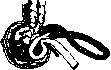
Figure 10. Reverse
incarceration of the small intestine in hernia of the cecum.
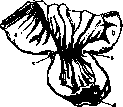
and less elastic the constricting ring, the more severely blood circulation is disrupted in the incarcerated organ and the faster its necrosis occurs. With a very narrow ring, necrosis can occur within the first 12 hours, and usually proceeds as dry incarceration, i.e. no fluid (hernial water) is observed in the hernial sac, as is usually the case. Acute, subacute, chronic and latent forms of incarceration are distinguished. The latter form is the most dangerous, because, proceeding with the patient's good general condition, it misleads not only the patient but also the doctor. This form is often given by cases of incarceration of the intestinal wall (parietal hernias). The chronic form of incarceration is observed with huge hernias with wide hernial gates and with irreducible hernias, when the large intestine is most often subjected to incarceration.- Symptoms occurring during incarceration can be divided into 3 groups: 1) general Fig
Incarceration
phenomena (FEVER, INCREASED temperature, DISORDERS of the digestive tract), 2) local phenomena (pain, tension, swelling, vomiting, cessation of peristalsis, inability to reduce the hernia), 3) phenomena from the side of the incarcerated organ (for intestinal obstruction). In the case of incarceration of the small intestine: a-afferent segment; b-efferent; cdc-incarcerated
1) general phenomena (fever, collapse of the loop; disorders of the urinary apparatus, prostration, collapse); 2) local phenomena (increase in the hernial tumor, tension, soreness, absence of cough impulse); 3) complications (peritonitis, fecal phlegmon of the coverings of the hernial tumor). The general rule that a physician must follow when observing a patient with signs of intestinal incarceration is reduced to three basic principles: 1) it is less dangerous in terms of prognosis for the patient to assume incarceration where it does not exist (it is worse to overlook incarceration or to mistake it for some other ailment, especially if the method of treatment will be different); 2) if the diagnosis of hernia incarceration remains in doubt, it is necessary to lean more toward incarceration than against it; 3) it is necessary each time in ileus to carefully investigate all the usual places of hernia appearance. In establishing the diagnosis of hernia incarceration, carefully collected anamnestic data are of great importance. In recognizing incarceration, two circumstances may be encountered: 1) when there is incarceration of an organ, but the signs do not fully correspond to the acute form of incarceration, 2) when there are symptoms of intestinal incarceration, but in reality there is none. The first group can be divided into the following sections. I. Cases where the peculiarities of symptoms depend either on the intestines themselves that have been subjected to incarceration, or on changes in the intestines located in the hernial sac. These include: 1) partial incarceration of intestines: a) incarceration of the intestinal wall, b) incarceration of the vermiform appendix of the cecum, c) incarceration of Meckel's diverticulum, d) incarceration of fatty appendages of the large intestine. 2) Incarcerations within the hernial sac: a) incarceration by a bridge, adhesions, etc., located inside the hernial sac, b) new growths of the hernia, c) bending, twisting of the intestine inside the hernial sac. II. Cases where the peculiarities depend on the hernial sac: a) multicameral hernial sacs, b) hernial sacs with a diverticulum. III. Cases where the peculiarities consist of multiple hernias: a) one irreducible, the other incarcerated, b) one hernia hides another, c) inguinal and femoral hernia on the same side, of which one is incarcerated and the other is not. IV. Cases where the peculiarities consist in the fact that the intestine is subjected to incarceration inside the abdomen (internal intestinal incarceration), while the patient suffers from an external hernia. V. Cases where the peculiarities consist in the fact that at the same time inflammation and incarceration of an organ occur: a) incarceration and inflammation of the vermiform appendix of the cecum, b) incarceration and inflammation of the appendages of the uterus. The second group of cases can be divided into two subgroups: I. When the hernia is not in a normal state: 1) incarceration of the omentum, 2) coprostasis of the hernia, 3) inflammation of the hernia, 4) incarceration in the hernia of other organs besides the intestine. II. Hernia in a normal state: 1) inflammation of lymphatic glands (subperitoneal) at the sites of the hernia, 2) inflammation of the testicle; its appendage and spermatic cord in inguinal hernia, 3) inflammation of the peritoneum, 4) vomiting in pregnant women, 5) phlegmon near the hernial sac. It should be noted that in incarceration of the omentum and female genital organs, all the symptoms of intestinal incarceration may be present, and only surgery sometimes clarifies the matter. Injuries to hernias. Injuries to hernias are divided into 2 categories: so-called spontaneous ruptures and traumatic injuries. Injury to a hernia can occur under the following conditions: 1) when rupture of the intestine occurs in free hernias under the influence of only a sharp increase in intra-abdominal pressure, 2) when an organ in the hernia ruptures from bruises of the abdominal wall far from the hernia, 3) when rupture of the contents of the hernia occurs from direct trauma to the hernia. The hernial sac and the external coverings of the hernia can rupture as if spontaneously under the influence of all sorts of pathological processes in them, which thin them out and deprive them of the normal elasticity of the tissues. When a hernia is injured, there can be two kinds of complications: 1) inflammation of the peritoneum, 2) bleeding. Sad consequences from either can be avoided only by early surgery. - New growths of hernias. Distinguish: 1) new growths of the contents of the hernia, 2) new growths of the hernial sac, 3) new growths of tissues and organs adjacent to the sac. New growths can be both benign and malignant.
Abdominal wall. Treatment of hernias. Strangulated hernias. In strangulation of a hernia, immediate intervention is necessary, either bloodless or surgical. In the vast majority of cases, surgical intervention should be preferred. Manual reduction of strangulated hernias can be applied somewhat more broadly in children, since in young age, with great elasticity of tissues and their high vitality, the matter does not quickly lead to necrosis of the hernia contents. One argument in favor of attempting reduction in children is that the existing hernial protrusion in early age may disappear with the child's growth due to changes in anatomical relationships within the abdominal cavity and in the structure of the inguinal canal. But even in children, the delay of surgical intervention should be limited to a few hours, and manual manipulations during reduction must be very delicate. Attempts at reduction of strangulated hernias in adults are permissible with severe changes in the cardiovascular system and lungs, which worsen the prognosis for any kind of surgical intervention. But even under such conditions, bloodless reduction is permissible only in the first hours after the onset of strangulation. Surgical intervention becomes increasingly necessary as the time since strangulation increases, especially after failed attempts at reduction. An operation performed in the initial stage of strangulation has great advantages compared to bloodless reduction. The advantages consist in that after examining the contents of the hernia and incising the constricting ring, reduction is performed with greater safety than with blind bloodless reduction. The operation makes it possible to help with the changes that have occurred in the prolapsed organs due to strangulation; and finally, the operation can be completed with measures for radical cure of the hernia. In late stages of strangulation, when symptoms of beginning necrosis of the hernia contents and inflammation of the tissues surrounding the hernia sac with signs of general intoxication appear, attempts at reduction become impermissible, and surgical intervention becomes absolutely necessary. For bloodless reduction, the following conditions must be observed: reduction should not be prolonged, should be cautious, without applying great force, and should immediately give way to surgical intervention in case of failure. Various auxiliary means have been proposed to facilitate reduction of hernias, with which one tries to relax the tension of the abdominal wall muscles and the constricting ring. First of all, it is necessary to empty the bladder and clear the intestines with enemas. Placing the patient in a warm bath helps reduction; at the same time, subcutaneous injection of morphine or atropine is prescribed. To reduce the volume of the hernia and partly to stimulate peristalsis, cold is used, most often in the form of ether application. For this, on the hernial protrusion, protecting the surrounding tissues with vaseline, 10 cm3 of ether is poured every 10 minutes. During manual reduction, the patient is placed in a position with the pelvis elevated and legs bent toward the abdomen (also to relax the abdominal walls). In this position, a certain traction is created from the abdominal cavity in relation to the prolapsed organs. In children, this position is replaced by hanging the child upside down, holding by the legs. Often in children, in this position, the hernia reduces without any other manipulations. Manual reduction is performed as follows: the fingers of the left hand are placed near the hernial opening to prevent displacement of the hernia contents to the side; with the right hand, uniform pressure is applied to the hernial tumor in the direction of the hernial canal. Reduction is easier if it is possible to start with the diverting segment of the prolapsed intestinal loop. In the diverting loop, pathological changes in the wall and narrowing of the lumen are less pronounced, so it is easier to remove intestinal contents from this segment, which leads to a decrease in tension and volume of the prolapsed intestine. In inguinal hernias, the diverting segment of the intestine is most often located at the back, and patients usually start reduction from the back. They apparently come to this order of reduction of intestines on the basis of experience. If reduction is proceeding successfully, the hernial tumor gradually decreases as segments of intestine with characteristic gurgling retreat into the abdominal cavity. Pulling on the tumor forward and to the side, digital expansion of the constricting ring (which is sometimes done for reduction) should be considered dangerous, as this can lead to rupture of the overstretched intestine with altered walls. After reduction, it is necessary to examine the hernial gates, and the hernia sac should be empty and lie in front of the hernial opening, and this opening should be freely passable. The general condition of the patient after successful reduction should improve rapidly. When reducing strangulated hernias, one must always keep in mind the serious complications that can occur during reduction. First of all, it should be noted that the process of necrosis of strangulated organs can occur with different speed, which is very difficult to account for. It can be noted that different parts of the intestine, due to differences in blood supply, necrotize faster or slower. The vitality of the segment closest to the mesentery is preserved longer, while the wall of the intestine opposite the mesentery dies more quickly. Therefore, in cases of parietal strangulation, gangrene of the intestine occurs especially quickly. The smaller the hernia, the narrower and less yielding the hernial ring, the sooner gangrene occurs. Therefore, strangulations of femoral hernias are especially dangerous. Sometimes, with large hernias, twisting of the strangulated loop can occur, and after reduction, symptoms of intestinal volvulus may develop. It should be kept in mind that manipulations with strangulated intestines can lead to bruises of their walls, on which necrotic areas can subsequently form, followed by perforation and peritonitis. In the intestinal wall, corresponding to the constriction ring, scar strictures can subsequently form, often requiring surgical intervention. In addition to the mentioned complications, one should remember the possibility of so-called false reduction. Under this name, individual cases are described where during reduction not only was the strangulation of the hernia not eliminated, but complications were added that worsened the condition. With complex hernia sacs (two-chambered with diverticula), displacement of the sac contents from the superficial part to the deeper part is possible. The entire hernia sac can be displaced into the layered preperitoneal tissue. Rupture of the hernia sac and escape of its contents into the surrounding tissues is possible. If this rupture occurs near the neck of the sac, its contents may end up in the preperitoneal tissue. Individual cases of very severe injuries are described when the constricting ring tore away from the hernia sac and even simultaneously from the surrounding tissues, and the contents of the hernia, still strangulated, moved into the free abdominal cavity. These circumstances require keeping the patient under observation for the next few hours after reduction. If the general condition and symptoms from the abdominal cavity do not improve, it becomes necessary to proceed with an operation to eliminate the possible complications described above. Surgical treatment of strangulated hernias consists in exposing the hernia sac and, after opening it, examining its contents, after which the constricting ring is incised. The hernia sac should not be opened too close to its neck, as in this place during strangulation, intestinal loops are often closely adhered to the wall. The constricting ring should be incised after examining the contents of the hernia sac so as not to allow gangrenous loops of intestine into the abdominal cavity. The ring can be incised either with a special knife (herniotome) from the inside or from the outside along a grooved probe. Subsequently, the operation is planned differently depending on the condition of the strangulated organs. If deep nutritional disorders have not yet occurred, the contents of the sac are reduced into the abdominal cavity and a radical operation for the hernia is performed. In the presence of gangrene of the intestinal wall, various methods of surgical assistance are possible. If the patient's general condition is satisfactory, resection of the affected segment of intestine should be performed, followed by a radical operation. But very often the patient's general condition is so severe that the surgical intervention must be as short and simple as possible. With long-existing strangulation, the tissues surrounding the hernia sac may be inflamed, infiltrated with purulent, ichorous fluid. Due to this, in a typical operation, it is extremely difficult to protect the free abdominal cavity from infection. For such hernias, various methods of surgical assistance have been proposed, and there are still debates about the advantages of individual methods. In particularly severe phlegmonous hernias, when the patient is in collapse, a proposal has been made (Bobrov) to limit oneself to a simple incision of the hernial tumor.
In cases of recovery after such intervention, the gangrenous portions of the hernia's contents slough off in the coming days, and an anus praeternaturalis forms at the site of incarceration, which is subsequently closed by appropriate measures. Such an operation does not always create conditions for good emptying of the intestinal loop, therefore in these cases some surgeons prefer to create an anus praeternaturalis outside the area of incarceration, through a separate incision in the abdominal wall. After the patient recovers, freed from intestinal intoxication, an operation is undertaken to remove the incarcerated loop of intestine. Subsequently, staged surgical closure of the initially created ani praetern. is performed. Proponents of primary resection emphasize the unfavorable aspects of the aforementioned methods. A simple incision of a phlegmonous hernia does not eliminate the incarceration; the creation of an ani praetern. necessitates a series of operations later to eliminate the incarceration and the ani praetern. Each of these operations in turn presents a danger, increasing the overall mortality rate. Statistical figures speak in favor of the benefits of primary resection. According to Shilovtsev's statistics, out of 408 cases of resections by Russian authors, there was 45% mortality, and out of 117 cases of ani praetern., 76% mortality, for a total of 525 cases with 52% mortality. It should be thought that these figures indicate not so much the advantages of primary resection as that cases of varying severity are operated on by one method or another. And of course, among resections there are more mild cases, while ani praetern. is most often applied in very severe general condition. Each surgical method has its indications, and despite a preference for one method or another, each surgeon in individual cases must apply both plans of surgical treatment. For some incarcerated gangrenous hernias (umbilical, ventral, and femoral hernia in women), Grekov recommends en bloc resection according to the type of resection of malignant tumors. The operation consists of the following. Retreating 1-2 fingers from the hernial tumor, within healthy tissues, an oval incision is made outlining the hernial tumor. The incision penetrates to the aponeurosis. Then the aponeurosis is circumferentially incised around the hernial pedicle, the peritoneum is opened, and under the guidance of a finger the incision is continued all the way around. Thus the entire hernial tumor, without opening it, is separated from the abdominal wall and remains connected to the body only by its pedicle, containing the intestine and part of the omentum. The omentum and intestine are resected, the ends of the intestine are joined, and the incision in the abdominal wall, depending on the circumstances, can be completely closed. Free hernias. Treatment of free femoral hernias can be conservative or surgical. Conservative treatment consists of wearing a well-fitted truss. A truss can contribute to the cure of femoral hernias only in children. By holding the hernia, the truss leaves the unobliterated processus vaginalis empty and thus eliminates the obstacle to its obliteration. At the same time in children it is necessary to eliminate everything that increases intra-abdominal pressure (phimosis, intestinal disorders, coughing, etc.). It is permissible to prescribe a truss in the case when the child's parents do not agree to an operation. In adults a truss does not lead to a cure, but it cannot be dispensed with in cases when an operation cannot be performed, for example, in very depleted diabetics, severe cardiac patients, etc., or in the case when patients do not agree to an operation. In view of the great danger associated with surgical interventions in the pre-antiseptic era, methods were previously proposed for closing the hernial orifice and obliterating the hernial sac by artificially induced tissue irritation. For this purpose, tincture of iodine, concentrated solution of table salt, 70% alcohol, paraffin were injected into the circumference of the hernial orifice and into the hernial sac. At present, for the treatment of femoral hernias, several dozen operative methods have been proposed, and to this day reports continue to appear of ever new modifications. Such abundance of methods indicates that the methods proposed up to now do not cover all variations of the anatomical peculiarities of the femoral area of hernia carriers and do not eliminate the peculiarities of the pathogenesis of hernias. At present, surgical treatment of hernias is limited to action on the femoral area. It should be thought that failures in treatment are explained in part by the imperfection of the methods used to strengthen the walls of the femoral canal, and in part by the impossibility of eliminating those causes of hernia formation that lie outside the femoral canal and depend on the nature of labor, living conditions, age-related changes in the body's tissues, and constitutional peculiarities. The numerous methods of surgical treatment of femoral hernias can be divided into several groups. First of all, mention should be made of the methods proposed at the end of the 19th century and that did not become widespread: suturing the internal opening of the femoral canal from the abdominal cavity side (Lawson Tait, Assaky). At present the methods of Czerny, Kocher, Roux, Girard, Bassini, and Bobrov are most widely used. The course of the operation at the beginning is almost the same in all the aforementioned methods. With an incision parallel to the femoral ligament, the aponeurosis of the external oblique muscle is exposed. In the lower part of the wound, the hernial sac is exposed by layer-by-layer incision of its coverings until the avascular wall of the sac itself appears. The sac is isolated from the surrounding tissues and opened. If there are adhesions of the sac wall to the omentum or intestine, they are incised. After this, the contents of the sac are returned to the abdominal cavity, the neck of the sac is closed with a suture or ligated, and the sac is excised. When isolating the neck from the medial and posterior sides, especially with direct hernias, care must be taken not to injure the urinary bladder. When incising the hernial sac, caution is also necessary, because on the one hand, in hernias from slipping, the lateral wall of the sac may be replaced by part of the large intestine or its mesentery (see Figure 12); in bladder hernias, the medial part of the wall may be occupied by the wall of the urinary bladder. These peculiarities should be suspected when the wall of the sac is not of uniform thickness everywhere. In such cases, the incision of the sac should be made away from these thickenings. Upon discovering these peculiarities, it is better to replace ligation of the neck with suturing it from the inside, so as to avoid the possibility of catching the vessels of the mesentery or the wall of the urinary bladder with the ligature. When isolating the distal part of the sac, especially with large old hernias,
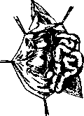
Figure 12. Right-sided sliding hernia.
here it should be cut through and the peritoneal end closed with a suture. The peculiarities of the various methods of herniotomy consist in the further course of the operation. Czerny proposed after excision of the hernial sac to place three to four sutures on the external opening of the femoral canal in order to narrow it. Roux recommended after excision of the sac to narrow the femoral canal by placing sutures that would capture not only the aponeurosis but also the muscles. Thus all layers of the abdominal wall are pulled up to the femoral ligament. Kocher, strengthening the wall of the femoral canal, just as in Roux's method, indicated that it is necessary 1) to make the direction of the femoral canal oblique and 2) to eliminate the funnel-shaped protrusion of the peritoneum in the
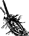
}'/%
Figure 13. Operation for femoral hernia according to Kocher. Suturing the exposed hernial sac.
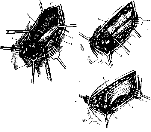
Figure 14. Operation for femoral hernia according to Girard: 1 and 2 - internal and external laminae of the aponeurosis of the external oblique muscle; 3 - tunica vaginalis; 4 - Poupart's ligament; 5 - m. obliquus abdominis internus.
difficulties may be encountered due to dense adhesions of the sac with the surrounding tissues, in particular with the elements of the spermatic cord. Separation of these adhesions leads to disruption of the integrity of numerous small vessels and to injury of the spermatic cord, and after the operation extensive hematomas and infiltrates are observed in the scrotum. To avoid this, considering the isolation of the distal portion of the sac inadvisable, it is proposed (Razumovsky, Anschutz) not to isolate the hernial sac, but to isolate it only at the neck of the internal opening of the inguinal canal. For this purpose, Kocher proposed the following: the hernial sac is not excised; through the aponeurosis of the external oblique muscle and the muscular wall outward from the internal opening of the inguinal canal, a forceps is passed in the direction of the external opening of the inguinal canal; the tip of the isolated hernial sac is grasped with the forceps, pulled through the made opening in the muscles and aponeurosis, and fixed with sutures; the excess of the sac is excised (see Figure 13). Bobrov proposed by incising the aponeurosis of the external oblique muscle to open the inguinal canal and cleanse it of excess fatty tissue. After excising the hernial sac as high as possible, the inguinal canal is closed, with all layers of the abdominal wall being sutured to the inguinal ligament. Girard modified this method in that after suturing the inner edge of the inguinal triangle to the inguinal ligament, he proposed doubling the aponeurosis of the external oblique muscle, placing a sheet of aponeurosis from the inguinal ligament onto the one that covers the sutured muscles (see Figure 14). In Bassini's method, until the moment of excision of the hernial sac, the operation is carried out in the same way as in Bobrov's method. Further, the spermatic cord is elevated and displaced to the side. The muscles (internal oblique and transverse) are sutured to the inguinal ligament under the spermatic cord. The spermatic cord is placed on the sutured muscles and over it the incised aponeurosis is sutured (see Figure 15). To the present time, a considerable number of observations have accumulated in the practice of large surgical departments, which indicate that none of the methods of herniotomy guarantees against recurrences and the number of recurrences is almost the same with any of the common methods of operation, amounting to approximately 10% in indirect H. (3.5-16%) and rising to 26% in direct ones. It was stated above that the pathogenesis of H. is very complex and is characterized, in addition to features in the structure of the inguinal canal, by constitutional features of the organism, working and living conditions. Therefore, by local action on the inguinal canal, it is not always possible to guarantee against recurrence. Nevertheless, the number of recurrences can be partly reduced if one operates not by template, but by analyzing the peculiarities of each individual case. It is difficult to take into account all possible variants in the structure of the inguinal canal of hernia carriers, but still some landmarks can be outlined in choosing one or another method of operation for various H. In H. in children, the non-closure of the processus vaginalis is of primary importance. The walls of the inguinal canal continue to form during the period of growth, just as the anatomical features inside the abdominal cavity and in the structure of the abdominal wall in general change. Therefore, in herniotomy in children, one can limit oneself to closing the stump of the excised neck of the hernial sac. It is possible to add to this the suturing of the external opening of the inguinal canal. In particular, displacement of the spermatic cord should not be done, since in young age it is easy to injure it, which can cause a disturbance in the nutrition of the testicle with subsequent atrophy. In adults, when indirect inguinal H. occur, in most cases the weakness and fraying of the aponeurosis of the external oblique muscle are primarily noted. The defect in the posterior wall of the inguinal canal is initially small, slit-like. Only in the later period does the posterior wall of the inguinal canal suffer, and the indirect H. becomes similar to the direct one (Sozon-Yaroshevich). Thus, in the initial stages of indirect H., with a strong posterior wall of the inguinal canal, the surgical intervention should be directed at strengthening the anterior wall. For this purpose, the methods of Roux, Girard are applicable. In direct H., primarily the strength of the posterior wall of the inguinal canal is lost. At the same time, flabbiness
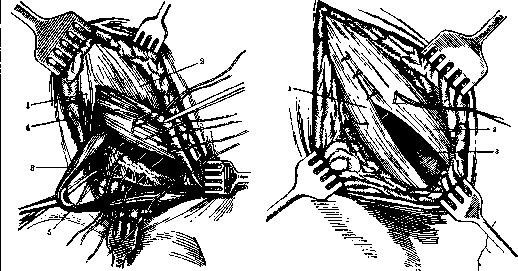
Figure 15. Operation for inguinal hernia according to Bassini: 1 and 2-outer and inner sheets of the aponeurosis of the external oblique muscle; 3-funiculus spermaticus; 4-musculus obliquus abdominis internus; 5-Poupart's ligament.
and insufficiency of muscles in the high inguinal triangle. Consequently, in these cases, efforts should be directed at strengthening the posterior wall. For this purpose, the Bassini operation was widely used until recently, in which, by suturing the muscles to the inguinal ligament with displacement of the cord, it was hoped to create a strong posterior wall from muscles. But observations during repeat operations and experiments on animals (Seelig and K. S. Chouke, Khesin) showed that muscles sutured to the inguinal ligament lose their contractile properties and turn into scar tissue. And the higher the inguinal triangle, the greater the distance between the edge of the muscles and the inguinal ligament, the sooner one can expect the muscle to turn into a scar and the stronger this scar will be stretched. Mobilization of the muscles of the inguinal triangle by incision of the anterior wall of the rectus muscle sheath according to Venglovsky not only does not resolve the difficulties but also promotes the disintegration of muscle fibers. Brenner proposed strengthening the posterior wall by suturing the m. cremaster with the m. obliquus int. and below with the m. rectus. Schwartz recommended taking a flap from the m. rectus with the base below, Sauerbruch-the same, with the base above. One edge of the muscle is sutured to the internal oblique muscle, the other edge-to the inguinal ligament. For the same purposes, the sartorius muscle was also used. In individual cases (Trendelenburg, Kraske), a bone-periosteal flap from the pubic bone was transplanted. Vreden proposes to move a flap from the anterior sheath of the rectus muscle, suturing it to the inguinal ligament. All these methods and their numerous modifications have not become widespread partly because of their complexity, partly because of their unreliability, since transplanted muscles soon turn into scar tissue. Thus, the problem of recreating the posterior wall of the inguinal canal in direct H. still remains unsolved. Therefore, in operations for direct H., one has to use the same methods as for indirect H. Theoretically, some advantage must be recognized for Kocher's method, since with it it is possible to make the direction of the inguinal canal more oblique. The aforementioned experiments (Seelig's and Chouke's) showed that strong adhesions are obtained only between homogeneous tissues (fasciae and aponeuroses). The only indisputable fact in the treatment of hernias remains the high amputation of the sac and aponeurotic plastic surgery (Martynov). Therefore, Martynov sutures the inguinal canal in such a way that the medial (upper) flap of the divided aponeurosis of the external oblique muscle is sutured to the inguinal ligament, and over this the lateral (lower) flap of the same aponeurosis is strengthened. As material for sutures, some, like Kocher, Duplay and many others, stand for silk, others-for catgut. Some apply autoplastic sutures from aponeurosis. Some recurrences should be attributed to an irrational regimen in the immediate postoperative period. Nowadays no one recommends wearing a truss in the postoperative period. It is necessary in the next 2 months to avoid great strains of the abdominal press, as this can lead to stretching of the line of suture. On the other hand, long physical rest can lead to a weakening of the vital activity of tissues from inactivity. For these reasons, at present, a progressive scale of muscle exercises is proposed for the early resumption of the functions of the muscle-aponeurotic wall (Gorinevskaya). Without being able to influence the constitutional peculiarities in the body of hernia carriers, in the postoperative period, in addition to caring for the healing of the wound, it is necessary to eliminate everything that leads to rapid wear of muscles and aponeuroses in order to prevent recurrence. For this, it is necessary, by rationally dosing physical labor, to eliminate its excess. At the same time, it is necessary to free the patient from chronic intoxications, first of all from alcohol, and to improve nutrition. Femoral H. In those cases when surgical treatment of femoral hernia cannot be performed, a truss is prescribed, which differs from the truss for inguinal H. by a shorter, strongly curved downward neck and a more rounded pelotte. It is more difficult to fit a truss than in inguinal H., as it is more easily displaced, especially in obese women, and due to the proximity of large vessels it can lead to circulatory disorders in the leg. In the operation for strangulation, it should be remembered that the hernial sac, enveloped in fat, is usually surrounded by very thin membranes, and when it is incised, the contents of the sac can be injured more easily than in inguinal H. Further, sometimes the a. obturatoria does not pass along the lateral edge of the femoral ring but along the upper and inner. In this case, the hernial gates are surrounded by vessels on almost all sides, and their dissection from the inside in strangulation of H. can be complicated by profuse bleeding. For radical surgical treatment, very many methods have been proposed; despite this, in large H. after operations, recurrences are often observed (up to 15%, according to Herzen). An indispensable condition for the success of the operation is the high dissection and ligation of the neck of the sac, so as not to leave a protrusion of the peritoneum in the area of the femoral ring. When dissecting the neck, one should remember the proximity of the wall of the urinary bladder, which often protrudes into the field of operation and can be damaged. After removal of the sac, various methods are used to close the femoral ring. The simplest is suturing the fasciae pectineae to the inguinal ligament (see fig. 16). To eliminate tension in the sutures, it is recommended to make an incision in the fasc. pectinea below the line of sutures (Martynov). Kocher proposed to make a skin incision above the inguinal ligament. After dissection of the sac, in the aponeurosis of the external oblique muscle, near its lateral leg, a small incision is made, through which a forceps is passed behind the inguinal ligament; the hernial sac is grasped by its apex and pulled through the opening in the aponeurosis, where it is fixed. The excess of the sac is excised. The hernial opening

Figure 16. Radical operation for right-sided femoral hernia. After ligation and amputation of the hernial sac, the femoral vein is pulled outward with a blunt hook and silk threads are placed on the Poupart ligament and Cooper's ligament with the pectineal fascia.
is closed by sutures connecting fasc. pectinea with the inguinal ligament. In many modifications, suturing the inguinal ligament to Cooper's (resp. to the periosteum of the pubic bone) has been proposed. This is mainly what the Bassini operation amounts to, which is conducted as follows: by an incision below and parallel to the inguinal ligament with dissection of the fasc. cribrosae, the hernial sac is exposed. After the usual removal of the sac, the inguinal ligament is sutured to Cooper's, and the semilunar edge of the oval fossa-to the aponeurosis of the pectineal muscle. Individual surgeons noted that the inguinal ligament, sutured to Cooper's, subsequently separates from it, and in the area of the femoral ring, an easily stretchable scar tissue is formed. In subsequent modifications, they sought either to eliminate tension from the inguinal ligament or to make the fusion with the pubic bone more durable. Fabricius proposed to separate the inguinal ligament from the symphysis pubis before suturing. Rou and Pauchet proposed to nail the inguinal ligament to the pubic bone with U-shaped staples. Herzen proposed to drill the horizontal part of the pubic bone and through these holes to pull the inguinal ligament to the pubic bone with metal sutures. For the same purposes, Cavazzani proposed to pass sutures not through the bone but under the branch of the pubic bone, at the upper border of the obturator foramen. A number of authors tried to close the femoral ring plastically with various tissues. Thus, Trendelenburg proposed to use a periosteal-bony flap from the pubic bone, Chaput transplanted cartilage from a rib; the femoral ring was also closed by suturing the sartorius muscle to the inguinal ligament, Polya inserted this muscle into the femoral ring itself. The pectineal muscle was used for the same purposes by Lannelongue and Brenner. Prokunin also developed a method of closing the femoral ring with the pectineal muscle, which he passed through the femoral ring and fixed to the aponeurosis of the external oblique muscle above the inguinal ligament.
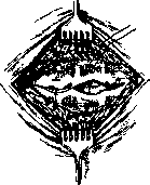
Figure 17. Operation for umbilical hernia according to Mayo's method. Aponeurosis tissue.
Usually, with these methods of plastic surgery, the muscular tissue soon regenerated into scar tissue, and the matter came down to the formation of a scar closure of the femoral ring of greater or lesser strength. The multitude of methods indicates the unsatisfactoriness of individual modifications for all cases. Obviously, in treatment, it is necessary to vary the plan of surgical intervention depending on the age of the patients, the size of the hernial opening, and the resistance of the tissues. It should be taken into account that the need for heavy physical labor with great strain on the abdominal press in the first period after the operation can cause a recurrence. Therefore, a long rest (up to 2 months) after the operation should be recommended. Umbilical Hernias. The treatment of umbilical hernias can be either conservative or radical. The conservative method is accompanied by success only in children's umbilical hernias and consists in wearing a bandage made of adhesive plaster. For this, after reducing the contents of the hernia in a horizontal position, a coin wrapped in cotton or a piece of cardboard is placed on the umbilical ring and this covering is attached with adhesive plaster. It is not advisable to use hemispherical pads as a bolster, which, pressing into the umbilical ring, not only do not contribute to its closure, but lead to its expansion. Even better, making 2 parallel folds in the vertical direction near the navel, connect them with a strip of adhesive plaster (leukoplast) stretched across the entire surface of the abdomen; then a strip of plaster is applied in a vertical direction and finally two strips crosswise. Thus, a star is obtained. The bandage is changed after 10 days. The use of trusses in adults is pointless and difficult, since it is very difficult to secure a truss on a flabby wall without great compression of the abdomen. Surgical treatment consists in exposing and removing the hernial sac and suturing the defect in the abdominal wall. In typical cases, the operation is performed as follows (Mayo's method). With two semi-oval transverse incisions to the aponeurosis, the hernial protrusion is surrounded. The aponeurosis around the hernial sac is dissected for 4-5 cm in all directions. The hernial sac is opened and, after eliminating possible adhesions with abdominal organs, it is removed. The hernial opening is expanded in both directions to the peritoneum. Suture on the peritoneum. P-shaped sutures are applied to the aponeurotic flaps in such a way that the lower flap lies under the upper one. Additional sutures on the edge of the upper flap (see Figure 17). With small hernias, the opening is closed with a purse-string suture, over which several sutures narrow the white line (Lexer, see Figure 18). With large hernias, a more complex method of closing the abdominal cavity is proposed (Sgaraser). After processing the hernial sac, the anterior leaf of the rectus muscle sheath is transversely incised on both sides; the muscles are bluntly separated from the posterior leaf. Then the peritoneum is longitudinally sutured together with the posterior leaf of the rectus muscle sheath; similarly
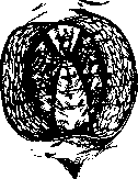
pu-
Figure 18. Operation for an umbilical hernia by Lexer's method. After circularly suturing the umbilical ring with silk sutures, the rectus muscles are approximated above and below the navel. The peritoneum and the posterior leaf of the rectus muscle sheath are sutured longitudinally in a second layer. Then the anterior leaves of the rectus muscle sheath are sutured transversely. Large umbilical hernias, especially in multiparous women, are often combined with stretching of the white line and general flabbiness of the abdominal wall. Extensive, often multi-chambered hernial sacs contain, in addition to the omentum, intestinal loops, often densely fused to the wall of the sac. Recurrences after operations in such cases are very frequent (up to 40%, according to Eiselsberg). The desire to increase the resistance of the white line led to the proposal of muscle plastic surgery (Dauriac, Volkovich, Salishchev, etc.). These proposals did not find supporters. The methods of aponeurotic plastic surgery (Sapezhko and others) are more popular. Martynov uses the following operation for this purpose. After excising the excess skin, the aponeurosis of the rectus muscles is exposed to the right and left. An incision is made along the entire length of the wound, running approximately 1-2 cm toward the middle from the edge of the left rectus muscle and opening the abdominal cavity. Thus, two unequal peritoneo-aponeurotic flaps are obtained. The edges of the aponeurosis on the right and left are strongly pulled to the side, - then the edges of the rectus muscles protrude. A series of sutures are applied to the edges of the muscles, the muscles are brought together to contact, but without opening their sheaths. The free edge of the aponeurosis of the right rectus muscle is applied from the front and sutured along its entire length to the anterior surface of the aponeurosis of the left muscle (see Figure 19).-Requires separate mention is the operation for embryonic umbilical hernias.* In those cases where the embryonic defect in the abdominal wall is replaced by a translucent plate with remnants of the amnion on the outer surface, surgical intervention must be urgent immediately after birth, as the indicated plate quickly necrotizes. The operation consists in removing the embryonic plate and closing the defect by layered suture of the abdominal wall. With large defects, due to the impossibility of pulling the muscles, it is necessary to ogra
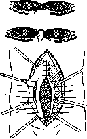
Figure 19. Modified Sapezhko's method.
by suturing the peritoneum and skin. The immediate postoperative mortality in such children is very high (36%, according to Perrin). Hernias of the linea alba are operated on by the same methods as umbilical hernias. In view of the usually small size of these hernias, the Mayo-type operation (see above) is applicable for most cases. As for the postoperative regimen and prevention of recurrences after operations, everything that was said regarding inguinal hernias should be kept in mind for umbilical hernias and hernias of the linea alba. Postoperative hernias, hernias of Spiegel's line, hernias of the rectus sheath, and lumbar hernias (in Petit's triangle or Lesgaft's triangle) require either wearing special bandages or surgical intervention. The operation consists in the layered suturing of the individual layers of the abdominal wall. Hernias of postoperative scars, due to the variety of their forms depending on their position and size, require a non-typical plan of operation in individual cases. In extensive hernias of the median line, it is sometimes necessary to use an aponeurotic graft to reconstruct the linea alba by the methods mentioned above. In extensive defects in the muscular wall, muscular plastic surgery or transplantation of aponeurosis (with fascia lata) may also be needed. Practically, it is important not to undertake radical surgery soon after the formation of postoperative hernias. It is more advantageous to wait (preferably not less than a year), counting on the fact that the infection in the scar, which caused the hernia, will be eliminated during this time. When operating at early stages, there is always the danger of opening a latent infection and, after getting suppuration in the wound postoperatively, not having a guarantee against recurrence. -3 apiratory hernia. In view of the difficulty of recognition and rarity of these hernias, operations were performed mainly when they were strangulated. The usual approach is through laparotomy. This approach is rational also because it is not always possible before the operation with certainty to determine the cause of ileus. The release of the intestinal loop from the constricting ring should be done with great care and under direct vision, since with these hernias, due to the narrowness and rigidity of the constricting ring, gangrene of the strangulated loop occurs very quickly. The incision of the obturator membrane should be made medially, since vessels and nerves are located laterally and downward. The closure of the hernial orifice is accomplished by suturing into it a flap of the pectineus muscle. - Ischiatic hernia. In view of the extreme rarity of these hernias, their recognition and surgical treatment were performed mainly when they were strangulated. The operation presents great difficulties, since one has to go through powerful layers of muscles and adipose tissue to reach the hernial orifice. The skin incision according to Wullstein is made between the ischial tuberosity and the coccyx. The operation in the area of the hernial orifice should be carried out carefully, since large vessels and nerves pass through this area: in hernia above the piriformis muscle - art. et n. glut. super., in hernia below the piriformis muscle - art. glut. infer., art. pudenda commun., and n. ischiadicus. To close the hernial orifice, either transplantation of fascia (in hernia above the piriformis muscle according to Wilms) or suturing the piriformis muscle with the sacro-tuberosum ligament (in hernia below the piriformis muscle according to Doberauer) is used. Recurrences are often observed after operations. - Perineal hernias. The operation for these hernias consists in suturing the pelvic floor muscles after removal of the hernial sac. In large defects, muscular plastic surgery with the gluteus maximus muscle is sometimes used. - Diaphragmatic hernias. For all diaphragmatic hernias, surgical treatment is necessary due to the possibility of strangulation. Individual surgeons indicate different approaches to the diaphragm for hernias. Sauerbruch and Seidel consider it rational to go from the thoracic cavity side, making an incision along the 7th intercostal space. Finsterer, Marwedel, and others propose to go from the abdominal cavity side, for which an incision is made from the xiphoid process to the midaxillary line along the edge of the costal arch. The skin incision is continued upward for 6 cm, and through this incision the VI-IX ribs are transected extrapleurally. When the upper flap is turned back, good access to the diaphragm is created. Kirschner proposes to open both the thoracic and abdominal cavities with one incision. Kirschner's technique creates great convenience when operating for strangulated hernia. Various methods are used to close the defect in the diaphragm. Most commonly, simple joining of the edges of the defect with sutures after their freshening is applied. To facilitate the application of sutures, Sauerbruch suggests blocking the phrenic nerve on the neck by injecting novocaine. In some cases, adjacent organs (liver, spleen, lung) were sutured into the defect for the purpose of closing it. Sometimes closure by plastic surgery with muscles (iliopsoas for posterior defects, muscles of the anterior abdominal wall for anterior defects) or free transplantation of fascial sheets was used. The immediate postoperative mortality remains high: 15% for free hernias and 73% for strangulated ones (Quenu).
A. Prokin. Hernia of the brain (hernia cerebralis, cephalocele) - a congenital developmental defect, characterized by the partial protrusion of the brain and its meninges (covered by skin) through an opening in the skull that communicates with the cranial cavity. By location, brain hernias are divided into anterior (h. frontales), posterior (h. occipitales), and lateral (h. laterales). Anterior hernias exit through the horizontal plate of the ethmoid bone (herniae sincipitales), posterior hernias - above or below the protuberantia occipitalis (herniae occipitales). Three main types of brain hernias are described. According to frequency of occurrence, Russian statistics place anterior hernias first, while Western European statistics place posterior hernias first; the rarest are lateral hernias. By structure and content, three types of brain hernias are distinguished: 1) hydrocephalocele - watery brain hernia - the most common form; 2) hydro-encephalomeningocele - true brain hernia, when the sac is in its main part filled only with brain tissue; 3) meningocele (syn. hydromeningocele) - a type of brain hernia without elements of brain tissue and sometimes without communication with the ventricular cavity, while the wall of the hernia also contains the dura mater. Some authors believe that all the described types represent genetically only different degrees of the same developmental defect. - Regarding the etiology and pathogenesis of brain hernias, there are several theories and hypotheses. Some of them have been abandoned, others are insufficiently proven. The most acceptable and new theory must be recognized as the explanation of the origin of brain hernias as a developmental defect during the initial laying of the primary brain plate and during the secondary closure of this plate into the neural tube. Anomalies in the initial laying explain the rarest forms of partial doubling of the spinal cord and so-called hernias of the vertebral bodies (spina bifida anterior) and the base of the skull (cephalocele basalis). All other forms are explained by anomalies in the closure of the brain plate into a tube. This theory has found confirmation in the facts experimentally obtained by Hertwig: in embryos with paired rudiments of the medullary plate, which partially fused, anomalies were obtained similar to defects in humans known as brain hernias and spina bifida. This theory makes it possible to explain the origin of all types of brain hernias, including rare hernias of the base of the skull. The clinical picture of brain hernia is clear and characteristic. The child has a congenital tumor in a specific place on the head (root of the nose, lower part of the occiput, etc.), which usually increases with the child's growth. The size of the tumor varies. The consistency depends on the type of brain hernia: elastic, firm, giving a fluctuation or more compact, and sometimes the hernia is covered with a thin layer of skin, filled with fluid, as if ready to burst. The clinical significance of brain hernias is not particularly great, since such patients mostly die early. In simple meningoceles, the brain may remain completely normal, but often combinations with hydrocephalus and microcephalus are found. In pure meningoceles and hernias in the forehead and nose area, normal intelligence is often observed. In hernias containing brain tissue, one or another developmental delay is almost constant. Atrophy of the optic nerves, strabismus, severe degrees of mental retardation, and other local and general phenomena are most often observed. Sometimes hernias increase rapidly; in these cases, rupture of the hernia sac with immediate fatal outcome or subsequent infection may occur, in other cases death occurs from signs of increased intracranial pressure. Differentially diagnostically, pure meningoceles and encephalocystoceles often transilluminate, fluctuate, and can be reduced, sometimes with signs of brain compression (slowing of the pulse, vomiting, clouding of consciousness, convulsions). Pure meningoceles often have a pedicle. Pure encephalocystoceles are dense and can be confused with neoplasms of the external coverings (lipomas, fibromas). The methods of treatment are conservative and so-called semi-conservative - in the form of punctures, pressure bandages, ligation of the tumor pedicle - cannot be considered satisfactory. Only operative methods are appropriate, and the operation should aim not only to eliminate the brain hernia but also to prevent recurrence. The latter is achieved by the use of bone plastic surgery. The most well-known methods are those of Lysenkov and Volkovich, the essence of which is the closure of the defect in the skull with bone plates attached from the outside, and the ligation of the pedicle with a ligature. The newest method was proposed by Herzen and differs from the previous ones in that 1) a ligature is not applied to the pedicle to avoid increased pressure and 2) the plate, taken from the frontal bone, is applied to the defect not from the outside, i.e., not in front of the defect, but from the inside, i.e., between the dura mater and the frontal bone. The immediate result of surgical treatment - in cases where patients survive - is good, but the long-term results are not very encouraging, since patients in case of long-term survival remain underdeveloped, mentally retarded, and often acquire hydrocephalus. Dense, sclerosed, small tumors should be operated on, as well as cases where there is a threat of rupture of the tumor. Firmly established absolute contraindications are exencephalia, hydrocephalus internus, paralysis, and other malformations that make the child non-viable. In other cases, when deciding on an operation, strict individualization must be carried out.
Related articles
Mentioned in
Cite this page
“HERNIAS.” Soviet Medical Encyclopedia. English translation of Bolshaya Meditsinskaya Entsiklopediya, 1st ed. (Moscow, 1928–1936), ed. N. A. Semashko. https://sovietmedicalencyclopedia.pages.dev/article/hernias/