Goiter
Historical document, translated for reference. It reflects medical knowledge of the 1920s–30s and is not medical advice.
Summary
This article defines goiter as a non-inflammatory, non-malignant enlargement of the thyroid gland and discusses its epidemiology. It details the prevalence of endemic, epidemic, and sporadic forms of the disease across various global regions, including the USSR, and touches upon early etiological theories.
Encyclopedia article (1928–1936)
Goiter (struma), in common parlance, means an enlargement of the thyroid gland, but this is incorrect, since in essence, goiter should only be used to refer to those permanent proliferations of the tissues and elements of the thyroid gland which are not associated with inflammatory processes, hemorrhages, or the formation of malignant tumors in the gl. thyreoidea. Epidemiology. Known since ancient times, goiter was considered a hernia of the airways until almost the 19th century, when it became clear that this tumor has a direct connection with the thyroid gland. Major works and studies by a number of authors (Lücke, Virchow, Billroth, Reverdin, H. Bircher, Th. Kocher, Wölfler, Langhans, and others), which appeared at the end of the 19th century, provided much that was important and valuable in the study of goiter. It is now definitely known that goiter is observed almost everywhere. But while in some places this disease appears rarely, and individual individuals fall ill during certain periods of life, in other places a significant part of the population is affected by goiter. In a number of regions, goiter has been known for a long time, almost since ancient times, it affects the majority of people living there and is noted even in newborns in the form of hyperplasia of the gl. thyr. Often, similar changes in the gl. thyr. are also noted in animals in these places. Such a form of goiter is known as endemic and occurs predominantly in mountainous areas. Another form of goiter is known as epidemic and appears in some place where a significant number of people gather, mainly young people (soldiers in barracks, students in schools, dormitories, etc.). de Quervain believes that pure epidemics of goiter are extremely rare; most often, it is obviously a matter of a concentration in one place of people who have moved to an area affected by goiter. Finally, the third form, the so-called sporadic goiter, is found everywhere on the globe, is not associated with a specific locality, and is observed in all races and nationalities, sometimes more frequently, and sometimes more rarely. At the present time, it is definitely known that endemic goiter is widespread among the population living predominantly in the valleys of mountainous areas and along the courses of large rivers. On plateaus and near sea coasts, the endemic form of goiter is rare. Data from recent years, however, allow us to think that in these places, too, endemic goiter, although differing in its course from that in mountainous areas, is also encountered. The most heavily affected places, the so-called goiter centers, are considered in Europe to be the regions of the Alps, Southern Germany, and Austria, and mainly Switzerland, where in some localities almost 90% of school-age children suffer from goiter. In Bern, for example, Wegelin discovered the presence of goiter in 70% of newborn children. In the USSR, foci of endemic goiter are known in the region of Lake Ladoga, in the Urals—in the Tagil, Perm, Sverdlovsk, Kungur, and Zlatoust districts. In the upper Volga basin, the goiter endemic is described in the modern Tatar and Chuvash Autonomous Republics, in the Mari and Votyak Autonomous Oblasts, and in the Vyatka Governorate. In Siberia, goiter is known in the Baikal region, along the Lena River, in the Irkutsk, Tulun, Kirensk, and Minusinsk districts, in the southern part of the Tomsk district, and in the Altai—in the Barnaul and Biysk districts. Goiter is also known in the Buryat-Mongolian Republic (Troitskosavsk). In the Far East, goiter is known in Transbaikalia along the Urov River, a tributary of the Argun, near the Chinese border. In the Caucasus, endemic goiter is described in Svaneti, Karachay, Balkaria, and others. In Central Asia, endemic goiter is known in Kokand, in the Samarkand region, and in the area of the city of Old Bukhara. In Belarus, in the Minsk region, foci of endemic goiter have also been described. For Moscow, there are no sufficiently accurate statistical data. However, some idea of goiter prevalence can be gleaned from the summary data from prosectorium journals, which register biopsied and surgically removed material, and it turns out that for 1926-27, out of a total of 13,540 examinations, 151 goiters were noted (the statistics do not cover the clinics of the 1st Moscow State University). At the present time, the Scientific Medical Council of the People's Commissariat of Health has created a commission for the study of goiter within the USSR. According to data presented by Eugen Bircher to the International Conference on the Study of Goiter in 1927, the distribution of foci by countries and states appears as follows: in the southwestern part of Switzerland, endemic goiter is less widespread, but in the Bern and Freiburg cantons, the prevalence of goiter in the population is quite significant, as it is in the cantons of Lucerne and Aargau, where previously there was significantly more cretinism than in the eastern cantons (Zurich, Thurgau, and St. Gallen). From the eastern part of Switzerland, the goiter endemic spreads from the northern slope of the Alps through Bavaria and Tyrol, descending partly to the north to the valleys along the Danube; in addition, a strip of endemic goiter also stretches from the Alps through Württemberg. South of the Alps, a strong endemic is known in Piedmont, descending to northern Lombardy. In Germany, the goiter endemic is widespread in the part through which the Alps pass: in Württemberg, in the Black Forest, in Baden, and along the Rhine valley. According to recent data, the endemic in Württemberg and Baden has weakened significantly, and at the same time, the number of cretins has decreased significantly. Furthermore, endemic goiter is known in Bavaria, Hesse-Nassau, Saxony, and other nearby locations. Recently, a goiter endemic has also been discovered in Silesia. Significant goiter prevalence in the population in Austria, the same as in Switzerland, is found in Tyrol, but there are no accurate statistical data on this matter yet. In the vicinity of Vienna, a significant spread of endemic goiter has been noted in the post-war period. There is also knowledge of foci of endemic goiter in Romania, near the Carpathian Mountains. In France, foci of endemic goiter are known in Savoy, spreading to the south, to the northeast toward the Rhone valley, and to the west, connecting with another center of goiter prevalence in Puy-de-Dôme. Furthermore, a significant spread of the goiter endemic is known near the Pyrenees Mountains. A characteristic feature of endemic goiter in France is the fact that the spread of the endemic does not reach such proportions as in other countries. Along with the mentioned, long-known, and sufficiently studied foci of endemic goiter, there are also endemic places in which goiter differs somewhat from the aforementioned forms in its character, course, distribution, etc. Such should be considered the goiter endemics in England, Scandinavia, and Holland, to which one can also attribute the endemic goiter in the northern part of Germany. In these places, cretinism is extremely rare, but on the other hand, phenomena of hyperthyroidism are quite often observed, up to clear signs of Basedow's disease. In Asia, endemic goiter is known in the northern part of China, in the eastern part of Tibet, along the slopes of the Himalayas, in the region of Punjab, the Bengal Plateau, Bombay, and Madras. Isolated foci of the endemic are also known in Asia Minor, in particular in Palestine. In Africa, endemic goiter is known in the Sierra Leone region in the area of the Riff, Alashre, and Morocco. Small foci have been discovered in Senegambia and in the upper reaches of the Niger. In the USA, endemic goiter is also found in mountainous areas. In the states located near the northeastern lakes (Wisconsin, Michigan, Dakota, Minnesota), the endemic is significantly more pronounced. Endemic goiter is also known in South America, but there are no accurate data on this matter yet. Endemic goiter has also been described in Australia and, recently, in New Zealand, where an enlarged gl. thyr. was discovered in almost 61% of schoolchildren. Characteristic of endemic goiter, according to data from Wegelin, Kocher, and others, is its nodularity, while in places free from goiter, a generalized (diffuse) form of goiter is more often observed. In those places where endemic goiter is widespread, symptoms of tracheal compression and signs of reduced function of the gl. thyr. are more often observed, as well as cretinism, the spread of which is closely linked to endemic goiter. In places free from the goiter endemic, the latter is rarely observed in newborns and in children before the onset of puberty. At the same time, foci of the endemic with phenomena of hyperthyroidism and even signs of Basedow's disease are also noted. Obviously, goiter endemics are not identical and differ from one another. In places free from the goiter endemic, enlargement of the gl. thyr. is encountered most often in women, which is usually associated with a number of endogenous causes (menstruation, pregnancy, etc.). Conversely, in endemic regions, the ratio between the sexes is somewhat smoothed out, and the number of men suffering from goiter is also quite significant, which is best confirmed by the following figures: Hamburg 1:13, Königsberg 1:11, Basel 1:3, and Bern 1:1.6. Etiology. The etiology of goiter continues to remain poorly understood to this day, despite the enormous number of works devoted to this issue. By 1876, there were already up to 40 theories of the origin of goiter, and in this regard, one can say, a kind of confusion was created. Some considered the cause of the emergence of goiter to be atmospheric air, the moon; others—atmospheric electricity, social and economic causes, alcoholism, etc. The so-called soil and water theories can be considered more well-founded. G.
Bircher, having long studied the distribution of endemic goiter in the area of his activity and throughout Switzerland, expressed the position that this disease is in complete dependence on the composition of the soil, appearing predominantly in mountainous areas, being connected with certain biological formations, especially with marine deposits of the Paleozoic period, the Triassic, and the Tertiary epoch. In 1885, he found goiter in 59% of schoolchildren in Rupperswil, and in 25% of young soldiers. In the summer of the same year, he managed to secure a supply of different water for this area, and in 1886, goiter was noted in 54% of schoolchildren, in 1889 in 25%, and in 1895 in 10%. The fact that goiter did not disappear completely, H. Bircher explained by the circumstance that part of the residents continued to use water from old reservoirs. Until 1895, endemic goiter with frequent phenomena of cretinism was significantly widespread in this area, but after this year, there were no more cretins and almost all children began to attend school. To indicate and explain the connection between goiter disease and soil composition has not yet been possible. Later studies, conducted in 1914 by Dieterle, Hirschfeld, and Klinger in the same area together with geologists, refuted all the positions expressed by H. Bircher. Geologically, this area does not fully correspond to his initial indications; goiter is noted when using water from various sources of Jurassic freshwater formations. Endemic goiter is present here, and a pronounced goiterous condition is noted in a significant part of the population. For a long time, almost since antiquity, it was known that residents who used water from a certain source or stream almost always contracted goiter, and in those cases when it was possible to change the water supply, the goiter decreased and even disappeared. Clinical, experimental, and sanitary-hygienic observations give reason to think that water can indeed be the cause of the goiterous condition of the population. Such a view is very widespread and popular among the residents of goiterous regions, where such sources bear the name "goiter wells" ("Kropfbrunnen"). The opinions of researchers on the question of what exactly in the water contributes to the spread of goiter, in general, diverge. Some, for example, are inclined to think that the presence of inorganic or organic compounds, or a lack or excess of them in the water, leads to the formation of goiter. Others are inclined to consider the presence of specific microorganisms or toxins in the water as the cause of goiter. Colloidal compounds in the water, radium emanation, according to some researchers, contribute to the appearance of goiter. Finally, both theories, the water and the soil one, can easily be combined, since the composition of the water depends to a certain extent on the geological formation and the soil through which it flows. The notion that the water of mountain rivers is a breeding ground for goiter is not a strictly verified and established fact, since it is known that in lowland places where there are no mountain streams at all, goiter is also sometimes observed. In one of the works that appeared in recent years, E. Bircher, referring to the works of Italian, Romanian, Austrian, French, German, and other authors, believes that drinking water is still the main cause of the occurrence of goiter, although on the other hand, there is no reason yet to consider water alone as the cause of goiter. Already in the middle of the 19th century, Prevost and Chatin knew the importance of iodine in goiter, which gave them reason to prescribe this preparation internally or externally. A number of severe complications observed in this regard in the form of hyperthyroidism forced the subsequent abandonment of the widespread use of iodine in diseases of the thyroid gland. Only recently, when more attention began to be paid to the wider spread of goiter, was the position expressed that the thyroid gland is in close dependence on the body's consumption of iodine, especially when its supply does not correspond to needs. The thyroid gland, being a vital organ, contains, as has been precisely established by recent works (Fellenberg), certain, albeit small, amounts of iodine, which is a necessary element for the living organism. At the same time, attention was drawn to the fact that in places affected by goiter, iodine metabolism is the lowest, and as one moves away from these foci to healthier places, it increases in parallel with the increase in the amount of iodine in the soil, water, air, and food products. All this seems to give reason to believe that a lack of iodine in the surrounding nature below a certain minimum (0.00004) of daily consumption is the cause of the gradual increase and growth of the thyroid gland, which, by increasing, as it were, adapts to the lack of iodine (Hunziker). The iodine theory of the origin of goiter seems quite attractive, especially when initially accounting for the results obtained from the widespread administration of minimal doses of iodine in the form of preventive measures. But facts and observations do not allow for full agreement with this theory, since the spread of goiter is also known in places located close to the sea, even in coastal areas. In countries (France and Italy) that consume sea salt containing iodine in sufficient quantities, there are also goiterous foci. Even less is the iodine theory capable of explaining the appearance of goiter in such places as Hamburg, Danzig, Lake Ladoga, the Mari Autonomous Oblast, etc. The observed illnesses of almost all residents of certain homesteads, as well as the simultaneous illness of domestic animals in the same place, gave reason to think about the infectious origin of goiter. Observations that persons with an enlarged thyroid gland, arriving from goiterous areas to healthy ones, soon recovered, and conversely, those arriving from places free of goiter to affected places quickly became ill, served as a kind of confirmation of this theory. Furthermore, it is known that residents of goiterous places who use raw water contract goiter, while those who drink boiled water in the same place remain healthy (Kocher, Bircher). In this regard, the experiments of Wilms and E. Bircher are of undoubted interest, who as early as 1908 managed to see hyperplasia of the thyroid gland, and then goiter in rats that were given water taken from an area affected by goiter. When heating the water to 70° or passing it through a filter, its effect remained unchanged, but when heated to 80°, the water lost its harmful influence on the thyroid gland. All this at one time gave reason to think about the presence of some toxic principle in the water; however, it has not yet been possible to isolate the causative agent of goiter by bacteriological and serological means, which makes the theory of the toxic-infectious origin of goiter insufficiently substantiated. The so-called Chagas disease, of infectious origin, occurring in tropical countries, transmitted via Trypanosoma Cruzi and similar to goiter, cannot be identified with goiter, but must be classified as inflammatory processes of the nature of thyroiditis. The assumption that frequent and large hikes in the mountains, lifting heavy loads in the mountains, and other similar causes contributing to some extent to congestive phenomena in the upper part of the body could be the cause of goiter is not confirmed. Among other factors influencing the development of goiter, heredity is also pointed out; it is definitely known that in many families, goiter exists over a number of generations (K. Bauer). Observations by Siemens, Pfaundler, and Weitz on identical twins speak in favor of this same view: the latter are either both affected by goiter (and to the same degree) or both are free from it. In any case, this question has not yet been resolved and remains open. The appearance of goiter is undoubtedly connected with a certain disturbance of the normal functions of the entire endocrine apparatus; it is known, for example, that during menstruation the thyroid gland swells, as it were, and during puberty or during pregnancy it increases and can even turn into a typical goiter. In men, the appearance of goiter also coincides with the period of puberty. All the cited theories, despite the certain attractiveness of some of them, still do not provide an exhaustive answer to the question about the etiology of goiter. Much remains unclear and awaits further research and study. Pathological-anatomical changes. The initial moments of the development of goiter are closely connected with pathological-anatomical changes in the thyroid gland, in which a number of functional disturbances are noted simultaneously. The constituent parts of the thyroid gland can undergo changes: the parenchyma, the vascular apparatus, and the connective tissue elements separately or all together, as a result of which the balance in the tissue of the gland turns out to be disturbed in the end. Changes in the parenchyma of the thyroid gland associated with the development of goiter sometimes consist of quantitative hyperplasia, expressed in the multiplication of slightly changed elements of the gland tissue, and sometimes represent hypoplastic changes, expressed in a decrease in the parenchyma of the thyroid gland. In the follicles, changes of the first kind can manifest themselves in the form of proliferation and growth of epithelial elements and the accumulation of a larger amount of colloidal substance (see separate table, figure 7).
The epithelium of the follicles, normally cuboidal, turns into columnar with multilayered clusters and forms papillary outgrowths inside the vesicles; the colloid substance becomes more liquid; along with this, the new formation of follicles is often noted. A more intensive function of the cellular apparatus is associated with a more abundant blood supply, as a result of which the vascularization of the thyroid gland is expressed much more strongly in these cases. The enlargement of the follicles can go far beyond the normal limits, up to the formation of cysts, in which the colloid substance is contained in a thicker state, as a result of which its absorption is slowed down to some extent. The epithelium lining the follicles in such cases noticeably flattens and may eventually disappear completely. The connective tissue located between the follicles sometimes proliferates and turns the goiter into a dense, so-called fibrous one. Sclerosis of the connective tissue, hyaline degeneration, petrification, formation of bone, cartilage gives a picture of sharp induration. Hemorrhages and the associated necrosis of certain parts of the thyroid gland lead to the formation of cysts. The changes described above in the constituent elements of the thyroid gland tissue are found in all possible combinations. As extreme points, between which all sorts of transitional forms are possible, one should consider, on the one hand, hyperplasia with proliferation of the epithelium, with an accumulation of lymphoid elements in the interstitial tissue (see Basedow's disease), and on the other hand, proliferation of connective tissue with atrophy of the follicular apparatus, observed in endemic forms of cretinism. These extremes are encountered relatively rarely; more often, all sorts of combinations of hypoplastic and hyperplastic changes are present both in the parenchyma and in the connective tissue and vascular apparatus of the thyroid gland. Goiter is usually divided as follows: diffuse goiters and nodular goiters. In diffuse goiter, the entire tissue of the thyroid gland is uniformly affected, while its contours remain little changed. The multiplication of normal elements of the thyroid gland without increased formation of colloid substance is called diffuse parenchymatous goiter (struma diffusa parenchymatosa) (see separate table, figure 4). The proliferation of connective tissue leads to atrophy of the follicles, as a result of which the entire gland noticeably decreases. Such a form of goiter is called diffuse fibrous goiter (struma diffusa fibrosa). A richer blood supply to the thyroid gland during the period of development of certain forms of struma leads to the formation of vascular goiters (struma vasculosa), where arteries and veins are subject to significant changes, with changes in the arteries being more pronounced in some cases, and in the veins in others. An increase in the colloid substance in the follicles and the often observed multiplication of follicles with the simultaneous formation of colloid substance in them gives a picture of diffuse colloid goiter (struma diffusa colloides). Nodular goiter (struma nodosa). Research by Aschoff, Wegelin, and others has now established that there are small adenomatous primordia (Adenomkeime) in the thyroid glands, capable of proliferating under the influence of any causes into a nodular form of goiter. Histological examination of still slightly changed nodules allows us to assert that in all these cases we are dealing with a true tumor of a benign nature, with an adenoma, and not with a limited hypertrophy of the thyroid gland tissue. In their structure, adenomas are similar not only to the tissue of a mature thyroid gland, with clearly expressed follicles filled with colloid masses, but in many cases it is possible to see tissue resembling various moments of the embryonic development of this gland. These nodules, or tumors, are separated from the rest of the tissue by a connective tissue capsule and can reach the size of an adult's fist or even a child's head in their growth. The blood supply to these nodules is different from that of the rest of the gland tissue, which explains the tendency to circulatory disorders and the associated degenerative changes in their tissue. In this case, instead of the dying parenchyma cells, connective tissue grows, often hyaline-degenerated, and then the degenerative process spreads further to the remaining parts of the nodule. The initial parenchymatous or colloid nodule—struma nodosa parenchymatosa or colloides—turns into fibrous and becomes dense—struma nodosa fibrosa. Subsequently, lime deposition is possible, and in rarer cases, even ossification of the nodule tissue. In colloid forms of goiter, such uniform fibrous degeneration is rarely observed. More often, hemorrhages are noted, associated with circulatory disorders, accompanied by malnutrition and necrosis of the tissue. With such hemorrhages into the tissue of degenerated nodules or into cysts formed as a result of necrosis, the formation of cholesterol and the deposition of calcareous salts on the walls of these tumors are observed. Calcification of colloid goiter nodules can also be observed without a tumor or nodule. It should be pointed out here that all sorts of combinations of all these forms of goiter are often observed—diffuse-nodular, colloid nodules along with calcified ones, etc. The morphological picture of the pathological-anatomical process must reflect the functional state of all tissue elements, which is especially important when studying the pathological function and clinical picture of goiter. Thus, hyperplasia of the thyroid gland with proliferative growth of cells corresponds to the clinical picture of hyperthyroidism and vice versa—hypoplasia of the epithelium with signs of degeneration—to the picture of hypothyroidism. In this regard, the desire of Aschoff and his students to classify all possible forms of goiter according to pathological-anatomical changes and in parallel with the clinical picture is of undoubted interest. Types of goiter: Pathological-anatomical picture: Clinical picture: A. Hyperplasia of the thyroid gland (struma diffusa). 1. Struma diffusa parenchymatosa neonatorum (diffuse parenchymatous goiter of newborns) 2. Struma diffusa colloides macrofollicularis (diffuse macrofollicular colloid goiter) (see separate table, fig. 7) a) Proliferating (proliferans) b) Non-proliferating (non proliferans) 3. Struma diffusa colloides microfollicularis (colloid microfollicular goiter) (normal thyroid gland, hist. location - Wegelin) (see separate table, fig. 8) 4. Struma diffusa parenchymatosa simplex (adultorum) (simple diffuse parenchymatous goiter) (juvenile age) Absence of colloid substance. Uniformly enlarged thyroid gland with good blood supply, large lobules and follicles. Wart-like proliferation of the parietal epithelium. Cellular elements in a resting state. Uniformly enlarged thyroid gland, small follicles, absence of proliferation of parietal cells. Clear lobulation, follicles still immature. (Norms, as in newborns). Absence of functional phenomena. Juvenile form of goiter. Hyperthyroidism from mild to more severe forms. There are no functional disorders. Absence of clinical symptoms, sometimes weakly expressed hypothyroidism. Functional disorders are absent. E. Hyperplastic-adenomatous form. 1. Struma diffusa (colloides aut parenchymatosa) et nodosa (colloides aut parenchymatosa) [diffuse (colloid or parenchymatous) and nodular (colloid or parenchymatous goiter)]. In the parenchymatous, micro- or macrofollicular tissue of the thyroid gland, small and larger colloid or follicular nodules are scattered. Either proliferation of the epithelium, or regressive-degenerative processes, or Basedowification of the entire tissue or individual nodules is noted. No clinical symptoms. Hyperthyroidism with proliferation of epithelium in large follicles of the thyroid gland tissue. More severe hyperthyroidism with Basedowification. B. Nodular goiter (struma nodosa). 1. Struma nodosa colloides (colloid nodular) a) Proliferans (proliferating) b) Non proliferans (non-proliferating) 2. Struma nodosa parenchymatosa simplex (parenchymatous nodular goiter simple). With the proliferation of cellular elements, phenomena of hyperthyroidism. Symptoms are absent. Absence of clinical symptoms or hypothyroidism. Formation of cysts. Thus, the initial parenchymatous or colloid nodule may turn out to be noticeably changed, but still, at the core, we are dealing with a benign tumor of the thyroid gland, with an adenoma. All the listed forms in the form of struma nodosa, fibrosa, cystica, haemorrhagica, calcificans are, according to Aschoff, only involutionary forms of the same benign tumor, or nodule. Clinical picture. Diffuse goiters generally retain the shape of a normal thyroid gland, occupying the anterior surface and both lateral sides of the neck. The general enlargement of the thyroid gland can be different, from the most insignificant, barely noticeable forms to more sharply expressed ones. Nevertheless, with this form of goiter, the enlargement of the thyroid gland does not reach significant sizes and it very rarely increases more than three times. The right lobe seems to swell slightly more (see separate table, fig. 3). Nodular goiters are found in the lateral lobes and along the midline, in the isthmus of the thyroid gland, descending downwards to the jugulum. If the nodules are small in size, then, being multiple and scattered throughout the thyroid gland, they can simulate a diffuse form of goiter. In the presence of several nodules in the thyroid gland, one can palpate a bumpy, irregularly shaped goiter.
Colloid nodular goiters are spherical or ovoid in shape (see separate table, figures 5 and 6), while cysts are almost always spherical. The consistency of all the listed forms of goiter can be soft, tensely elastic, firm, etc., depending on the changes that have occurred in the goiter tissue. Vascular goiters are softly elastic, easily compressible, and pulsate. In the absence of inflammatory changes, a goiter, especially a nodular one, is usually mobile in relation to the surrounding tissues. More limited mobility is noted during the rapid growth of a colloid nodule in young subjects with strong, elastic musculature and tissues. Formed adhesions and synechiae of an inflammatory nature noticeably limit the mobility of the goiter. The latter circumstance may also provide grounds to suspect a malignant tumor of the thyroid gland. With the gradual growth of a goiter, the vessels supplying the thyroid gland become larger and more accessible to palpation, which simultaneously serves as a characteristic of the increase in its function. In vascular goiters, a buzzing murmur is heard over the thyroid gland. An undoubted influence on the course of the disease is exerted by the abnormal position and mobility of the goiter, as well as the development of a goiter in an accessory thyroid gland. Among the factors contributing to the intrathoracic position of a goiter, the following deserve attention. 1. The site from which a goiter begins to develop is the lower pole of the left lobe, which is located somewhat lower under normal conditions. 2. With a lower position of the entire thyroid gland (Kocher's thyroptosis), observed in short necks, the low-lying parts of the goiter descend even lower. 3. Plunging goiter (Tauchkropf of the Germans, goitre plongeant of the French). This is the name given to a mobile goiter that follows the rhythm of respiration, disappearing into the thoracic cavity during inspiration and appearing above the jugulum during expiration. The most severe symptoms of compression of neighboring organs are observed precisely in this form of goiter (see separate table, figures 1 and 2). 4. A goiter that has grown in the usual place may turn out to be connected with a part of the thyroid gland located in the thoracic cavity and also growing into a goiter—a so-called accessory goiter. 5. Finally, a goiter may develop in only an accessory part of the thyroid gland located in the thoracic cavity.
Next follow forms of goiter that encircle the organs of the neck like a ring or appear between them as an accessory goiter. Retroesophageal, retropharyngeal, retrotracheal goiters (struma retrooesophagea, retropharyngea, retrotrachealis), etc., are distinguished, from the names of which it is easy to imagine the location of the goiter. Also described are accessory, so-called congenital goiters, which can be encountered throughout the entire space of the primary location of the thyroid gland, i.e., from the base of the tongue to the aortic arch and laterally to both trapezius muscles. In this regard, goiters that have grown in the region of the thyroglossal duct, most often at the base of the tongue and near the hyoid bone, deserve the greatest attention. Besides the general phenomena and symptoms associated with impaired function of the thyroid gland, expressed in phenomena of hyper- or hypothyroidism, great importance is attached to the rate of growth, size, weight, consistency of the goiter, position, formation of adhesions during inflammatory processes, and the extent to which all these factors contribute to pressure on neighboring, vital organs. If nothing hinders the development of a goiter, it grows without encountering obstacles in its growth into a so-called pendulous goiter. A different picture is presented when the growth of a goiter encounters obstacles in its path, which can be facilitated by the anatomical relationships between the growing goiter and other organs. In the neck, a goiter may turn out to be pressed either against the spine or against the trachea. Furthermore, the insertion of a goiter as if into the bony ring of the superior aperture of the thoracic cage is often observed, and then the pressure on neighboring organs is expressed sharply in view of the fact that the esophagus, respiratory apparatus, nerves, and blood vessels pass here, especially in those cases where there are adhesions of an inflammatory nature between them and the goiter.
The functions and significance of the compressed organ have certain consequences for the entire organism. Among them, compression of the trachea by a goiter with all its severe consequences stands in the first place. A trachea compressed by a goiter may undergo the following changes: 1) displacement, 2) narrowing of the lumen, and 3) changes in the wall itself. In some cases, displacements of the trachea (and larynx) can be quite significant, but without any clinical phenomena. A very severe clinical picture has to be observed in cases of narrowing of the tracheal lumen, compressed by a goiter either from one or from both sides, and sometimes circularly. The trachea can thus turn out to be compressed in the frontal or sagittal direction. Even more severe complications, up to a fatal outcome, are observed with changes in the tracheal wall (tracheomalacia). A whole series of researchers (Eppinger, Müller, and others) studied the pathological changes of the tracheal wall in such cases, but Kocher believed that there is hardly a single cause for such changes. Besides chronic forms of gradual tracheal compression, sudden attacks of severe asphyxia are also observed, ending even in death from compression and closure of the lumen of the windpipe, due to acute hemorrhage into the goiter (Wölfler) or in connection with an acute inflammatory process of the altered thyroid gland.
The movement of a so-called plunging goiter can strongly compress the trachea during respiration and thereby noticeably impede breathing. A more severe clinical picture of difficult breathing usually develops in these cases during strong movements, climbing a mountain, etc., when wheezing breathing with attacks of suffocation appears. But patients get used to this state and feel generally tolerable until the moment when deeper respiratory movements, caused by intense physical or emotional agitation, wedge the plunging goiter between the jugulum sterni and the trachea. In general, a kind of valve is formed, which closes and opens during respiratory movements. The character of the suffocation depends on the higher or lower position of the plunging or intrathoracic goiter, since in some cases the trachea may turn out to be compressed at the moment of inspiration (inspiratory valvular stenosis), and in others—at the moment of expiration (expiratory valvular stenosis). In both forms of stenosis, simultaneously with the patients' constant complaints of difficult breathing and shortness of breath, functional disorder of cardiac activity is often observed. If for any reason the trachea continues to remain chronically compressed and the disorder of the respiratory apparatus increases, then the heart muscle also undergoes deeper changes, amounting to hypertrophy and dilatation of the right heart (the so-called mechanical goiter heart). Along with this, the disorder of cardiac activity may also depend on the toxic effect of the thyroid gland secretion on the nervous apparatus of the heart. The clinical picture of heart disease in these patients is expressed by tachycardia and idiopathic hypertrophy, which is almost always observed in Basedow's disease. The adaptability of the organism subsequently decreases, attacks of suffocation recur more frequently and more severely, congestive phenomena develop simultaneously due to the weakening of cardiac activity, pulmonary emphysema appears, the face becomes bloated, the pulse becomes frequent and irregular, especially during movement. Severe attacks of suffocation (goiter asthma), sometimes ending in death, often appear in the absence of prior obvious signs of difficult breathing. Patients sometimes wake up suddenly at night from a lack of air, gasp for breath, toss about in bed, and die. In those cases where the attack ends safely, the patients feel generally tolerable until the next attack.
Disorder of swallowing is not observed often, since the esophagus is capable of slipping away from the pressure of the goiter. Difficulty in swallowing is observed in retroesophageal goiters, as well as in goiters located in the superior aperture of the thoracic cage and, finally, in circular goiters encircling the esophagus. In such cases, the patients' complaints boil down, according to Wölfler, to pain during swallowing, phenomena of dysphagia, depriving them of the ability to swallow solid food, and in very rare cases—to the inability to swallow even liquids. Chronic disorder of the blood circulation of the laryngeal mucosa is often accompanied by hoarseness of the voice, which can also be a consequence of the compression of one recurrent nerve; the latter is observed rarely, only in those cases when the goiter is fused with the surrounding tissues and the recurrent nerve. Compression of the recurrent nerve is accompanied by paralysis (cadaveric position) of the corresponding vocal cord. Pareses and paralyses of the recurrent nerve cause hoarseness of the voice and raspiness, which are compensated by the increased activity of the other vocal cord. In bilateral paralysis of both recurrent nerves, rarely observed in benign goiter, the voice disappears completely, and patients are deprived of the ability to cough. More formidable phenomena are observed in paralysis of the m. crico-arytaenoideus posticus, in which the vocal cords are as if inspired, which causes severe shortness of breath, and often complete cessation of breathing. Significant growth of a goiter, its fusion with surrounding tissues, can in individual cases cause compression of the cervical part of the sympathetic trunk.
Clinically, this is marked by paralysis of the vasomotor and secretory fibers, as a result of which sweating decreases and the capillaries dilate paralytically. Even more striking is the so-called Horner's syndrome on the corresponding side, i.e., narrowing of the palpebral fissure, the pupil, and enophthalmos of the entire eyeball. A completely opposite clinical picture is observed in those cases where there is only irritation of the sympathetic nerve. Diagnosis. Significant difficulty is presented by the determination of goiter in the initial stages of its development, when in essence there is not yet a great difference between a normal and an altered thyroid gland (see separate table, fig. 4). This is all the more difficult because (in the opinion of Eggenberger) it is difficult to give a macroscopic-anatomical definition even of a normal thyroid gland. In Switzerland, for example, the normal weight of an adult's thyroid gland is generally considered to be 20-30 g, in contrast to places free from goiter (Bordeaux), where such a weight is found only in enlarged thyroid glands. At the same time, the accurate recognition of initial forms of goiter can be of great importance in the study of endemic goiter, in the investigation of goiter foci, to which much attention has recently been paid in the USSR and in all countries. The Munich Commission for the Study of Goiter proposed to consider as goiter any thyroid gland enlarged beyond what is appropriate for age, which noticeably changes the contours of the neck and causes general or local phenomena. There is no reason to consider physiological swelling of the thyroid gland during the period of transition or puberty as goiter. Bürkle de la Camp does not agree with the latter; in his opinion, changes in a physiologically swelling thyroid gland in places affected by goiter are, in comparison with similar changes in glands in places free from goiter, pathological, turning it subsequently into a typical goiter. In such cases, the question could be more accurately resolved if it were possible to perform a functional study of the state of the thyroid gland by examining gas exchange. The latter is accessible to clinical institutions and is completely unfeasible in mass screening. Here, besides a general sanitary-hygienic examination of all aspects of life and living conditions of the population, it is necessary to accurately measure the thyroid gland, mainly in children, so that based on the data obtained, it would be possible to diagnose the presence of mass goiter.
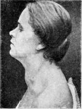
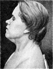
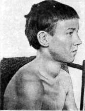
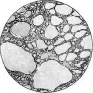
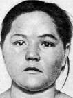
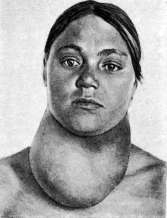
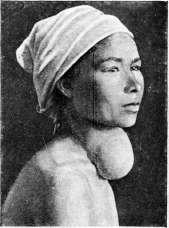
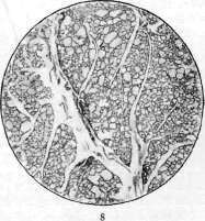
Figure 1 and 2 of a 19-year-old. Retrosternal goiter (1 during exhalation; 2 during inhalation). 3. Parenchymatous goiter in a 12-year-old boy (initial stage). 4. Diffuse parenchymatous goiter in a young woman. 5. Struma diffusa et nodosa colloides. 6. Struma nodosa cystica. 7. Macrofollicular diffuse goiter (according to Sudeck). 8. Microfollicular diffuse goiter (according to Aschoff).
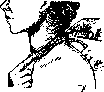
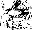
Of undoubted interest in this regard is the instruction of the Swiss Commission for the Study of Goiter on the measurement of the thyroid gland, especially in schoolchildren. Of all the methods of measuring the thyroid gland on the living, this method can be considered the most objective, and therefore the most appropriate (figs. 1, 2, 3, and 4). 1. The size of the neck circumference is determined by a measuring tape or an ordinary centimeter tape passed around the neck through the jugulum and the vertebra prominens (fig. 1). A second measurement should be performed in those cases where the greatest circumference of the neck is located above this line. With a healthy thyroid gland, the neck circumference length in men and women aged 7 to 16 years is 25-35 cm. The limits are the figures 24-39 cm. 2. By antero-posterior transverse measurement, i.e., by placing the leg of a caliper (pelvimeter) over the vertebra prominens and the jugulum, it is possible to determine the swelling of the neck depending on the enlargement of the thyroid gland (figure 2). This size at the age of
from 7 to 10 years is on average 7-9.5 cm in healthy children, and the extreme limits of these measurements are 6.5-11 cm. These two methods allow one to measure only the size of the neck circumference, which in essence does not give an accurate idea of the degree of enlargement of one or another lobe of the thyroid gland.
Hunzicker believes that even in mass screening it is necessary to measure the heights of the individual lobes and the entire width of the gland (fig. 3). The upper poles of both lobes are easily palpable; the lower ones, due to their low position, cannot always be palpated (fig. 4). In these cases, the height of the thyroid gland lobe is measured from the upper pole to the clavicle. The width is determined by measuring with a ruler or calipers the most distant lateral points of the surface of the thyroid gland. In this, inaccuracies are possible due to the fact that, when searching for the extreme lateral points, one presses the gland sometimes harder, sometimes weaker. Thus, the height and width of the entire gland form a rectangle, according to which one can project the thyroid gland. In mass screenings of children (including schoolchildren) and recruits, the Swiss Commission considers it most appropriate to divide the thyroid gland into the following 5 categories: 0-thyroid gland is not palpable; I-thyroid gland is slightly palpable; II-thyroid gland.

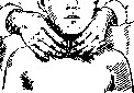
Figure 4. Palpation of the thyroid gland. The thyroid gland is easily palpable but does not protrude noticeably; III-"thick neck"; IV- clearly expressed goiter; V- noted in the presence of nodules. Preventive measures are of great importance in the fight against goiter, but on a large scale they are appropriate in the so-called endemic goiter foci. To speak of absolutely studied and precisely verified specific measures is still premature, since for this we must know exactly the cause of the origin and existence of this disease. There are a number of observations, facts, and theories, but none of them fully illuminates the question. This circumstance forces us to reduce preventive measures primarily to improving the general sanitary-hygienic living conditions of the population, paying special attention to childhood. Light, dry dwellings, good water supply by switching water pipes to verified healthy sources, nutritional conditions, the nature of nutrition, and even the method of food preparation (Th. Kocher, Ko He, Langhans, Wegelin, etc.) will undoubtedly reduce the number of goiter patients. The history of the fight against this disease in Switzerland gives bright examples of favorable results in this regard. The idea that the appearance of goiter depends on the lack of iodine necessary for proper metabolism was very tempting. It is possible that in goiter foci there is a lack of the necessary amount of iodine in food, water, and air, and therefore the introduction of the missing iodine per os is a reasonable preventive measure. This method is essentially not new, since in the middle of the 19th century, iodine treatment for goiter was widely practiced in France. Soon this treatment had to be abandoned, because due to the large doses prescribed by doctors, many patients showed signs of hyperthyroidism, up to mild or severe forms of Basedow's disease. At the end of the 19th century, Kocher and Roux began to prescribe iodine internally for goiter again, but this method did not become widespread. In the last 10-15 years, iodine has been used again, but as a preventive measure in endemic goiter areas, initially in America, then in Switzerland, Germany, and more recently in the USSR. The method of administration is now somewhat different, since the doses of iodine compared to the original ones are minimal. The prevention being carried out in Switzerland in the form of adding small doses of iodine to the table salt consumed (as Wagner von Jauregg recommended as early as 1898) gives obviously favorable results. Since in these places children are already born with an altered thyroid gland, mothers are recommended to take iodized salt during pregnancy. In this way, it will be possible to ensure that children's thyroid glands are healthy. For now, preventive measures are aimed at school-age children. Swiss doctors, with enormous experience, prescribe different doses of iodine: A. Kocher-0.1-0.3 mg daily for 3-4 weeks; Klinger-4-5 mg; de Quervain and Oswald-1-2 mg; Guntziker gives 1/6 of this dose per week and considers this quite sufficient. The prescription of even minimal doses of iodine in endemic goiter has been approved by the Swiss commission, but not unanimously. A number of authoritative doctors (Galli-Valerio, Messerli, Eug. Bircher) disagreed with this decision and spoke against such widespread use of iodine in view of several cases of pronounced hyperthyroidism observed in recent years that developed after taking iodized salt. Reports (Wagner v. Jauregg's, Muggia's, Silberschmidt's, etc.) at the International Goiter Conference in 1927 allow one to trust the preventive prescription of minimal doses of iodine in goiter. Nevertheless, this question will finally be decided after a long time, after long-term observations of the population living in endemic goiter foci. Treatment. The prescription of iodine for goiter can be considered not only as preventive but also as a purely therapeutic measure for already formed goiter. The colloid accumulated in the follicles becomes more liquid under the influence of iodine and is more easily removed through the lymphatic pathways, as a result of which the thyroid gland decreases. The introduction of iodine is effective in diffuse and nodular forms of goiter, in which the tissue has retained its functional ability. If the degeneration of the thyroid gland tissue has reached significant proportions, then iodine is unlikely to have any effect on such a form of goiter. Iodine is also ineffective in pronounced hypothyroidism, where the introduction of thyroid gland preparations is more appropriate, the effect of which can be best verified by studying gas exchange. Of other drugs prescribed internally, one can mention quinine, phosphorus, bromides, calcium compounds, etc. Galli-Valerio, McCarrison, Messerli recommend giving patients agents that disinfect the intestines, for example Benzonaphthol, Thymol, etc. Treating ordinary goiter with radiant energy is pointless, since besides adhesions that complicate subsequent surgical measures, hardly anything will be achieved. Surgical treatment. A significant number of goiter patients receive no help from iodine treatment, and in cases of hyperthyroidism it requires the special attention of a physician. If the goiter in its growth begins to compress neighboring organs, first of all the respiratory tract, affects the functional activity of the heart, or, reaching significant sizes, deprives the patient of working capacity, then the question arises of surgical removal of the goiter. Initial attempts at surgical treatment of goiter are known in the form of cauterizations and ligation of the thyroid gland directly through the skin [Purmann; 1716]. Hamilton (1865) acted in the same way, but first incised the skin. Ligation of blood vessels was first used on humans in 1814 (Buzzard, Walter). At a later time, the indications and technique of blood vessel ligation were thoroughly developed by Billroth and his students. Enucleation of limited nodes, described by Porta, was detailed in 1876 by Socin, who recommended its wide application. Extirpation of the goiter, already used by surgeons in the early 19th century, was developed in all details by Billroth and Kocher. Subsequently, in the study of the general condition of the body after removal (strumipriva tetany, etc.), besides the mentioned scientists, Mikulicz, Eiselsberg, their students, and a number of other prominent surgeons also participated. In Russia, the first operation for goiter was performed by Mislavsky in 1850 in the Urals. Modern methods of surgical treatment of goiter are considered: 1) excision, exsection of the thyroid gland (according to Kocher) after preliminary ligation of both vessels, 2) enucleation of individual colloid or cystic nodes (according to Socin), 3) bilateral resection (according to Mikulicz) and 4) enucleation with resection (according to Kocher). 1. Unilateral exsection of goiter according to Kocher consists in the fact that after exposing the changed part of the thyroid gland, on one side both thyroid arteries and all large veins are isolated and ligated, the isthmus is separated from the trachea, compressed with the appropriate instrument; after this a significant part of the thyroid gland is removed, leaving a thin layer of it to avoid injury to the recurrent nerve and parathyroid bodies. The wound is sutured, and a drainage tube is inserted for one to two days. 2. Enucleation of goiter, recommended by Socin, is performed as follows. With the same Kocher-type collar incision, the thyroid gland is exposed, a node or cyst is found, covered by the thinnest part of the gland, which is cut over it, after which the goiter node is separated with a finger or Kocher's probe. Bleeding at this time is quite significant, but is easily stopped by tamponade. Nevertheless, very severe bleeding is often observed, requiring additional ligation of blood vessels. Despite the ease of this operation, it is performed less and less often in recent times, especially since the methods used now are excellently developed and achieve their purpose. 3. Resection according to Mikulicz consists in that only the superior thyroid artery is ligated. The inferior one is not isolated, and thus the recurrent nerve and parathyroid bodies are not exposed. The removed part of the thyroid gland is excised with a wedge-shaped incision, which is then sutured with catgut sutures. The same is done on the other side. With this method, complications from the recurrent nerve or parathyroid bodies are extremely rarely observed. Bleeding, however, is often significant, interfering with work and sometimes requiring additional ligation of the inferior thyroid artery. 4. When removing large nodular colloid and cystic goiters, as well as in recurrences, Kocher recommended combining enucleation with exsection or resection, excising part of the gland over the node and then enucleating the latter. Bleeding is generally insignificant, and the remaining part protects against damage to the recurrent nerve and parathyroid bodies.
A significant portion of German and Swiss surgeons (Enderlen, Hotz, Kirchner, Kreuter, Florcken, de Quervain, Sauerbruch and many others), performing bilateral excision or resection of goiter, ligate all three and even four vessels on both sides, with some of them transecting the isthmus (Enderlen, Sauerbruch and others), while others (Rehn, Schloffer, Payr and others) leave it untouched in cases where it is unchanged. Complications associated with ligation of all 4 vessels were almost never noted. It should be taken into account that all the aforementioned surgeons worked in areas where goiter is endemic, where pathological-anatomical changes reach significant sizes. Nevertheless, not all surgeons agree with such ligation of vessels, and Hildebrand in 1923 stated that one can limit oneself to unilateral excision of goiter with ligation of vessels only on the corresponding side, without fearing recurrences. The operation for goiter is at present anatomically worked out in all details and almost always, with few exceptions, proceeds without blood loss. During and after the operation for goiter, a number of complications may occur, which are severe in their course and dangerous for the patient's life. At the moment of mobilizing the goiter, injury to one or several large vessels is possible, bleeding from which floods the entire operative field and requires appropriate rapid measures. Even more dangerous at this moment is the entry of air into the gaping vessel lumen and the associated air embolism, which can lead to a fatal outcome. Asphyxias observed before and during the operation require rapid orientation and intervention. The trachea may be compressed by the changed thyroid gland. In such cases, already the incision of skin and muscles, exposure and outward dislocation of the gland (Schloffer) immediately eliminate severe attacks of suffocation. If the walls of the trachea are severely changed, then after removal of the goiter they may collapse, at that moment closing the access of air to the lungs. The elevation of the collapsed tracheal wall, sutured with a thread fixed to the sternocleidomastoid muscle, or quickly performed tracheotomy can still save life. At the moment of ligation of the inferior thyroid artery, injury to the recurrent nerve is possible, noted during the operation performed under local anesthesia, by hoarseness and loss of voice. Among the possible complications in the postoperative period, mention should be made of bleeding from the severed superior thyroid artery or large veins when ligatures slip off them. Severe complications include the tetany sometimes observed in the postoperative period, associated with the removal of parathyroid bodies. A number of surgeons (de Quervain, Madlener, Schloffer, Halsted, Riedel-Help) suggest ligating only the goiter, maximum 3½ arteries (only a branch of the 4th artery-the inferior thyroid artery), believing that ligation of all 4 vessels disrupts the nutrition of parathyroid bodies and their functions. Clinical observations of tetany after operations on other organs (Melchior, Enderlen) give reason to think that there may be other causes for this, in the form of 'readiness for tetany' ('Tetaniebereitschaft'), to which certain individuals are particularly predisposed. Furthermore, the following facts are undoubtedly of interest. In 3,500 excisions of goiter, Urban never observed tetany. Of 106 goiter specimens operated on in Hacker's clinic and examined microscopically, Knaus in 21 found parathyroid bodies, while tetany was observed only once. According to Lebsche's data based on Sauerbruch's material (2,000 cases), tetany in Zurich was observed extremely rarely, while in Munich by the same surgeons in 1.3%. These observations give the right to associate postoperative tetany not only with the simultaneous removal of parathyroid bodies. Obviously there are still some, as yet unknown, causes. The treatment of tetany consists either in transplantation of parathyroid bodies or in the administration of appropriate preparations internally. Among medicinal preparations, Calc. lact. is prescribed 4-6 times 2.0-3.0 per os or intravenous infusion of a 5% solution up to 200 cm³. German surgeons praise Afe-nil (a compound of calcium chloride with urea) and recommend administering 10 cm³ intravenously. Pain on swallowing in the first postoperative days, associated with stretching of the esophagus, soon passes without a trace. In older patients, postoperative pneumonia is possible, to which patients are particularly predisposed due to insufficient ventilation of the lungs, associated with difficult expectoration. Suppuration of the wound is possible, as in other operations, but here there is a danger of spread of infection into the mediastinum and the associated mediastinitis. In the first 2-3 days after the operation, an elevated temperature is usually observed, which does not always depend on infection. In these cases, the temperature is associated with the absorption of the gland's secretion during and after the operation. In cases where the remaining part of the thyroid gland is insufficient to perform the necessary functions for the body, a severe picture of cachexia strumipriva develops, the main symptoms of which are a sharp decrease in mental functions, loss of energy, and apathy. Subsequently, these patients become dull-witted with decreased sensitivity, weakly reacting to external stimuli, clumsy and slowly moving individuals. Mortality after the operation for goiter at present is negligible and averages ⅓-1% (de Querven). This percentage includes mainly those severe cases of huge, retrosternal goiters, in which patients come to the operating table already with noticeably weakened cardiac activity. As for the long-term results, in a significant number of cases, almost 25% (Kocher), a recurrence of goiter was observed. The frequently observed recurrence is inclined to be associated with the technique of intervention-hemithyroidectomy, enucleation, etc. In modern methods of bilateral resection with simultaneous ligation of vessels, many surgeons tend to see a means against recurrences. At the same time, they consider it advisable to carry out postoperative preventive measures in the form of administration of iodine, etc. (Breitner). In any case, prolonged postoperative observations are necessary in order to be able to solve the question of how much the number of recurrences has decreased after more radical methods of operating.
Related articles
Mentioned in
Cite this page
“Goiter.” Soviet Medical Encyclopedia. English translation of Bolshaya Meditsinskaya Entsiklopediya, 1st ed. (Moscow, 1928–1936), ed. N. A. Semashko. https://sovietmedicalencyclopedia.pages.dev/article/goiter/