Corpora Geniculata
Historical document, translated for reference. It reflects medical knowledge of the 1920s–30s and is not medical advice.
Summary
This article describes the anatomy, embryology, and functional significance of the corpora geniculata (lateral and medial geniculate bodies) in the diencephalon. It details their roles as primary subcortical centers for visual and auditory pathways, respectively, and discusses associated clinical pathologies.
Encyclopedia article (1928–1936)
CORPORA GENICULATA (geniculate bodies), formations related to vision and hearing, located in the diencephalon in the region of the metathalamus. There are two Corpora geniculata: Corpus geniculatum laterale and Figure 1. Brainstem: 1-thalamus; 2-pulvinar; 3-brachium quadrigeminum superius; 4 and 5-corpora quadrigemina (colliculus superior et inferior); 6-frenulum veli medullaris anterioris; 7-trigonum lemnisci; 8-lingula cerebelli; 9-fila lateralia pontis; 10-brachium conjunctivum; 11-ventriculus quartus; 12-corpus restiforme; 13-taenia ventriculi quarti; 14-nervi glossopharyngeus et vagus; 15-tuberculum cuneatum; 16-clava; 17-tuberculum cinereum; 18-funiculus gracilis; 19-funiculus cuneatus; 20-stria terminalis; 21-corpus geniculatum laterale; 22-radix lateralis tractus optici; 23-corpus geniculatum mediale; 24-radix medialis tractus optici; 25-tractus opticus; 26-pedunculus cerebri; 27-brachium quadrigeminum inferius; 28-sulcus lateralis mesencephali; 29-nervus trochlearis; 30 and 31-nervus trigeminus (portio minor et major); 32-pons Varolii; 33-brachium pontis; 34-nervus acusticus; 35-nervus facialis; 36-nervus abducens; 37-oliva; 38-nervus hypoglossus; 39-nervus accessorius; 40-radix anterior nervi cervicalis I; 41-funiculus lateralis. (From Spalteholz.)
Corpus geniculatum mediale.-Corpus geniculatum laterale, s. externum (Figure 1) (external geniculate body) is located on the lower surface of the pulvinar (Figure 2), lateral to the base of the cerebral peduncle and the corpus geniculatum mediale, and medial to Wernicke's field; in cross-section, it has the shape of a playing-card heart, with the base turned upward, backward, and slightly inward, and the apex downward, forward, and outward; it continues into the tractus opticus (Figure 3).
4t^, pulvinar; 2 - corpus geniculatum laterale, surrounded by a capsule of myelin fibers of the tractus opticus; 3 and 4 - and has a very characteristic
structure: it is formed by the alternation of gray and white laminae, which have the shape of a curved line. In the gray laminae are large (principal) cells, among which small ones are scattered. Monakow distinguishes three parts in the Corpus geniculatum laterale: the part of the optic tract, the main part, and the cortical part. According to Monakow, the fibers of the tractus opticus terminate near the small cells, which communicate via their processes with the main ones, which give rise to the radiatio optica; crossed and uncrossed fibers lie densely against one another; the fibers of the macular bundle enter into connection with many cells. According to Henschen, however, the fibers of the tractus opticus communicate directly with those cells that give rise to the radiatio optica; the crossed fibers are located along the midline, and the uncrossed ones form the medial and, partly, the lateral
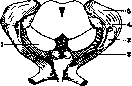
Figure 3. Termination of visual fibers in the corpus geniculatum laterale: 1-pedunculi cerebri; 2-tractus opticus; 3-corpus geniculatum laterale; 4-corpus geniculatum mediale; 5-pulvinar.
capsules. Henschen believes that there is a projection of the retina onto the corpus geniculatum laterale, i.e., the fibers of the tractus opticus, coming from specific areas of the retina, terminate in corresponding places of the Corpus geniculatum laterale; the same projection exists for the macular bundle. The radiatio optica (Gratiolet's bundle, see) originates from the Corpus geniculatum laterale, the initial part of which is called Wernicke's field and which, through the pars retrolenticularis capsulae internae, is directed toward the occipital lobe. A small part of the fibers from the cortex of the occipital lobe (visual area) terminates in the corpus geniculatum laterale. The corpus geniculatum mediale has an elongated shape, lies between the corpus quadrigeminum anterius and the Corpus geniculatum laterale; the brachium of the corpus quadrigeminum anterius separates it from the pulvinar, and the brachium of the corpus quadrigeminum posterius separates it from the outer nucleus of the thalamus opticus, the nucleus semilunaris, and the lemniscus medialis. It is formed by a nucleus of gray matter, the cells of which are very large, stellate, with numerous processes; the fibers of the lemniscus lateralis (3rd auditory neuron), passing through the brachium of the corpus quadrigeminum posterius, terminate in it. The cells give rise to fibers which, upon exiting the Corpus geniculatum mediale, join together, forming the 4th auditory neuron, which goes through the pars sublenticularis capsulae internae to the auditory area of the temporal lobe of the hemisphere; centrifugal fibers, terminating in the Corpus geniculatum mediale, originate from this area.
The Corpora geniculata develop from the primary anterior cerebral vesicle, the cavity of which turns into the third ventricle, while the walls, thickening, form the diencephalon. In addition to the thalamus opticus, the metathalamus, to which the Corpora geniculata belong, is formed from the upper part of the lateral wall. They receive blood from the arteria cerebri posterior through the arteriae pedunculares externae. Since the tractus opticus terminates and the radiatio optica originates in the Corpus geniculatum laterale, i.e., the transmission of visual sensations from the peripheral neuron to the central one occurs, it is therefore classified as a primary or subcortical visual center. A lesion of the Corpus geniculatum laterale causes visual impairment in the form of hemianopsia homonyma bilateralis (see Hemianopsia). The lemniscus lateralis, the 3rd auditory neuron, terminates in the Corpus geniculatum mediale, and the 4th, or central, neuron originates there, which is directed to the cortex of the 1st temporal gyrus; consequently, the transmission of auditory sensations from the peripheral neuron to the central one occurs in it. Its lesion leads to hearing impairment, mainly on the opposite side, and to weakening on the same side due to the incomplete decussation of auditory fibers in the pons Varolii. The Corpora geniculata are almost never affected in isolation, but are involved in the process (hemorrhages, tumors) during diseases of the surrounding formations.
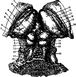
E. Kononova. CORPORA QUADRIGEMINA (lamina quadrigemina, quadrigeminal plate) are located in the midbrain, on its posterior surface [the anterior is formed by the cerebral peduncles (pedunculi cerebri)], covered from above by the cerebral hemispheres, and therefore, to see the Corpora quadrigemina, it is necessary to remove the hemisphere. The Corpora quadrigemina consist of four tubercles, or colliculi: two anterior, or upper (corpus quadrigeminum anterius, s. superius), and two posterior, or lower (corpus quadrigeminum posterius, s. inferius) (see figure). Bundles of fibers, so-called brachia of the Corpora quadrigemina, go from the colliculi: from the corpus quadrigeminum anterius to the corpus geniculatum laterale, and from the corpus quadrigeminum posterius to the corpus geniculatum mediale. The main part of the corpus quadrigeminum posterius is formed by an oval-shaped nucleus ("nucleus of the lower colliculus"), formed by rather large ganglionic cells and lying among the fibers of the lemniscus lateralis, which form a kind of capsule around it. Lateral to the lemniscus lateralis, between it and the brachium of the corpus quadrigeminum posterius, the quadrigeminal plate from behind and above: 1-corpus pineale; 2-commissura posterior; 3-colliculus superior; 4 and 5-brachium quadrigeminum superius et inferius; 6 and 7-corpus geniculatum mediale et laterale; 8-pedunculus cerebri; 9-colliculus inferior; 10-frenulum veli medullaris anterioris; 11-trigonum lemnisci; 12-nervus trochlearis; 13-brachium conjunctivum cerebelli; 14-fila lateralia pontis; 15-tuberculum anterius thalami; 16-taenia chorioidea; 17-lamina affixa; 18-stria terminalis; 19-corpus striatum; 20-pulvinar; 21-corpora quadrigemina; 22-sulcus lateralis mesencephali; 23-nervus trochlearis; 24-lingula cerebelli. (From Spalteholz.)
is located the gray matter (corpus parabigeminum of Flechsig), which connects above with the medial geniculate body and apparently serves as the site of termination of the auditory fibers (lateral lemniscus). Along the periphery is located the stratum zonale, and around the capsule is gray matter, which has no relation to the posterior corpora quadrigemina. Both posterior colliculi are connected to each other by gray matter, in which transversely crossing fibers pass. A portion of the fibers of the lateral lemniscus (both auditory fibers and fibers of the Gowers' tract) terminates in the nucleus; a small portion of the fibers passes through the commissure to the opposite posterior corpus quadrigeminum. From the posterior corpus quadrigeminum, fibers are directed through its brachium to the medial geniculate body, and from there to the auditory region of the temporal lobe; from this region, centrifugal fibers also go to the posterior corpus quadrigeminum through its brachium. The anterior corpus quadrigeminum has a layered structure; seven layers are distinguished in it: three gray and four white. From outside to inside are located: 1) a thin layer of fibers, the stratum zonale, 2) a superficial gray layer of small cells, 3) a layer of optic fibers, 4) a middle gray layer with rather large cells, 5) a layer of the lemniscus, 6) deep gray matter, and 7) deep white matter, or a layer of arcuate fibers; more internally from it is located the gray matter of the Sylvian aqueduct with the nucleus of the cerebral root of the trigeminal nerve. Fibers of the optic tract enter the anterior corpus quadrigeminum through its brachium, occupy the third layer in it, and give off branches to the upper and lower layers; in the fifth layer, fibers of the lateral lemniscus (spino-tectal fibers and fibers of Gowers' tract) terminate; in this same layer are located cortico-tectal fibers, coming from various parts of the cortex, mainly from the occipital lobe, and penetrating into the anterior corpus quadrigeminum through its brachium, and partly through the tegmentum of the cerebral peduncle (partial destruction of the cortex, mainly of the visual region, also causes degeneration in the anterior corpus quadrigeminum). The deep layers give rise to efferent fibers; a part of them is directed to the tegmentum of the cerebral peduncle of its own side, and the other, larger part, as part of the tectospinal tract, or predorsal bundle, crosses to the opposite side, forming Meynert's fountain decussation; in the spinal cord, this bundle occupies the antero-internal part of the anterior column (at the anterior median fissure), descends to the lower thoracic segments, and terminates in the anterior horns of the gray matter. On its path, it gives fibers to the nuclei of the oculomotor nerve, to the red nucleus, and to the substantia reticularis; by this path, optic fibers communicate with the nuclei of the oculomotor nerve and with the spinal cord. Efferent fibers also include the tectopontine tract (Münzer's bundle) and tectoreticular fibers; the former, crossing the cerebral peduncle, terminates in the gray matter of the base of the pons Varolii; through it, optic fibers communicate with the cerebellar cortex (by means of the pontine nuclei and the middle cerebellar peduncle); the latter terminates in the substantia reticularis of the cerebral peduncle and the pons Varolii. The corpora quadrigemina, together with the cerebral peduncles, develop from the midbrain vesicle and form the midbrain, or mesencephalon; the cavity of this vesicle narrows due to the thickening of the walls and turns into the Sylvian aqueduct. In cold-blooded animals and in birds, the corpora quadrigemina represent a strongly developed organ—the optic lobe. As the cerebral hemispheres develop, they lose their significance, shrinking in their relative mass and in functional respect. The corpora quadrigemina receive their vessels from the plexus formed by branches of the superior cerebellar artery and the posterior cerebral artery. Physiology of the corpora quadrigemina. The posterior corpus quadrigeminum has a connection with the auditory system; a portion of the fibers of the lateral lemniscus terminates in it, as well as fibers from the temporal lobe from the auditory center, which indicates its relation to the auditory function; but on the other hand, destruction of the posterior corpus quadrigeminum is not accompanied by complete loss of hearing, which suggests that it is not the main station of the auditory pathway and does not take part in the conduction of auditory sensations, but plays the role of the main sensorimotor subcortical center, i.e., a center of complex auditory reflexes in the form of movements of the head, vocal cords, etc.; this conclusion is also confirmed by experimental studies. In the anterior corpus quadrigeminum, the optic tract terminates, but there is no connection with the cortex in the form of centripetal fibers; thus, in humans, the anterior corpus quadrigeminum apparently does not take part in the transmission of visual sensations; on the other hand, it is connected with the nuclei of the oculomotor nerve; thus, through the mediation of the corpora quadrigemina, the optic nerve communicates with these nuclei and transmits light stimulation to them, thanks to which the nuclei of the oculomotor nerve are reflexively excited by impulses from the anterior corpus quadrigeminum, which causes corresponding movement of the eye and constriction of the pupil. On the basis of this, one can think that the anterior corpus quadrigeminum is a reflex visual center for conjugate eye movements. The termination in it of centrifugal fibers from the visual center of the cortex indicates that its function is under the control of the cortex and that it can be excited by impulses not only from the periphery but also from the center. Due to the fact that the corpora quadrigemina are surrounded by functionally important structures, it is very difficult in diseases to isolate their symptoms from the symptoms of other structures, i.e., to establish a pure syndrome of disease of the corpora quadrigemina; from the general picture, these symptoms have to be isolated based on physiological data.
E. Kononova. CORPUS CALLOSUM, s. commissura pallii, belongs to the commissural systems of fibers and serves to connect both hemispheres of the brain. It represents a layer of myelin fibers, situated in the depth of the interhemispheric fissure and forming the roof of the third and part of the lateral ventricles of the brain. Three parts are distinguished in it: 1) the central part, the body, or truncus corporis callosi; 2) the anterior curved part (knee, genu of the corpus callosum), located 3 cm from the frontal pole; this part gradually thins and ends in the beak, or rostrum; 3) the posterior thickened part, the splenium, or bulb, located 5 cm from the occipital pole (Fig. 1). The length of the corpus callosum is 7–9 cm; the thickness is on average 1 cm, posteriorly 2 cm. The corpus callosum is covered from above by a thin layer of gray matter and longitudinal fibers—the indusium griseum, forming 4 longitudinal thickenings, or strips—striae longitudinales: 2 run along the midline—the nerves of Lancisi, or striae longitudinales mediales, s. internae, and 2—outward, on the sides, one on each side—striae longitudinales laterales, s. externae, s. taeniae tectae (Fig. 2); in places in these strips, the amount of gray matter increases—the nucleus of the nerve of Lancisi. At the posterior end of the corpus callosum, the indusium griseum and the longitudinal striae pass into the fasciola cinerea, and then into the fascia dentata; at its anterior end, from the rostrum of the corpus callosum, the fibers of the indusium, forming the so-called pedunculus corporis callosi, enter the 'carrefour olfactif' of Broca and partly into the subcallosal gyrus of Zuckerkandl. From the si-
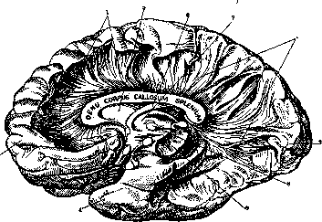
Figure 1. Internal surface of the right hemisphere (gyri removed to show the distribution in the hemispheres of the radiatio corporis callosi): 1—radiatio corporis callosi; 2—frontal pole; 3—occipital pole; 4—temporal pole; 5—septum pellucidum; 6—fornix; 7—thalamus opticus; 8—anterior white commissure; 9—cerebral peduncle.
des, the upper surface of the corpus callosum is covered by gyri—the gyri fornicati, from which it is separated by a furrow—the fissure of the corpus callosum. The lower, or ventral, surface of the corpus callosum forms the roof of the lateral ventricles on the sides, and along the midline in the anterior parts it is fused with the septum pellucidum, and in the posterior parts with the fornix and with the psalterium; the fornix separates it from the III ventricle. The cells giving rise to the corpus callosum, according to some authors, belong to the small and medium pyramids of the superficial layers, according to others—to the pyramidal cells of the deep layers. The fibers of the corpus callosum entering the hemispheres are called the radiatio corporis callosi; they run transversely in all directions and connect the cortex of both hemispheres with each other, and according to some data, the cortex with the internal capsule, external capsule, and neostriatum (Fig. 3). The corpus callosum connects not only homologous but also heterologous gyri of both hemispheres. All parts of the brain are connected by the corpus callosum with the exception of the temporal pole, the Ammon's horn, and the olfactory lobes. From various gyri, fibers go through the nearest parts of the corpus callosum; for example, from the frontal lobe—through the genu, from the occipital and posterior parts of the temporal lobe—through the splenium, etc. Fibers going from the corpus callosum into the frontal and occipital lobes and surrounding
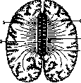
Figure 2. Horizontal section through the upper surface of the corpus callosum. Radiatio corporis callosi: 1—striae longitudinales mediales (nerves of Lancisi); 2—striae longitudinales laterales (taeniae tectae); 3 and 4—radiatio corporis callosi (frontal and parietal parts); 5—radiatio corporis callosi (temporal and occipital parts).
the horns of the lateral ventricles are called forceps: forceps anterior into the frontal lobe and forceps posterior into the occipital lobe. This latter, due to the calcar avis located in the posterior horn, is divided into the forceps major and the forceps minor; fibers from the outer and inner surfaces of the occipital lobe pass through the former, and from the lower surface through the forceps minor. The fibers of the corpus callosum give off collaterals to various parts of the hemisphere of their own side: these are no longer commissural, but association branches, serving to achieve the simultaneous action of different parts of the cerebral cortex. The corpus callosum is pierced by fibers—fibrae perforantes corporis callosi—belonging to the olfactory system. The corpus callosum receives blood mainly from the anterior cerebral artery, which, having passed to the inner surface of the hemisphere, runs parallel to the corpus callosum and sends branches to it all the time; the posterior parts of the corpus callosum also receive blood from the posterior cerebral artery. The corpus callosum develops later than other parts of the brain: its first primordium begins anterior to and above the foramen of Monro, where transverse bundles appear, which grow from the deep parts of the hemispheres toward each other; gradually the corpus callosum increases anteriorly and posteriorly by the further fusion of both halves; according to some authors, the site of development of the corpus callosum is the lamina terminalis. And in the phylogenetic sense, the corpus callosum turns out to be a late formation: it is not yet present even in lower mammals; its development proceeds in parallel with the development of the cerebral hemispheres. The physiological significance of the corpus callosum, which serves in general for the synergistic activity of both cerebral hemispheres, has not yet been clarified in all respects: experimental studies on animals by stimulating the corpus callosum with an electric current or by cutting it have not yielded significant results. Stimulation of the upper surface of the corpus callosum is transmitted to the motor centers; upon stimulation of its anterior parts, movements are observed in the eyes and head; upon stimulation somewhat further posteriorly—in the forepaws, then in the trunk, etc. After preliminary extirpation of the motor centers, it is no longer possible to induce movements by stimulation. Cutting the corpus callosum does not cause noticeable disorders in animals. Diseases of the corpus callosum in humans do not always produce clear symptoms, sometimes they even pass completely without a trace and are discovered only at autopsies. But nevertheless, there are parallel observations which indicate that a lesion of the middle parts of the corpus callosum causes awkwardness in the movements of the left hand, which is explained by the lack of influence on the right hemisphere from the left, to which the general control of motor acts belongs. A pure syndrome of a lesion of the corpus callosum, in the opinion of some authors, is characterized by mental and motor disorders. The mental disorders are unclearly expressed: weakening of the connection in thoughts, certain oddities in actions, disorder of orientation in space, changes in character, irritability, carelessness; in general, there is nothing characteristic in the aggregate of these symptoms. In addition to these mental disorders, movement disorders are observed, resembling apraxia (mainly in the left half of the body). The absence of a complete description of the corpus callosum syndrome is explained by the rarity of isolated disease of the corpus callosum; the structures surrounding it, which produce their own symptoms, are almost always affected. Among the pathological processes observed in the corpus callosum, it is necessary to note: 1) agenesis of the corpus callosum, or its complete absence, or an arrest in development; 2) hemorrhages, softenings, involving either the entire corpus callosum or its individual parts in various combinations; sometimes the connection between both hemispheres is completely destroyed; 3) scleroses, atrophies; 4) tumors (observed more often than other processes); gliomas and sarcomas are in first place, then endotheliomas, etc.; abscesses have also been described; 5) secondary degenerations. In the literature, there are indications of attempts to localize a disease in one or another part of the corpus callosum during life on the basis of observed symptoms, but these attempts have not been fully verified and require further confirmation.
Related articles
Mentioned in
Cite this page
“Corpora Geniculata.” Soviet Medical Encyclopedia. English translation of Bolshaya Meditsinskaya Entsiklopediya, 1st ed. (Moscow, 1928–1936), ed. N. A. Semashko. https://sovietmedicalencyclopedia.pages.dev/article/corpora-geniculata/