Cerebral Peduncles
Historical document, translated for reference. It reflects medical knowledge of the 1920s–30s and is not medical advice.
Summary
The cerebral peduncles are structures forming part of the midbrain, consisting of two bundles connecting the pons to the cerebral hemispheres. They contain important pathways for motor, sensory, and autonomic functions, and include key structures like the substantia nigra and red nucleus.
Encyclopedia article (1928–1936)
CEREBRAL PEDUNCLES (pedunculi, s. crura cerebri), structures that are part of the midbrain (see Mesencephalon). They appear as two trunks extending from the pons to the cerebral hemispheres; due to their divergence, a triangular space is formed between them, the interpeduncular space, closed by the perforated posterior lamina of gray matter, through which vessels penetrate into the brain substance. Between the peduncles emerges the third pair of cranial nerves (n. oculomotorius). The cerebral peduncles are visible only from the base of the brain, where they are partially covered by the optic tracts and temporal lobes. Near the cerebral peduncles is one of the large cisterns. The cerebral peduncles develop from the midbrain vesicle.--The cerebral peduncles can be divided into two parts: the base (basis, pes pedunculi) and the tegmentum, between which is located the substantia nigra Soemmeringi. Posterior to the tegmentum is the aquaeductus Sylvii, connecting the third and fourth ventricles, and above it are the corpora quadrigemina, called the tectum. The structure of the base of the cerebral peduncles is the same throughout their length, while the structure of the tegmentum changes depending on the level of the section (figs. 1 and 2). In the base of the cerebral peduncles, in the middle part (middle 3/5), pass the pyramidal fibers going from the cortex of the central gyri to the anterior horns of the spinal cord, and the corticonuclear pathways to the motor nuclei of the cranial nerves; the latter occupy a more internal position. In the inner third of the cerebral peduncles passes the frontopontine system, and in the outer part the temporopontine and occipitopontine systems (see Corticopontine pathways). In the upper parts of the cerebral peduncles, two bundles separate from the pyramidal pathway (pes lemniscus prorandus and pes lemniscus superficialis), which go to the lemniscus (see Lemniscus lateralis, medialis). In the middle part of the tegmentum in the lower parts of the cerebral peduncles (at the level of corp. quadrig. posteriora) (figs. 2 and 3) is located the decussation of brachia conjunctiva; posteriorly are the fasciculus longitudinalis posterior, and on the sides are the lemniscus medialis and lateralis, located at an angle to each other; lemn. lateralis partially ends in corp. quadrigeminum posterius; at the outer edge of lemniscus medialis is located corp. parabigeminum, and more peripherally brachium corp. quadr. posterius. Posteriorly and medially to the lemnisci is located substantia reticularis. Aquaeductus Sylvii is surrounded by gray matter, in the anterior part of which is located the nucleus of n. trochlearis; the roots of this nerve form a decussation above the Sylvian aqueduct and then go downward, curving around the side of the cerebral peduncle. Laterally from the root of n. trochlearis in the gray matter lies the cerebral root of n. trigemini.
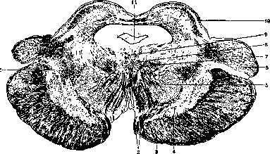
Figure 1. Section through the upper parts of the cerebral peduncle (through corpus quadrigeminum anterius) (see figure 3, AD); 1-lemniscus medialis; 2-n. oculomotorius; 3-base of the cerebral peduncle (corticopontine fibers); 4-substantia nigra; 5-nucleus ruber; 6-corpus geniculatum mediale; 7-fasciculus longitudinalis medialis; 8-lateral nucleus of n. oculomotorii; 9-central nucleus of n. oculomotorii; 10-corpus quadrigeminum anterior; 11-aquaeductus Sylvii.
In the upper parts of the cerebral peduncles (at the level of corp. quadr. anter.) the picture changes somewhat: instead of the decussating brachia conjunctiva, nuclei rubri appear, in which the brachia conjunctiva end. From nucl. ruber originates the fasciculus rubrospinalis, forming in the central part of the tegmentum the decussation of Forel; to the nucleus ruber approach fibers from the frontal lobe of the brain (details-see Nucleus ruber). Posterior (dorsal) to the decussation of Forel is the decussation of fasc. tectospinalis, or the fountain-like decussation of Meynert. Posteriorly and medially from nucl. ruber is located fasc. longitudinalis posterior, and posteriorly and medially from the latter are the nuclei of n. oculomotorii (see Oculomotorius n. and Westphal-Edinger nucleus);
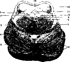
Figure 2. Section through the lower parts of the cerebral peduncle (through corpus quadrigeminum posterior) (see figure 3, AB): 1-radix mesencephalica n. trigemini; 2-locus caeruleus; 3-lemniscus medialis; 4-fasciculus rubrospinalis; 5-pyramidal fibers; 6-fibrae propriae pontis Varoli; 7-decussatio brachii conjunctiva; 8-fasciculus longitudinalis medialis; 9-nucleus n. trochlearis; 10-pars descendens n. trochlearis; 11-aquaeductus Sylvii; 12-corpus quadrigeminum posterior; 13-commissura corporis quadrigemini posterioris.
the roots of this nerve pass through nucl. ruber and exit between the peduncles, part of the root fibers forms a decussation along the midline. Laterally lie the lemnisci, with lemn. lateralis significantly reduced in size. Above aquaeductus Sylvii are the anterior hillocks or corpora quadrigemina anteriora.--The arteries of the cerebral peduncles mainly come from art. cerebri posterior, partially from art. choroidea anter., communicans posterior and art. cerebelli superior; these arteries divide into 4 groups: aa. centrales (for nucl. ruber and nuclei n. oculomotorii), aa. radiculares (for the roots of n. III), periphericae and aa. corporis quadrigemini. Physiology. The cerebral peduncles have enormous importance as conductors of various systems of fibers-motor, sensory, autonomic. The middle part (3/5) of the base of the cerebral peduncle is occupied by motor fibers, which are distributed to centers; most laterally go fibers from the centers of movement of the leg and trunk, then from the centers of the arm and finally from the centers of movement of the face, larynx, etc. to the nuclei of the cranial nerves (so-called corticonuclear fibers); in the inner and outer parts of the peduncle pass fibers from the cerebral cortex to the pons (fronto-, temporo-, occipitopontine systems), connecting through the pons the cerebral hemispheres with the cerebellum. In the tegmentum of the cerebral peduncles are located lemniscus medialis and lateralis - conductors of deep sensitivity, touch and hearing. Through the tegmentum pass fibers from the cerebellum to the brain (brachia conjunctiva); on the path of these fibers are located very important physiologically the red nuclei, belonging to the extrapyramidal system. In the cerebral peduncles is located substantia nigra, which is one of the autonomic centers, and above the tegmentum of the cerebral peduncles lie the nuclei of cranial nerves III and IV, united for coordinated movements by fibers passing in fasc. longitudinalis posterior and in fasc. tectospinalis. Some authors consider the central gray matter as a center regulating sleep (see Encephalitides). Pathology. Anat.-physiol. data indicate how different the symptom complex should be with different localization of one or another pathological process-in the base or in the tegmentum, on one side or on both. Lesion of the base of the peduncle gives alternating paralysis (hemiplegia alternans oculomotoria), known as Weber's syndrome and characterized by paralysis of the limbs and face on the side opposite to the lesion and paralysis of n. oculomotorius on the side of the lesion (see Alternating syndromes); when the process crosses the midline, bilateral ophthalmoplegia is observed, with localization of the process in the outer parts of the base ophthalmoplegia is not observed. With lesion of the tegmentum, an alternating syndrome is also observed-Benedict's syndrome: paralysis of conjugate eye movements (lesion of fasc.
14 13
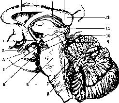
Figure 3. Median section of the brain: 1-corpus mamillare; 2-optic nerve; 3-hypophysis; 4-oculomotor nerve; 5-sulcus oculomotorii; 6-pontis Varoli; 7-fourth ventricle; 8-medulla oblongata; 9-aqueductus Sylvii; 10-lamina quadrigemina; 11-thalamus opticus; 12-splenium; 13-corpus pineale; 14-brachium conjunctivum; 15-corpus fornicis; 16-lateral ventricle; AB and AD-lines of brain sections in figs. 1 and 2. longitudinalis post.), sleep disorders (drowsiness or insomnia), ophthalmoplegia on the side of the lesion, while on the opposite side-cerebellar phenomena (depending on the involvement of the nucleus ruber, brachia conjunctiva), sensory disturbances (involvement of lemnisci). Sometimes in non-specific lesions of the cerebral peduncles, the Argyll-Robertson symptom is observed; with foci in the area of the nucleus ruber, two alternating syndromes are observed: the superior red nucleus syndrome, or Foix's syndrome, and the inferior syndrome-Claude's syndrome. Among the pathological processes observed in the cerebral peduncles, vascular (softenings, hemorrhages), tumors (for example gliomas), tubercles, syphilitic gummata, traumatic lesions, vulgar inflammatory processes, etc., should be noted. Softenings and hemorrhages have the same etiology, pathogenesis, onset, and development as in the brain (see Brain, Apoplexy); they can be limited, diffuse, or very extensive depending on the affected artery. The symptomatology depends on the localization of the process in the base or in the tegmentum (see above); in addition to focal symptoms, general cerebral symptoms-headaches, dizziness, vomiting, paresthesias, delirium, drowsiness, etc., are observed. Treatment and prevention are the same as for brain diseases. Tumors can be single or multiple, ranging in size from a small pea to complete destruction of the cerebral peduncles; sometimes the tumor is encapsulated, in other cases it grows into the brain tissue, destroying it. In addition to focal symptoms depending on the localization of the tumor, general cerebral symptoms (stagnant papilla, headaches, vomiting, convulsions, changes in respiration and pulse) are also observed. Diagnosis is very difficult due to the influence of the tumor on surrounding structures. Therapy is possible only if the process is of syphilitic nature. Traumatic lesions of the cerebral peduncles are very rare due to their deep location; they are observed in gunshot wounds, in various head injuries, etc. In various intoxications and infections, inflammations localized in the area of the cerebral peduncles are mostly of a hemorrhagic nature; they have been isolated by Wernicke as a special form under the name polioencephalitis superior haemorrhagica (see Polioencephalites).
Related articles
Mentioned in
- Alternating Syndromes
- Area
- Auditory Pathways
- Brain (cerebrum, a comprehensive term for the entire)
- Cerebral Aqueduct
- Corpora Geniculata
- Darkshevich's Nucleus
- Fennel (medicinal or Florentine, fennel, Foeniculum)
- Gowers' Tract
- Internal Capsule
- Lane's Ligaments
- Mesencephalon and Polien G. Mezernitsky
- Oculomotorius Nervus
- Polioencephalitis
- Rhombencephalon
- Substantia Nigra
- Substantia Perforata
- Substantia Reticularis
- Trochlear Nerve
- Varolius' Bridge
Cite this page
“Cerebral Peduncles.” Soviet Medical Encyclopedia. English translation of Bolshaya Meditsinskaya Entsiklopediya, 1st ed. (Moscow, 1928–1936), ed. N. A. Semashko. https://sovietmedicalencyclopedia.pages.dev/article/cerebral-peduncles/