Decussation
Historical document, translated for reference. It reflects medical knowledge of the 1920s–30s and is not medical advice.
Summary
An overview of the anatomical phenomenon of decussation within the nervous system, where nerve fibers cross from one side of the body to the other. The article details complete and partial decussations, their physiological significance in maintaining partial function during localized lesions, and their importance for topical neurological diagnosis.
Encyclopedia article (1928–1936)
DECUSSATIO (Latin), decussation, a term used principally in neurology to designate the meeting and intertwining of fibers running from opposite sides. Already in the lowest stages of the animal organism, in invertebrates (worms), this kind of decussation is encountered; in higher vertebrates, it reaches its greatest diversity. Decussation is complete if the fibers pass entirely from one side to the other, and partial if a part of the fibers remains on its own side. Examples of complete decussation can serve all sensory fibers (decussation of the lemnisci, decussation of Gowers' tract), brachia conjunctiva (see Figure 1), the rubrospinal tract - Forel's decussation, the tectospinal tract - Meynert's decussation, and others. An example of partial decussation is the pyramidal fibers - decussation of the pyramids, the fibers of the optic nerve (see Figure 2) - optic chiasm, the fibers of the auditory pathway, and others. (For details, see the corresponding articles.) The question of the expediency of the partial decussation phenomenon becomes more or less clear when studying the physiological functions of the crossed fibers (pathways). It can be established quite precisely that with a selective lesion of fibers running on the ipsilateral side, the carried function is still not completely lost thanks to the crossed fibers of the opposite side entering it. This explains, for example, the incomplete loss of vision in one eye upon a lesion of the homonymous optic tract. Thus, thanks to the existence of the partial decussation phenomenon, a partial preservation of one or another function, homonymous to the side of the lesion, is ensured, unless the process entirely captures both the crossed and uncrossed fibers on the given side. Both types of these fibers, depending on the selectivity of the pathological process, play the role of a reserve of healthy impulses for at least the partial preservation of the impaired function. Almost all pathways of the central nervous system undergo decussation, which occurs in various sections and levels of the brain. In some cases, decussation occurs simultaneously for all fibers of a given pathway (for example, brachia conjunctiva); in others, this decussation occurs gradually, over the course of various levels or many segments (for example, the decussation of the fibers of Gowers' tract or the direct pyramidal tract, observed along the entire spinal cord). Due to the existence of decussations, one hemisphere governs
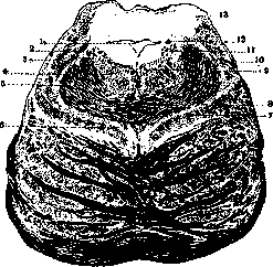
Figure 1. Cross-section of the pons at the level of the mamillary bodies: 1 - cerebral aqueduct; 2 - mesencephalic root of the trigeminal nerve; 3 - tectospinal tract; 4 - thalamo-olivary tract (Bechterew); 5 - brachium conjunctivum; 6 - rubrospinal tract (Monakow); 7 - medial lemniscus; 8 - decussation of the brachium conjunctivum; 9 - medial longitudinal fasciculus; 10 - lateral lemniscus; 11 - locus coeruleus; 12 - descending part of the trochlear nerve; 13 - inferior colliculus. (According to Rauber.)
the functions of the opposite half of the body, as a consequence of which, for example, a lesion in the right hemisphere of the brain causes paralysis of the left half of the body, and vice versa. The same applies to sensitivity. An exception is the cerebellum, the influence of which is exerted on the corresponding side thanks to the double decussation of its efferent fibers (decussation of the brachium
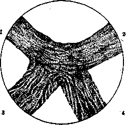
Figure 2. Section through the optic chiasm. Adult dog; removal of the right eye (Marchi method): 1 - left optic tract; 2 - right optic tract; 3 - left optic nerve; 4 - right optic nerve. (From Darkshevich.) conjunctivum and rubrospinal tract). The study of the decussations existing in the central nervous system has great practical importance for the topical diagnosis of pathological processes.
L. Brusilovski.
Related articles
Mentioned in
Cite this page
“Decussation.” Soviet Medical Encyclopedia. English translation of Bolshaya Meditsinskaya Entsiklopediya, 1st ed. (Moscow, 1928–1936), ed. N. A. Semashko. https://sovietmedicalencyclopedia.pages.dev/article/decussation/