Bulbar Palsy
Historical document, translated for reference. It reflects medical knowledge of the 1920s–30s and is not medical advice.
Summary
This article from the 1928–1936 Great Medical Encyclopedia describes the clinical manifestations and syndromes associated with bulbar palsy, detailing various unilateral and bilateral lesions of the medulla oblongata affecting cranial nerve nuclei and pathways.
Encyclopedia article (1928–1936)
BULBAR PARSY. In the limited space represented by the medulla oblongata (bulbus), the principal motor and sensory pathways running through this area cross. Here also are located the nuclei of such nerves vital to life as the vagus, glossopharyngeal, and hypoglossal nerves, and consequently the centers for respiration, swallowing, articulation, phonation, and cardiac activity. Bulbar palsy is clinically manifested either as a symptom complex or as an independent nosological entity (disease). Symptom complexes can be divided into two groups: a) unilateral, caused by a lesion of one side of the medulla oblongata, and b) bilateral, when the disease process affects both halves. Among the former are known: 1. Interolivary bulbar symptom complexes, when there is hemiplegia (without facial involvement) on one side, opposite to the site of the lesion, and paralysis of the tongue with its degenerative atrophy on the side of the lesion (opposite to the hemiplegia). In this case, the pathological lesion is located in the region of the pyramidal tract, above its decussation, affecting the nucleus of the hypoglossal nerve or its rootlets on the same side.
Retrolivary bulbar symptom complexes (see Figure 1), when the nuclei of the nerves located here are affected, and sometimes simultaneously the pyramidal tract and sensory pathways. Clinically, this symptom complex is expressed by: a) paralysis of the soft palate on one side and simultaneously paralysis of both vocal cords or one vocal cord (Aveline) on the same side; b) this latter symptom complex may be combined with symptoms of sympathetic nerve involvement, i.e., on the damaged side there is also miosis (with preserved light reaction of the pupil), enophthalmos, and narrowing of the palpebral fissure; c) a combination of symptom complex "a" with paralysis of the XII pair of nerves; d) a symptom complex of damage to the XI and XII pairs of nerves (Jackson); e) a symptom complex of paralysis of the recurrent laryngeal nerve, the XI and XII pairs of nerves, and the sympathetic nerve; f) a symptom complex of lateropulsis and hemiasynergia (due to a lesion of the restiform body), miosis, enophthalmos, and narrowing of the palpebral fissure on the side of the lesion (due to damage to the sympathetic nerve); on the opposite side: hemiplegia, hemianesthesia for pain and temperature sensitivity (syringomyelic type); in addition, nystagmus and vertigo (Rossolimo, Babinski-Nageotte);
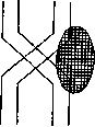
g) Deiters' nucleus symptom complex (Bonnier): vertigo due to impaired equilibrium, instability of the legs (lesion of the restiform body), paralysis of the VI pair of nerves, deafness, tinnitus, nystagmus, pain in the temple, glycosuria, albuminuria, polyuria, migraine, agoraphobia; h) hemiplegia cruciata s. alternans (see Figure 2): when the lesion is located at the site of the pyramidal decussation, paralysis of the arm on one side and paralysis of the leg on the opposite side are produced; i) hemiesthesia cruciata s. alternans: when the region of the spinal root of the V pair of nerves is damaged, anesthesia of the corresponding half of the face and anesthesia of the opposite side of the body are produced.
...
All the mentioned symptom complexes can be observed either in pure form or in various combinations, depending on the extent and localization of the lesion, during the course of many diseases, such as, for example, tumors nesting in the medulla oblongata or in the cerebellum, but putting pressure on the medulla oblongata. They can be observed as a result of hemorrhage, abscess, thrombosis, or embolism of the posterior inferior cerebellar artery, basilar artery, vertebral artery, supplying the bulb, and so on. They also occur as complications of syringomyelia (syringobulbia), tabes, but can also be independent diseases. Bilateral symptom complexes are caused either by damage to the nuclei of both sides or, in addition, to the conducting pathways. It is necessary to differentiate bulbar symptom complexes from polyneuritis of the cranial nerves and meningitis. In polyneuritis, there is a more complete involvement of the nerves than in the bulbar symptom complex. In meningitis, besides what has been said, other symptoms of a general character are observed (rigidity of the neck muscles, Kernig's sign, and so on). Disorders of cardiac activity, respiration (Cheyne-Stokes), glucosuria, polyuria point to a bulbar origin of the symptoms. Bilateral symptom complexes include: 1. Acute bulbar palsy (apoplectic). Most frequently it is encountered due to damage to the vessels supplying the medulla oblongata, i.e., the vertebral artery, basilar artery, and posterior inferior cerebellar artery. In this case, either hemorrhage, embolism, or thrombosis of their branches takes place. Frequently, following prodromal symptoms (dizziness, tinnitus, vomiting), stroke-like bulbar phenomena occur in the form of dysarthria and even anarthria, swallowing disorders, paralysis of the soft palate, and pharyngeal muscles. To this are often added paralyses of the limbs and cranial nerves of the type of crossed hemiplegia or crossed hemianesthesia, respiratory and pulse disorders, salivation, and elevated temperature. Here, cases in which lesions of the posterior inferior cerebellar artery form the basis deserve attention; their clinical picture consists of crossed hemianesthesia, paralysis of the soft palate and vocal cord on the side of the trigeminal nerve lesion, paralysis of the sympathetic nerve on the same side, and swallowing and equilibrium disorders. To this may be added crossed hemiplegia or even triplegia. In such cases, the lesion is nested in the region closest to the pyramid decussation. When the branches of the vertebral artery are damaged, motor paralysis and tactile and deep sensitivity anesthesia in all limbs, and paralysis of the tongue with its atrophy are observed. Course. In severe cases, death ensues as a result of respiratory and cardiac activity disorders. Not infrequently, patients survive, presenting residual phenomena. Pathoanatomy. The substrate is very diverse: hemorrhage, softening, abscess, infectious inflammation. Acute bulbar palsy differs from progressive palsy by the rapidity of development and subsequent course; alternating hemiplegia is not characteristic of progressive bulbar palsy. From pseudobulbar palsy, acute bulbar palsy differs by the absence of all cerebral symptoms encountered in pseudobulbar palsy; in addition, acute bulbar palsy is accompanied by paralyses of the cranial nerves of a degenerative character. 2. Bulbar palsy from compression. Clinically, it is expressed in the most diverse above-described combinations of symptoms of damage to the medulla oblongata. It develops gradually or in attacks, with general cerebral phenomena in the form of headache, dizziness, vomiting, and can be caused by pressure on the bulb from a cerebellar tumor, a tumor of the base of the brain, the cerebellopontine angle, a diseased cervical vertebra, as well as an aneurysm of the basilar artery and vertebral artery. In this latter case, fluctuation of bulbar phenomena and asymmetry of paralysis are characteristic to a certain degree. Sometimes, in aneurysm of the basilar artery, a peculiar respiratory disorder is encountered: when the head is tilted forward, respiration stops in the inspiratory phase. 3. Progressive bulbar palsy is a rare disease affecting people at the age of 50-60 years. The causes are unknown; cold, trauma, overwork, intoxication (lead) are cited. It is characterized by slowly developing disorders of speech, swallowing, chewing, and phonation, thanks to progressive symmetric paralysis with atrophy of the lips, tongue, palatal curtain, pharynx, larynx, and masticatory muscles. The earliest symptom is a speech disorder: the patient himself and those around him begin to notice difficulty in speech; especially after a more or less prolonged conversation, fatigue is striking, and words are articulated unclearly. First of all, the pronunciation of lingual letters suffers: d, t, l, r, n, s, sh, and then the ability to pronounce them is lost entirely. Further on, difficulty arises in the pronunciation of labial letters: i, e, f, m, also o, u, especially those where one needs to firmly press the lips together. Simultaneously or later, nasal speech appears, air often escapes through the nose, and the letters "b" and "m" sound like "mb" and "mp". A typical bulbar speech disorder sets in—dysarthria: words are unclear, slurred, with a more or less nasal tinge; the patient speaks as if he had a lump in his mouth. Individual words succeed better than long speech. At the same time or somewhat later, swallowing is disordered (dysphagia), liquid gets into the nose, the patient chokes when eating, and chewing is also severely disordered. Further, phonation and respiration disorders are added; the voice becomes monotonous, devoid of modulation. Breathing becomes difficult, dyspneic. Movements of the tongue are still possible, but articulation is already clearly absent. Gradually, movements of the tongue also become impossible, noticeable paresis of the lips sets in, the pharyngeal muscles are paretic, and phonation becomes impossible. The pharyngeal reflex is absent. Atrophy in the paralyzed muscles is of a degenerative character and is ascertained first of all in the tongue; fibrillary twitching and longitudinal wrinkles appear in it. The disease progresses: the mouth is open, the lower lip is drooping, salivation. Very infrequently, the facial nerve also takes part in the paralysis. Pathoanatomical degeneration of the motor nuclei of the bulbar nerves is observed. The course is chronic, progressive. The prognosis is unfavorable. The outcome is fatal as a result of respiratory disorders or from exhaustion or pneumonia as a result of food entering the respiratory tract. Characteristic is the symmetric lesion of the motor nuclei of the medulla oblongata, most of all the X and XII pairs, without tract symptoms. Progressive bulbar palsy must be distinguished from pseudobulbar palsy, which represents the result of damage to the cortico-muscular tract, is a central paralysis, and develops as a result of repeated cerebral strokes; in the latter, phenomena of forced laughter and crying occur, and in addition, pyramidal symptoms. From myasthenia gravis, progressive bulbar palsy differs in that in myasthenia there are sharp fluctuations in muscle strength in view of the marked fatigability of muscles; in it, the eye muscles are also often involved in the process (ptosis, strabismus), which is uncharacteristic of progressive bulbar palsy. Treatment is symptomatic: semi-liquid food, internal tonic remedies. 4. Asthenic bulbar palsy (Erb's disease, myasthenia gravis pseudoparalytica) is clinically manifested by weakness and pathological fatigability of the musculature. Initially, patients complain of more or less marked weakness of the corresponding musculature during conversation, chewing, swallowing. This weakness reaches complete loss of movement upon repetition. It also spreads to the remaining muscles of the body. These disorders can subsequently become persistent. However, muscle atrophy is not observed, but there is a specific myasthenic reaction, which consists in rapid muscle fatigability upon stimulation with a faradic current. Pathoanatomical lymphorrhagias in the muscles and medulla oblongata. The etiology is unknown. In diagnosis, attention is drawn to the dependence of muscle paretic states on fatigue (paresis changes several times during the day), to the presence of the myasthenic reaction. The prognosis is unfavorable: death from general exhaustion, pneumonia, asphyxia. Treatment: bed rest, slow chewing of food; internal general strengthening remedies; some use thyroidin, hypophysin, adrenaline, but usually these preparations do not yield results. 5. Pseudobulbar palsy is a symptom complex of a brain disease simulating true bulbar palsy, depending on damage to the cortico-muscular tract on its way to the nuclei of the bulbar nerves. It is encountered most frequently in bilateral lesions in the region of the internal capsule or subcortical ganglia. Clinically, it is expressed in the form of dysarthria, dysphagia, sometimes with disorders of phonation, respiration, and facial expression. Paralyses of the bulbar nerves in this case do not bear an trophic degenerative character: during emotions, reflex movements, the muscles innervated by the bulbar nerves function normally. These phenomena are accompanied by simple or bilateral hemiplegia with damage to the facial muscles on the same side.
Related articles
Mentioned in
Cite this page
“Bulbar Palsy.” Soviet Medical Encyclopedia. English translation of Bolshaya Meditsinskaya Entsiklopediya, 1st ed. (Moscow, 1928–1936), ed. N. A. Semashko. https://sovietmedicalencyclopedia.pages.dev/article/bulbar-paralysis/