Recurrent Nerve
Historical document, translated for reference. It reflects medical knowledge of the 1920s–30s and is not medical advice.
Summary
This article from the 1928–1936 Soviet medical encyclopedia details the anatomy, course, and branches of the recurrent nerve (nervus recurrens), a crucial motor nerve for the larynx. It describes the nerve's origin, its loops around the subclavian and aortic arches, and its terminal branches innervating specific laryngeal muscles. The text also covers the pathology of the nerve, including causes of paralysis and the clinical signs observed, such as the 'corpse position' of the vocal cords.
Encyclopedia article (1928–1936)
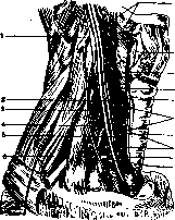
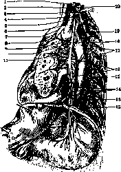
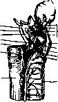
RECURRENS NERVUS (laryngous inferior, возвратный нерв), mainly a motor nerve innervating all muscles of the larynx except the m. cricothyreoidei (n. laryngeus superior); a branch of the p. vagi. It originates on the right before the appearance of the a. subclavia (Fig. 1), on the left in front of the end of the arcus aortae (Fig. 2). The right R. n. forms a loop around the a. subclavia, the left one around the arcus aortae; then both ascend between the pharynx and the esophagus to the larynx; penetrating the inferior constrictor muscle of the pharynx, they enter the larynx, where they divide into terminal branches—the anterior and posterior. On its way, R. n. gives off many branches (Fig. 3): rami tracheales, oesophagei sup., rami cardiaci sup. et inf., which participate in the formation of the plexus cardiacus; anastomoses to the ganglion cervicalis inf. The anterior 14 terminal branch ends in the m. crico-arytaeno-nideus lateralis, m. thyreo-arytaeno-10ideus, m. vocalis, mm. thyreo-epiglotticus and ary-epiglotticus. The posterior branch after anastomosis with the r. anastomoticus n. laryngei sup. ends in the m. crico-arytaenoideus post. et m. arytaenoideus. Pathology of R. n. Among the etiological factors, as in the disease of other nerves, intoxication, infection, trauma (in the form of compression of the R. n. by an enlarged thyroid gland, lymph nodes, aneurysm of the aorta, or a tumor in the surrounding R. n. formations) can be noted. In infection and intoxication, the R. n. is affected together with other branches of the p. vagi and with other cranial nerves; the paralysis is mostly bilateral. In acute and chronic diseases of the brain stem, the nuclei of the p. vagi, which give origin to the R. n., are also affected; the process is also mostly bilateral (see Poliomyelitis, Bulbar paralysis). In all organic lesions of the R. n., paralysis of the dilator of the glottis (m. crico-arytaenoideus posticus)—the Semen-Rosenbach law—sets in first, and only in the further course are other muscles innervated by the R. n. affected, thanks to the synergistic action of which narrowing, i.e., closure of the glottis, occurs. According to the Semen-Rosenbach law, dilators enter function first and only then constrictors. Since the R. n. is the main motor nerve of the pharynx, innervating both dilators and constrictors of the glottis, then in its diseases both muscle groups are paralyzed, of which only one (m. cricothyreoideus) is innervated by the n. laryngeus sup. and plays such a insignificant role in the movements of the vocal cords that its activity, in contrast to all other muscles, is not taken into account. As for the muscle dilating the glottis (m. crico-arytaenoideus post.), its: paralysis is the first sign of disease of the R. n. The Semen-Rosenbach law is now universally recognized for all progressive diseases concerning the trunk of the R. n., whereas in nuclear and root disease its significance is disputed (Korner). In the examination of the larynx, together with the paralysis of the muscles closing the glottis, incomplete separation during breathing of one or both vocal cords can be seen. In this case, the cord remains near the median line; it takes a position intermediate between adduction and abduction, the so-called corpse position, and no longer moves to the medial line during phonation, while the cord on the healthy side goes behind the median line, and the thyroid cartilages cross. At the moment of inspiration, the vocal cord on the affected side remains at rest, and only the healthy one takes the abducted position. In bilateral paralysis of the recurrent nerve, both vocal cords take the corpse position and during speech and breathing cannot be brought out of this position, due to which aphonia and significant disturbance of breathing are observed. In lesions of the brain stem, together with paralysis of the laryngeal muscles, paralysis of the soft palate and muscles of the pharynx is observed, due to which swallowing is also difficult. In the removal of pressure on the nerve or improvement of the inflammatory process, first the function of the muscles opening the glottis returns, then the muscles constricting it. - TREATMENT. In most cases of disease of the R. n., treatment is symptomatic, except for paralyzes of syphilitic origin, for which mercury treatment is applied. In bilateral paralysis of the laryngeal muscles, tracheotomy has to be performed.
Fig. 1. Course of the p. recurrens on the right side: 2-p. cervicalis IV; 2-p. cardiacus sup.; 3-p. phrenicus; 4-a. thyreoidea inf.; 5-ganglion cervicalis medium; 6-plexus brachialis; 7-ansa subclavia (Veis-senii); 8-n. cardiacus medius; 9-rami oesophagi; 10-n. recurrens; 11-rami tracheales; 12-n. laryngeus inf.; 13-ramus ant.; 14-mm. pest.; 25-ramus anastomoticus cum n. larynge inf.; 16-ramus externus et ramus internus (n. laryngei sup.).
Fig. 2. Location of the p. recurrens on the left side: 2-ganglion cervicalis medium; 2-n. cardiacus medius; 3-ganglion thoracale I; 4-ansa subclavia (Veis-senii); 5-n. cardiacus inf.; 6-n. cardiacus sup.; 7-ramus cardiacus sup. n. vagi; 8-plexus cardiacus; 9-ramus cardiacus inf. n. vagi; 10-ramus bronchialis ant.; 11-rami bronchiales post.; 12-n. splanchnicus minor; 13-n. splanchnicus major; 14-plexus oesophagi ant.; 15-truncus sympathicus; 16-rami oesophagi; 17-rami communicantes; 18-n. recurrens; 19-n. vagus sin.; 20-ganglion cervicalis inf.
tus of the glottis (m. crico-arytaenoideus posticus)—the Semen-Rosenbach law—and only in the further course are other muscles, innervated by the R. n., affected, thanks to the synergistic action of which narrowing, i.e., closure of the glottis, occurs. According to the Semen-Rosenbach law, dilators enter function first and only then constrictors. Since the R. n. is the main motor nerve of the pharynx, innervating both dilators and constrictors of the glottis, then in its diseases both muscle groups are paralyzed, of which only one (m. cricothyreoideus) is innervated by the n. laryngeus sup. and plays such a insignificant role in the movements of the vocal cords that its activity, in contrast to all other muscles, is not taken into account. As for the muscle dilating the glottis (m. crico-arytaenoideus post.), its: paralysis is the first sign of disease of the R. n. The Semen-Rosenbach law is now universally recognized for all progressive diseases concerning the trunk of the R. n., whereas in nuclear and root disease its significance is disputed (Korner). In the examination of the larynx, together with the paralysis of the muscles closing the glottis, incomplete separation during breathing of one or both vocal cords can be seen. In this case, the cord remains near the median line; it takes a position intermediate between adduction and abduction, the so-called corpse position, and no longer moves to the medial line during phonation, while the cord on the healthy side goes behind the median line, and the thyroid cartilages cross. At the moment of inspiration, the vocal cord on the affected side remains at rest, and only the healthy one takes the abducted position. In bilateral paralysis of the recurrent nerve, both vocal cords take the corpse position and during speech and breathing cannot be brought out of this position, due to which aphonia and significant disturbance of breathing are observed. In lesions of the brain stem, together with paralysis of the laryngeal muscles, paralysis of the soft palate and muscles of the pharynx is observed, due to which swallowing is also difficult. In the removal of pressure on the nerve or improvement of the inflammatory process, first the function of the muscles opening the glottis returns, then the muscles constricting it. - TREATMENT. In most cases of disease of the R. n., treatment is symptomatic, except for paralyzes of syphilitic origin, for which mercury treatment is applied. In bilateral paralysis of the laryngeal muscles, tracheotomy has to be performed.
Fig. 3. Muscles of the pharynx, larynx, innervated by the p. recurrens: 1-lig. thyo-thyreoideum lat.; 2-cornu sup. g. thyreoideus; 3-membrana 3na hyo-thyreoidea; 4-branch for m. *arytaenoideus transversus; 5-m. crico-arytaenoideus post.; 6-branch of p. йlaryngei inf. to m. crico-arytaenoideus post.; 7-oesophagus; 8-trachea; 9-branch to m. crico-arytaenoideus lat.; 10-m. crico-arytaenoideus lat.; 7-11-m. thyreo-arytaenoideus; 12-branch to m. thyreo-arytaenoideus; 13-cartilage thyreoidea; 14-n. laryngeus sup. (r. internus).
According to the Semen-Rosenbach law, dilators enter function first and only then constrictors. Since the R. n. is the main motor nerve of the pharynx, innervating both dilators and constrictors of the glottis, then in its diseases both muscle groups are paralyzed, of which only one (m. cricothyreoideus) is innervated by the n. laryngeus sup. and plays such a insignificant role in the movements of the vocal cords that its activity, in contrast to all other muscles, is not taken into account. As for the muscle dilating the glottis (m. crico-arytaenoideus post.), its: paralysis is the first sign of disease of the R. n. The Semen-Rosenbach law is now universally recognized for all progressive diseases concerning the trunk of the R. n., whereas in nuclear and root disease its significance is disputed (Korner). In the examination of the larynx, together with the paralysis of the muscles closing the glottis, incomplete separation during breathing of one or both vocal cords can be seen. In this case, the cord remains near the median line; it takes a position intermediate between adduction and abduction, the so-called corpse position, and no longer moves to the medial line during phonation, while the cord on the healthy side goes behind the median line, and the thyroid cartilages cross. At the moment of inspiration, the vocal cord on the affected side remains at rest, and only the healthy one takes the abducted position. In bilateral paralysis of the recurrent nerve, both vocal cords take the corpse position and during speech and breathing cannot be brought out of this position, due to which aphonia and significant disturbance of breathing are observed. In lesions of the brain stem, together with paralysis of the laryngeal muscles, paralysis of the soft palate and muscles of the pharynx is observed, due to which swallowing is also difficult. In the removal of pressure on the nerve or improvement of the inflammatory process, first the function of the muscles opening the glottis returns, then the muscles constricting it. - TREATMENT. In most cases of disease of the R. n., treatment is symptomatic, except for paralyzes of syphilitic origin, for which mercury treatment is applied. In bilateral paralysis of the laryngeal muscles, tracheotomy has to be performed.
к- Kiselev. REMAK Robert (Robert Remak, 1815-1865), one of the largest German neurologists. In 1838 R. graduated from the Berlin med. faculty. 20 €15
R. Remak.
Due to the prevailing antisemitism, R. was long denied the right to teach and only in 1847 began to give lectures, being the first Jew in Prussia to be a privatdozent. The main merit of R. consists in the study of the fine structure of the nervous system and in particular the vegetative. He showed that the fibers of the sympathetic nerve are devoid of a myelin sheath. Such non-myelinated fibers were named "Remak fibers" in honor of Remak by Kelliker. The fibrils constituting them were also called "Remak fibers." Another merit of Remak lies in the field of clinical neurology: he was the first to introduce direct current into electrotherapy.

Related articles
Mentioned in
Cite this page
“Recurrent Nerve.” Soviet Medical Encyclopedia. English translation of Bolshaya Meditsinskaya Entsiklopediya, 1st ed. (Moscow, 1928–1936), ed. N. A. Semashko. https://sovietmedicalencyclopedia.pages.dev/article/recurrent-nerve-2/