Aortic Aneurysm
Historical document, translated for reference. It reflects medical knowledge of the 1920s–30s and is not medical advice.
Summary
Aortic aneurysm occurs more frequently than aneurysms of other arteries and is almost always caused by syphilis. The article details the history, pathology, and clinical manifestations of this condition, noting that approximately 85% of cases result from syphilitic mesoaortitis.
Encyclopedia article (1928–1936)
AORTIC ANEURYSM occurs much more frequently than aneurysms of other arteries. In the clinic of internal diseases, aortic aneurysm holds a special position, since, with rare exceptions, it arises on a syphilitic basis and, along with the Wassermann reaction, with a high degree of probability serves as an indicator of syphilitic infection. - History. Aortic aneurysm was first described by Jean Fernel in 1554. Vesalius in 1557 was the first to diagnose aortic aneurysm during life. Since that time, aortic aneurysm has been studied in detail from pathological-anatomical and clinical aspects. The etiological connection with syphilis remained unknown, however, for more than 300 years. Only in 1879 did Welch first speak in favor of the connection between aortic aneurysm and syphilis based on a mass study of English troops, which found that in 66% of patients with aortic aneurysm, syphilis was in the anamnesis. Later authors confirmed Welch's hypothesis. Heller and Doehle in 1885 gave a detailed description of syphilitic mesoaortitis, which gained general recognition in 1903 after the exhaustive works of Benda and Chiari. The final proof of the specificity of the process was the discovery of the pale spirochete in the wall of the aorta in mesoaortitis and aortic aneurysm. With the discovery of the Wassermann reaction, the latter turned out to be positive in most cases of aortic aneurysm. Finally, in recent times, the question of aortic aneurysm has again been developed from the point of view of the influence of salvarsan therapy on the localization and course of syphilis, in connection with the fact that all statistics show an increase in recent years in aortic aneurysm and neurosyphilis along with a decrease in the number of diseases with bone and skin forms of syphilis. In general, it can now be considered established that approximately 85% of aortic aneurysms arise on the basis of syphilitic mesoaortitis (see Aorta, Aortitis - syphilitic). Pathological anatomy. Macroscopically, the less common fusiform (see Figure 1) and the more frequent saccular aortic aneurysms are usually distinguished. Aortic aneurysm is called dissecting (aneurysma dissecans) when blood penetrates into the thickness of the aortic wall itself and separates its layers. In size and external appearance, aortic aneurysms show a great variety of forms. The fusiform aneurysm represents a symmetrical expansion of the aortic wall in a certain area, but rarely reaches very large sizes; on its inner surface, thrombi are usually not present. The more common saccular aortic aneurysm represents an asymmetrical bulging of the aortic wall in the form of a sac. This form is almost exclusively of syphilitic origin. The sac can reach large sizes (have a diameter of 20 cm or more) and in such cases cause severe clinical manifestations already from one mechanical compression of neighboring organs. The aneurysmal sac connects with the cavity of the aorta by an opening of varying size. Sometimes this is a relatively narrow neck; sometimes a wide opening equal in diameter to the aneurysm, with beveled edges. In the first case, the entire aortic aneurysm is often filled inside with thrombotic masses, peculiarly tamponading the cavity of the aortic aneurysm. Due to this, the pulsation of the sac is significantly reduced and to a certain degree the possibility of its rupture is reduced. Thrombi organize relatively rarely. Cases of multiple aneurysms have been described, for example, four or more, in different parts of the thoracic and abdominal aorta.
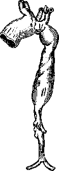
Figure 1. - Fusiform aneurysm (according to Kiilbs).
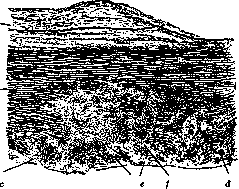
Figure 2. Gummous aortitis: a-sclerotic proliferation of the intima with insignificant inflammatory phenomena; b-elastic plates of tunica media; c-gummous small-cell infiltrate; d-giant cell; e-necrosis; f-fragments of elastic plates (according to Aschoff).
Fusiform and saccular aortic aneurysms (A. a.) may exist simultaneously. Sometimes a sac forms on a fusiform A. a. Of 181 cases of aneurysm studied by Cominotti, he found 37 of the ascending aorta, 49 of the aortic arch, 29 of the descending aorta, and 16 of the abdominal aorta. In non-syphilitic A. a., which occur rarely (about 15%), changes may be observed that are closely related to the underlying process on the basis of which such aneurysms form. These include ulcerative atherosclerosis, erosive processes, embolic, traumatic, etc., disorders of the aortic wall. Aortic dilation on the basis of developmental anomalies, e.g., in stenosis of the isthmus aortae, is more correctly classified as arteriectasias (see). Pathophysiology. The reason why focal changes in the aortic wall lead to the formation of A. a., and the conditions under which the aortic wall bulges, were attempted to be resolved experimentally. Soon, however, it became clear that neither incisions in the aortic wall followed by suture, nor mechanical damage to the muscular and elastic layers, e.g., by stretching, cause A. a. in dogs, rabbits, and other animals. A scar forms at the site of the wound. Only in cases where suppuration and necrosis occurred in the area of the wound did bulging of the aortic wall sometimes happen, which, however, did not subsequently show the progressive enlargement characteristic of human A. a. (Borst, Enderlen, etc.). Fabris also failed to produce A. a. by wounding in animal experiments. But he produced an aneurysm by injecting chemical substances into the arterial wall that caused necrosis, predominantly of the middle coat. The easiest way to produce focal, necrotic changes in the aorta of rabbits is by repeated small injections of adrenaline (see table for article 287-288, fig. 4). Adrenaline necroses often produce multiple A. a. in rabbits. The entire middle coat at the site of such an aneurysm may be necrotized with subsequent deposition of calcium salts. These experimental changes have no analogy with atherosclerosis. Therefore, Fischer proposed to call them adrenaline arterionecroses. They have theoretical interest because they show the important role of focal destruction of the middle coat in the formation of A. a. Subsequently, experimental aneurysms were obtained with solutions of KJ, salts of zinc, lead, mercury, etc. These experimental A. a. show that the primary, most important, condition is local, focal destruction of the middle coat with relative integrity of the intima and adventitia, i.e., approximately the conditions that occur in syphilitic mesoaortitis in humans (see figure 2). Simple incision of the intima, adventitia, or even all three layers together is insufficient for the formation of A. a. It is therefore understandable that atheromatous ulcers of the intima, as a rule, do not lead to A. a. The increase in arterial blood pressure, often previously mentioned as the main factor in the pathogenesis of A. a., is now denied by almost all. Adrenaline aneurysms (B. Fischer), individual descriptions of aneurysms in chronic adrenaline intoxication, etc., cannot be contrasted with the well-known clinical fact that in patients with A. a., blood pressure is either normal or reduced in most cases. The question of the participation of normal blood pressure as a mechanical factor in the bulging of the affected aortic area has caused fundamental disagreements. Previously it was considered almost obvious that the growth of A. a. occurs due to stretching of its wall under the pressure of blood flowing inside the A. a. According to such a view, the wall of the aneurysmal sac should rather appear atrophic. Benda rejects the old stretching theory. In the histological study of the structure of the A. a. wall, he found almost newly formed scar connective tissue. Benda believes that primarily, in the fresh stages of aortitis, not only the elastic and muscular elements, but also the surrounding connective tissue, are destroyed by inflammatory or traumatic processes. In their place, under the influence of irritation from blood pressure, young connective tissue begins to form compensatorily. The growth of the aneurysmal sac occurs due to the destruction of old connective tissue and the simultaneous formation of new tissue, mainly from adventitial cellular elements. Benda recognizes the connective tissue of the adventitia, like fasciae, as having great strength and low extensibility, quite sufficient to withstand blood pressure. Connective tissue, e.g., of syphilitic scars, is strong enough not to cause aneurysmal dilations. But a fresh gummatous process can cause them. Jores, generally supporting the theory of compensatory growth, considers it probable that a primary small A. a. may begin with stretching of the wall (ectasia) if it is affected by an acute process, e.g., syphilis, by analogy with the experimental aneurysms of Fabris, Fischer. A healthy, undamaged aortic wall is very strong. Oppenheimer, for example, needed to raise the pressure to 2,070 mm of mercury, i.e., almost 20 times the normal human pressure, to rupture an anatomically healthy aorta of a woman. Modern pathologists and physiologists believe that arterial hypertension is not a primary pathogenetic factor for the development of A. a. Such a factor is the focal destruction of the middle coat. Intra-aortic blood pressure not merely stretches the walls but serves as an irritating factor for the proliferation of the connective tissue of the aortic wall. However, stretching certainly occurs in the development, e.g., of large A. a., which often occurs in spurts. Periods of stationary state may be replaced by sudden, rapid enlargement of the aneurysmal sac under the influence of stretching, sometimes with ruptures and perforations. Etiology and pathogenesis. The detection of the pale spirochete of syphilis in the aortic wall in mesoaortitis and A. a. refuted previous assumptions about the para- or metasyphilitic origin of the process in the aorta. Aortitis is the result of the vital activity of the treponemes themselves, which have directly penetrated into the aortic tissue. The question of why spirochetes so often remain in the aorta for decades remains completely open. In non-syphilitic A. a., etiology and pathogenesis are varied. Erosive A. a. arise from the spread of a destructive process from neighboring organs to the aorta, e.g., in tuberculosis of the spine, lungs, mediastinal glands, in purulent mediastinitis, in ulcerated neoplasms of the esophagus, bronchi, lungs, stomach, etc. Erosive A. a. often end in fatal bleeding, e.g., into the stomach, esophagus, mediastinum. Atherosclerotic A. a. on the basis of ulcerative atheromatosis are not common because in such cases the process develops in the intima, which thickens. The middle and adventitia remain little changed and usually do not lose their elasticity. The development of A. a. on the basis of atherosclerosis occurs only in those rare cases when the process involves the middle coat, disrupting the continuity of its muscular-elastic layers. Embolic A. a. occur due to metastases in the so-called endaortitis lenta, sepsis. Parasitic aneurysms in humans, e.g., in echinococcosis, are extremely rare. Malarial and rheumatic aneurysms have been described. Traumatic A. a. with peri- and paraaortal hematoma are often found in wounds, e.g., gunshot wounds of the chest cavity. They mostly belong to the aneurysms formerly called false. In them, the sac is formed not by the wall of the aorta itself, but by the periadventitial connective tissue and tissues and organs adjacent to the aorta. Subsequently, the inner surface of such traumatic aneurysms is usually lined with endothelium originating from the intima of the aorta. In wounds, multiple aneurysms often occur in different parts of the aorta. Cases of arteriovenous aneurysms have been described, connecting, for example, the ascending aorta and the superior vena cava—aneurisma varicosum. Kraus noted a decrease in temperature in such patients. There are non-syphilitic A. a. that were preceded by severe trauma to the patient, but without direct injury to the chest cavity. Since the discovery of syphilitic mesoaortitis, many have completely denied indirect trauma as an etiological factor. However, Orth, Heller, and others, on the basis of histological studies, consider it proven in some cases that ruptures of the middle coat with hemorrhage into the thickness of the aorta and subsequent formation of an aneurysm occur without any participation of syphilis. Dissecting aneurysm, aneur. dissecans, has a peculiar pathogenesis. In it, blood penetrates into the thickness of the aortic wall and dissects its layers. Blood penetrates through primary ruptures of the intima and middle coat, which most often occur in the initial part of the ascending aorta. The cause was previously sought in increased arterial blood pressure. But it is noted far from always, which is confirmed by the frequent absence of left ventricular hypertrophy in aneur. dissecans at autopsy.
According to Iores, the cause lies in the insufficiently intensive growth of connective tissue elements in certain areas of the aortic wall. It is generally impossible to experimentally reproduce such an aneurysm mechanically on a human corpse, provided the aorta is healthy, but dissection occurs with an atheromatous aorta. Thus, it has been established that a diseased aortic wall is an essential condition for the formation of aneur. dissecans, in addition to ruptures of the intima. There is indication of the peculiar nature of this process. Babes and Minorescu, for example, observed a case where the aortic wall was dissected not only in the area of hemorrhage but also over a considerable distance beyond. Moreover, with slight effort, the aorta in the outer part of the middle coat easily separated into two layers. This case prompted the authors to consider the process in the aorta in aneur. dissecans as a peculiar independent disease, which they named aortitis dissecans. Undoubtedly, however, dissecting aneurysms occur that form due to local ruptures of the wall of an already existing aneurysm with hemorrhages, which are observed during the growth period of almost any A. a. They arise mainly in the peripheral parts of the sac, where it transitions into the aortic wall. Fissures and cavities connecting with the lumen of the aorta, sometimes through several openings, and containing liquid blood, gradually split the aortic wall and produce aneur. dissecans. Cases have been described where the dissection, starting in the ascending aorta, descended along the descending and abdominal aorta and reached the iliac and femoral arteries, so that the entire aorta, surrounded by a sheath of blood, lay in a case formed by the outer layers of its own wall. Statistics. Schrotter found aneurysms of arteries 220 times in 19,300 autopsies, Juda 48 times in 8,871, and Eppinger 22 times in 3,150. On average, therefore, 1-2%. A. a. occur (according to Chrisp's 551 cases), approximately, in 40% of all arterial aneurysms; of these, about 30% are of the thoracic aorta and about 10% of the abdominal aorta. Consequently, at autopsies A. a. occur in 0.4-0.8%, on average in 0.7% of all autopsies. Exact figures, of course, cannot be given. They will vary depending on race, nationality, sex, age, locality, etc., on the degree of spread of syphilis, and the methods and intensity of its treatment. Hart found syphilitic aortitis in 8% of 505 autopsies, Oberndorfer in 8.9%. Pletnev in 1,873 cases of heart and vascular diseases found: Patients Number Syphilis Aortic aneurysms total of which syphilitic of which with insufficient aortic valves Men Women 1,121 752 270 92 118 42 88 30 36 14 Combined statistical data characterize the ratio of the number of men to the number of women as 4:1. In terms of etiology, according to combined statistical data, 85% fall on syphilitic A. a.; 15% on non-syphilitic. According to Külbs, non-syphilitic (traumatic, inflammatory, erosive, etc.) occur more frequently at a young age, up to 30 years; syphilitic from 30 to 60 years. The time of appearance of clinical symptoms varies from 5 to 40 years after syphilis infection, in cases where it was established, i.e., on average, after 20 years. Clinic. Even A. a. of considerable size can occur without any symptoms and be discovered accidentally, for example, on X-ray or at autopsy. In E. Romberg's cases, only 65% of patients with A. a. complained of subjective phenomena. A latent period is probably characteristic of every A. a. After syphilis infection, many years usually pass without clinical manifestations. With the now common so-called chancre-free form of syphilis, the moment of infection remains entirely unknown. Furthermore, it is unknown how long mesaortitis lasts and when its aneurysmal phase begins. Therefore, the duration of A. a. always remains unknown. The clinical picture in that stage of A. a., when painful symptoms appear, is very diverse and depends not only on the nature and size of the A. a. itself, but also on the mechanical pressure on surrounding organs and tissues. Klever gives the following figures for clinical symptoms: Clinical symptoms of A. a. Diffuse A. a. Sac-like A. a. 75 42 82 58 42 25 50 42 25 50 12 42 25 75 Radiation of pains into the arms. Cough........... Pulsation of the chest Oliver-Cardarelli symptom. Systolic murmur..... Diastolic murmur.... Paralysis of the n. recurrens .... Wassermann reaction .... Clinical symptoms depend greatly on where in the aorta the aneurysm develops. According to Cominotti, in 131 cases of A. a., it was located in the ascending aorta-28%, in the aortic arch-38%, in the descending aorta-22% and abdominal aorta-12%. Rare forms of aneurysm of the ascending aorta, if they are located at its very beginning, are known under the name of aneurysms of the sinuses of Valsalva. They arise on the basis of ulcerative septic endocarditis, syphilis, atherosclerosis, injuries, and congenital anomalies. Aneurysms of the right sinus are more common, the wall of which adjoins the septum membranaceum and has the least external support. Rupture into the right ventricle may occur, accompanied by the sudden appearance of a systolic murmur in the region of cardiac dullness, a diastolic one on the aorta, and a 'cat's purr'. Such cases have been described in detail by Fr. Kraus. Aneurysms of the remaining part of the ascending aorta are more common. Sometimes X-ray for the first time reveals enhanced pulsation and bulging of the ascending aorta to the right. Often patients begin to experience pain in the chest in the form of either brief, acute attacks of angina pectoris that pass quickly, abrupt, occurring with movement, excitement, etc., or of the aortalgia type. In this case, the pains are less severe-dull, pressing. They slowly increase and subside; last for hours and days and are localized more often in the upper part of the chest. Pains of the first type probably arise on the basis of myocardial ischemia due to narrowing of the mouths of the coronary arteries by syphilitic aortitis. Aortalgic-p apparently due to irritation of nerve plexuses in the wall of the aorta itself or the nearest cervical sympathetic nodes. Diverse radiation of pains into the upper extremities, into the back-more often between the scapulae, into the neck, into the epigastric region, etc., occurs with both forms of pain. Small aneurysms are rarely accessible to percussion. A systolic murmur on the aorta is often heard, depending not on narrowing of the aortic valves, but on change in physical properties, on the rough surface of the aorta itself. The same explains the often accentuated and metallic shade of the 2nd tone on the aorta, which can appear without any increase in arterial blood pressure. Often the syphilitic process extends downward to the aortic valves and leads to their insufficiency. A diastolic murmur appears on the aorta, and a picture of heart defect develops with typical symptoms of aortic valve insufficiency, with expansion of the heart to the left and downward, capillary pulse, double Duroziez murmur on the femoral artery, increase in pulse blood pressure, etc. A patient with an aneurysm of the ascending aorta, due to the spread of aortitis to the valves, turns into a patient with a heart defect. Pulsus celer and the other symptoms of aortic valve insufficiency, however, may appear even with intact valves if the aneurysm is very large, and the backflow of blood during diastole occurs into the cavity of the aneurysm itself. If the aortic valves and the openings of the coronary arteries remain unaffected, the heart remains within normal limits even with very large A. a., since expansion of the aortic lumen does not create conditions for increased work of the heart. The right ventricle hypertrophies only in those rare cases when the pulmonary artery or a considerable part of the lungs is compressed. With large aneurysms of the ascending aorta, dullness on percussion appears to the right of the sternum in the 2nd, 3rd, etc. intercostal spaces, pulsation of the chest in the same area, displacement of the heart to the left and downward. Under the pressure of the aneurysm, the chest wall begins to protrude forward, and the cartilages, ribs, right clavicle, manubrium, and upper part of the sternum become thin and resorb. The aneurysm may appear under the skin as a pulsating swelling, sometimes firm to the touch from thrombotic masses (see Figure 3 and 4). With compression
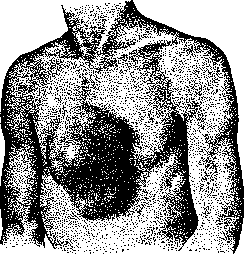
Figure 3. Aortic aneurysm (personal observation).
of the large veins-innominate, right subclavian, jugular, etc.-their engorgement and cyanosis of the corresponding area appear. Compression of the lymph vessels of the chest cavity can cause local edema of the right half of the chest, neck, and right arm. Compression of the trachea (according to Hampeln, occurs in about 20%) and the left bronchus (about 10%) gives symptoms of inspiratory dyspnea with sometimes audible at a distance stridorous stenotic breathing. In severe degrees of compression of the bronchi and lung lobes, atelectatic pneumonia and gangrene of the lung may occur. Compression of the bronchus may increase or decrease simultaneously with the systematic filling of the aneurysm and be accompanied by characteristic intermittent breathing. In the case of fusion of the aneurysm of the ascending aorta with the trachea or bronchus, the symptom of Oliver and Cardarelli may be observed, which is considered characteristic, mainly, for aneurysm of the arch.-Aneurysms of the aortic arch occur more frequently than others. They are richer in clinical symptoms than others, depending on the topography of the process. The mouths of each of the arteries branching from the arch-innominate, left carotid and subclavian-may narrow due to the pathological process. A difference in the filling of the corresponding arteries occurs, for example, in the carotid and radial arteries. In relation to the latter, this becomes especially clear if the patient is made to quickly raise both arms upward. The pulse, at this time, may disappear completely on the affected side. The second distinguishing feature (first described by Traube) is paresis or paralysis of the recurrent nerve (according to M. Schmidt, occurs in up to 80%; according to Hampeln-in 60%). The left recurrent nerve is almost always compressed, as it is this nerve that bends over the aortic arch. Paralysis of the left vocal cord is easily detected by laryngoscopy, and clinically manifests as hoarseness and a rough tone of the voice. Sometimes this is the first symptom of A. a. to appear. The right recurrent nerve is paralyzed only in rare cases of aneurysm of the innominate artery (with involvement of the right subclavian artery), over which this nerve bends. Due to the fact that the left bronchus lies under the aortic arch, the symptom of Oliver and Cardarelli is characteristic of aneurysms of the arches, i.e., displacement of the larynx with each systole, from transmission of the pulsation of the A. a. to the bronchus, trachea, and larynx. Compression and displacement to the right of the trachea and larynx, of the large veins, lymph vessels of the lungs, and gradual destruction of the manubrium and upper part of the sternum from pressure may also be observed here.-Aneurysms on the upper, convex surface of the arch can be palpated with the finger in the jugular notch early on. A. a. on the concave, downward-facing surface of the arch, in small sizes, may not be detected even under X-ray. They more often than others cause early and persistent hemoptysis, as well as paralysis of the recurrent nerve. The aneurysmal sac in the left part of the arch gives pulsation, bulging, and dullness to the left of the upper part of the sternum. Sometimes a systolic murmur is heard here as well. In large aneurysms, the pulse in the femoral artery may be noticeably delayed compared to the radial. Compression of the cervical sympathetic nodes gives unequal width of the pupils. Compression of the nerves gives various changes in skin sensitivity: anesthesias
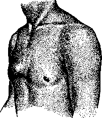
Figure 4. Aortic aneurysm (personal observation).
and paresthesias. On X-ray, there is a characteristic, of varying size, pulsating bulging to the left in the area of the aortic arch. Sometimes difficulty in swallowing occurs due to compression of the esophagus. Mesh-like aneurysms of the arch rarely involve the entire arch, which is more commonly observed in spindle-shaped aneurysms. The symptoms in such cases are even more varied. Finally, multiple aneurysms are observed.-Aneurysms of the descending aorta, giving symptoms common to all tumors of the mediastinum and thoracic cavity, especially often cause compression of the esophagus and difficulty in swallowing, as well as compression of the thoracic vertebrae. An early symptom in such cases is persistent pain of the neuralgia type in the back, chest, and sometimes in the left arm. The aneurysm can destroy the bodies of the vertebrae, compress the spinal cord and cause phenomena characteristic of transverse myelitis from compression. Paraplegia, bladder and rectal symptoms, bedsores, etc. develop. On X-ray, aneurysms of the descending aorta are best visible in the first and second oblique positions.-Aneurysms of the abdominal aorta occur least frequently. They are located almost exclusively in its upper part, under the diaphragm, at the sites of departure of tripodis Halleri and art. mesentericae superior. Clinically, attacks of abdominal pain of the angina abdominalis type often occur. At the same time, severe bloating of the abdomen occurs, with retention of gas and stool. The attacks last for minutes and hours. They resemble tabetic gastric crises or lead colic. Their basis is ischemia of the intestines due to narrowing of the mouth of art. mesentericae, which is why they are sometimes well relieved by nitroglycerin. Persistent pain in the back from compression of the nerve plexuses located in front of the spine is common. The pain can resemble renal colic. When the roots are compressed, corresponding girdle pains are observed. A common symptom is pulsation in the epigastric region; sometimes a tumor can be palpated in the left part of the hypochondrium. Systolic and diastolic murmurs have been described over the area of pulsation, conducted to the femoral arteries, and sometimes audible from the back at the lower thoracic and upper lumbar vertebrae. Aneurysms of the abdominal aorta are often filled with thrombotic masses. Sometimes the entire lumen of the abdominal aorta appears to be occluded by thrombotic masses. Clinically, complete occlusion of the abdominal aorta may not give any symptoms, as gradual thrombosis allows time for the restoration of sufficient collateral circulation.-Approximately from 15% (Hampeln) to 30% (Romberg) of A. a. ends in rupture. The bleeding accompanying rupture usually leads to death. However, cases are observed when the ruptures are small; hemorrhage occurs into a closed cavity, for example, into the pleural, and the patient survives. The sites of rupture of aneurysms were observed by E. Romberg and others in frequency, approximately, in the following order: trachea and bronchi, pleura, pericardium, esophagus, lungs, through the chest wall outward, into the abdominal cavity, into the retroperitoneal tissue, mediastinum, into the stomach, into the intestines, into the pulmonary artery, atria, right ventricle, superior vena cava. According to the material collected by E. Romberg, patients died, on average, from rupture in 35%, from heart failure in 25%, from narrowing of the coronary arteries in 20%, from extraneous causes in 20% of cases. Diagnosis. The initial phase of A. a. and syphilitic mzaortitis may not give any clinical symptoms and can be recognized only on the basis of X-ray. X-ray not only allows diagnosing A. a., but also with sufficient accuracy determine its size and location, which cannot be achieved to such an extent by any other clinical research method. When fluoroscopy and taking pictures, in addition to the frontal, it is necessary to examine patients in oblique positions, which is especially important for aneurysm of the descending aorta. [On a separate table (see art. 127-128) are presented X-ray photographs of aneurysms: 1-of the ascending aorta; 2-of the aortic arch; 3-of the descending aorta (frontal view); 4-of the descending aorta (in the first oblique position)]. In the initial stages, when the bulging is not clearly expressed, enhanced pulsation on a limited area of the aorta may indicate the beginning of A. a. Tumors of the lungs and mediastinium sometimes cannot be differentiated with accuracy even under X-ray. If the tumor is adjacent to the aorta, the pulsation of the latter will be transmitted to the neoplasm, although usually in one direction or another. On the other hand, aneurysmal sacs may not give any pulsations, being filled with thrombotic masses. This to a significant extent reduces the importance of the old sign of aneurysm-spread of pulsation in all directions-previously determined by palpation. A positive RW cannot have decisive value in such cases, as a syphilitic may also have a neoplasm. Palpation, especially above the clavicular spaces, can sometimes detect metastases. Repeated, prolonged hemoptysis is observed in both cases. Finally, on autopsy, in rare cases, the simultaneous existence of A. a. and a neoplasm is found. Aneurysms of medium size are recognized most accurately under X-ray, as with large ones it is sometimes difficult to say from which part of the aorta the aneurysm originates. In the X-ray diagnosis, all other things being equal, A. a. is indicated by even contours and the direct transition of the shadow of the aneurysm into the shadow of the vessels. Treatment. Patients with large A. a., especially with the threat of rupture, should be kept on strict bed rest. The same applies to patients with hemoptysis and frequent attacks of angina pectoris. Decompensated patients on the basis of accompanying insufficiency of the aortic valves are treated with rest and cardiac agents according to general rules. The main rule in A. a. of syphilitic etiology is possible early and energetic specific treatment. However, in cachectic, exhausted, elderly people, great caution is needed to avoid worsening of the general condition and weakness. They start with small doses and gradually increase. Caution is also required if there are changes from the kidneys. Treatment is generally carried out under frequent control of the urine composition. Usually they start with KJ. After 2 weeks, intravenous infusions of neosalvarsan are added in doses of 0.15-0.3-0.45, every 5-7 days; in total up to 4.0-6.0. Intramuscular injections of bismuth preparations, e.g., Bijochinol 3 cubic cm every third day; in total 30 cubic cm. In some cases, mercury treatment is carried out. Recovery in the anatomical sense does not occur, however, both specific treatment and properly conducted symptomatic treatment can in some cases slow down the progressive increase in A. a. and stop, and sometimes also noticeably reduce the aneurysm. Treatment in the latter cases probably affects the gummatous process in the wall of the aorta and increases its resistance to blood pressure. In some cases, under the influence of treatment, thrombi may be deposited in the cavity of the aneurysm. Remissions are often observed; during them the phenomena of compression of neighboring organs decrease, for example, paralysis of the vocal cord, neuralgic pains, etc., disappear. Under the influence of systematically conducted long-term treatment, many patients manage to prolong their life for several years.
Related articles
Mentioned in
Cite this page
“Aortic Aneurysm.” Soviet Medical Encyclopedia. English translation of Bolshaya Meditsinskaya Entsiklopediya, 1st ed. (Moscow, 1928–1936), ed. N. A. Semashko. https://sovietmedicalencyclopedia.pages.dev/article/aortic-aneurysm/