Endocarditis
Historical document, translated for reference. It reflects medical knowledge of the 1920s–30s and is not medical advice.
Summary
This article from the 1928–1936 Soviet medical encyclopedia defines endocarditis as inflammation of the heart's inner lining, emphasizing its morphological classification into valvular and mural forms. It details the historical understanding of the disease, from early descriptions of valve growths to the bacteriological theories established in the late 19th and early 20th centuries, and describes the pathological processes of warty and ulcerative endocarditis.
Encyclopedia article (1928–1936)
ENDOCARDITIS, inflammation of the endocardium, i.e., the inner lining of the heart. The greatest practical significance is held by lesions of the valvular apparatus of the heart—valvular endocarditis; lesions of the mural endocardium are designated as parietal (or mural) endocarditis. From a morphological standpoint, inflammation of the endocardium is distinguished by significant peculiarity, which is explained mainly by the fact that the endocardium has no vessels of its own, and the entire picture of inflammation proceeds under the conditions of so-called avascular inflammation. In other words, inflammation of the endocardium bears an almost exclusively histiocytic-productive character with one or another degree of alterative changes on the part of the valve substance. The purulent-infiltrative phenomena observed in the valves are explained in various ways. If such phenomena take place in hitherto unchanged (non-sclerosed) valves, then, given the lack of proof of the process spreading from the myocardium, i.e., from the base of the valve, the possibility of direct emigration of leukocytes from the blood bathing the valves is admitted. If, however, leukocytic infiltrates arise in already strongly changed, sclerosed, and vascularized valves (see below, recurrent endocarditis), then the emigration of leukocytes from the newly formed vessels of such valves is accepted as the most probable. Historical data. The word "endocarditis" first appeared in medical literature in 1834; with this term, Bouillaud named the inflammation of the valves discovered by him during rheumatism. Bouillaud already distinguished two forms: benign and malignant. But the changes on the valves which are observed in endocarditis were known to physicians even earlier. Senac (1749), Morgagni (1761), and others describe various cases where, during autopsy, soft growths were noticed on the valves, which they took for either syphilitic condylomata or traumatic injuries. Kreysig (1815) posed the question regarding the inflammatory origin of all lesions encountered on the valves particularly clearly, although Parry (1801) had recognized the inflammatory nature of heart and valve diseases even before him. In Russian medical literature, endocarditis is first mentioned in Sokolsky's work "Doctrine of Chest Diseases" (1838). Sokolsky was perfectly familiar with both the works of Kreysig and the views of Bouillaud, but believed that the "growths" on the valves were not caused by an inflammatory process, but "are formed as a result of small cracks or ruptures of ossified parts." The first complete description of acute endocarditis from a pathological-anatomical standpoint belongs to Rokitansky, who, having pointed out the variety of causes of endocarditis, tried to distinguish its individual forms. Further development of the question belongs to Virchow, who gave an excellent description of diphtheritic ulcerative endocarditis and pointed out for the first time its connection with septic-pyemic diseases. Winge and Heiberg (1869) first found bacteria in the deposits on the valves in endocarditis and thus laid the foundation for the doctrine, dominant in clinical practice even at the present time, that inflammation of the valves is caused by the settling and multiplication of bacteria on the valves. Subsequently, this was confirmed by Vysokovich and Orth, Weichselbaum, and others. The same Vysokovich proved experimentally that by introducing streptococcus or staphylococcus into the blood after mechanical traumatization of the valves, one can induce inflammation on the valves. Subsequently, the possibility of this without preliminary traumatization was confirmed by Meyer, Saltykow, and others. The doctrine of chronic endocarditis, known to clinicians for a long time under the name endocardite prolongée (Bouillaud) or end. recurrens (Jürgensen), was especially brought to the fore in clinical practice in recent years, ever since first Osler (1885) and, most importantly, Schottmüller (1909) described a special form of endocarditis ending in death, named by Schottmüller end. lenta, which forced a thorough revision of the entire question of endocarditis. In the USSR, Talalaev, Strazhesko, Kondratovich, Zimnitsky, Lang, Egorov, Bukhshtab, Gelshtein, Tsukershtein, and others took special part in the development of the entire doctrine of endocarditis. The initial morphological changes in endocarditis consist of the following three moments: damage (alteration) of the endocardium, histiocytic-cellular reaction, and thrombotic processes. The variety in the quantitative and qualitative side of these processes lies at the basis of the significant variety of endocarditis in general. Thus, in some cases, the presence of pronounced destructive (alterative) changes in the valves with a clearly noticeable loss of the substance of the latter is noted (ulcerative endocarditis); in other cases, thrombotic processes are found to be especially pronounced (thromboendocarditis); in third cases, neither one nor the other moment is prominent, and the entire process proceeds under the banner of productive inflammation with the development of sclerosis of the valve substance, its deformation, shortening, etc. (fibrous endocarditis). More often, however, one or another combination of the indicated moments or their succession in the course of the process takes place; thus, in the initial periods, alterative-thrombotic processes predominate, while later, sclerotic ones do, including the phenomena of organization of thrombotic masses. As for thrombus formation in endocarditis in general, it must be considered the rule, exceptions to which are quite rare. In other words, almost every endocarditis is to one degree or another a thromboendocarditis. From this, however, it does not follow that thrombotic phenomena represent the most important moment in the entire process. Such a moment should be considered the valvulitis itself, i.e., the lesion of the valve substance. The most frequent form of endocarditis is the so-called warty endocarditis, or verrucous (e. verrucosa). Macroscopically, in this case, one finds on the surface of the valve series of reddish or grayish deposits having the appearance of rounded, sometimes barely noticeable warts. The warts are usually arranged in a row along the line of closure of the valves, but can also lie randomly, for example, in parietal endocarditis. If the warts reach very large sizes (up to 1-2 cm or more), then one also speaks of papillary endocarditis. The wart itself, at the beginning of its formation, consists of three elements which normally go into the construction of a thrombus (platelets, fibrin, formed elements of the blood); later, connective tissue enters the composition of the wart, growing together with vessels into the substance of the thrombus from the side of the endocardium in the order of ordinary organization. The phenomena of organization are recognized by the sign that the wart itself becomes more compact; it either does not scrape off with a knife at all or scrapes off partially. Its color becomes gray, the consistency somewhat dense. Sometimes warts are not connected with thrombus formation, arising as a productive-cellular reaction of the endocardium. The following details are characteristic of the initial forms of endocarditis. On the part of the connective tissue fibers, a clear increase in their hydrophilicity is noted, expressed in swelling and loss of fibrillarity; the cells of the stroma swell and become rounded. Between the connective tissue fibers and cells, fibrin fibers appear, which, emerging onto the surface of the valve, usually merge with the mass of the thrombus. However, the question of the origin of fibrinous-fibrous structures in the valve tissue (they may not have any connection with thrombotic deposits on the surface) is not fully clarified; it is highly probable that a peculiar ("fibrinoid") transformation of the collagen fibers themselves takes place here, and not an exudation or "pressing in" of fibrin from the surface. The arrangement of the cells of the swollen endocardium is characteristic: the greater part of them lie with their long axis perpendicular to the surface of the valve, forming palisade-like structures, interleaved and covered by threads of fibrin (or fibrinoid). Endocarditis can be accompanied by hemorrhages into the valve substance; it is interesting that this phenomenon is encountered even in fresh endocarditis, i.e., in avascular valves. Ulcerative endocarditis (e. ulcerosa) is characterized by a vivid alterative moment, i.e., signs of destruction of the endocardial substance, which is easily detected macroscopically in the form of defects, perforations, etc., of one size or another. In the complex conditions of intracardiac hemodynamics, the loss of endocardial substance (valvular, chordal, or mural) gives a series of sequential effects in the form of tears and detachments of parts of the valve, chordae, papillary muscles, as well as in the form of valve aneurysms (especially in the anterior leaflet of the bicuspid valve), subsequently usually leading to their perforation [see separate table (p. 415-416), fig. 1 and 2]. Ulcerative endocarditis can also produce ulcers of the heart, sometimes also with perforative phenomena, especially in the upper part of the cardiac septum, e.g., immediately below the aortic valves. Usually, ulcerative endocarditis is accompanied by a lush development of thrombotic processes; in this case, the forming warts are distinguished by particular friability, crumble easily, and sometimes undergo softening and disintegration of the type of septic melting of the thrombus; their color then becomes dirty gray or greenish; sometimes the valves look as if they are completely covered with purulent-fibrinous deposits. Microscopically: more or less deep disintegration, leukocytic infiltrates; masses of bacteria, often in the form of entire colonies, are very characteristic.
Ulcerative endocarditis can arise on the surface of previously unchanged valves (acute ulcerative forms). But even more often they prove to be a complication of a chronically occurring endocarditis—recurrent ulcerative endocarditis (see below). Fibrous endocarditis is a concept that is not sufficiently defined. It is more correct to call those forms fibrous endocarditis where, from the very beginning, there are clearly expressed fibroplastic phenomena, i.e., in the entire picture, moments of histiocytic reaction with swelling and coarsening of the fibrous connective tissue of the valve substance predominate; the latter thereby becomes matte, leathery, and at the same time loses little in its general shape. Warty and thrombotic phenomena are completely absent in pure cases. Recurrent endocarditis refers to all those forms where one can prove the presence of a previous endocarditis (see below) and the presence at the same time of fresh changes (exacerbations), whether these are of a warty or ulcerative character (recurrent warty, recurrent ulcerative endocarditis). Recurrent endocarditis constitutes the main mass of endocarditis in general, i.e., it is the main form of their course. Healing processes in endocarditis, on the one hand, consist of the resorption and organization of thrombotic deposits with the formation of new vessels (see below), and on the other hand, there is a resorption of infiltrates in the substance of the valve, with the tissues of the latter undergoing more or less severe fibrous-cicatricial changes. The course of the fibers in the valves becomes disorderly, and numerous scar fields of keloid-like, almost homogeneous tissue arise. Organizational-thrombotic processes from the surface, sclerotic phenomena in the tissue of the valve itself, and finally—as a concluding moment—the shrinking of the latter in length and width (or height) lead to that deformation of the valvular apparatus, its shortening, and immobility, which are so characteristic of valvular defect. With previous ulcerative changes, all these moments are especially vivid: as a result of them, the entire valvular apparatus usually turns into a shapeless fibrous ridge or cord, and with abundant calcareous deposits, the entire given system of valves turns into a solid stony conglomerate. Especially abundant deposits of lime are observed in ulcerative endocarditis and, moreover, often in the early (presclerotic) phases of their development. An extremely characteristic and important detail of the healing process in endocarditis is the formation of new vessels in the substance of the valves and in organized thrombi. This new formation proceeds, on the one hand, by the development of offshoots from pre-existing vessels at the base of the valve, and on the other hand, there apparently takes place an autochthonous development of vascular loops in the mass of the changed valves, since upon artificial injection of the latter, a part of the vascular loops often remains uninjected. On the part of the bicuspid and tricuspid valves, one usually also observes changes in the chordal apparatus in the form of thickening, shortening, and adhesion of the chordae, which further limits the mobility of the leaflets; these changes of the chordae are sometimes preceded by one or another form of endocarditis (e. chordalis), including the possibility of ulcerative endocarditis with rupture of the chordae. Most often, sclerotic changes of the chordae proceed without thrombus formation in the order of their progressive sclerosis and thickening, as is observed in valves in pure forms of fibrous endocarditis. Upon microscopic examination, it turns out that the very thickening of the chordae falls on the proliferation of the subendothelial tissue of the chorda, while the axis of the latter remains without special changes. It is interesting that the very adhesion of the chordae does not occur as a result of their direct sticking together, e.g., with the help of interchordal thrombi and the organization of these thrombi, but in such a way that the hyperplastic and sclerotic processes descend subendothelially from the edge of the valve onto the chordae attached to it, gradually smoothing out the interchordal spaces in the process. Scleroses often spread to the ends of the papillary muscles. The first place in frequency is occupied by endocarditis of the bicuspid valve, usually leading to various degrees of its insufficiency and stenosis. The second place is occupied by endocarditis of the aortic valves. Aortic lesions are often combined with mitral ones. Endocarditis of the tricuspid valve is rarer. It should be pointed out, however, that endocarditis and defect of the tricuspid valve are comparatively often not recognized clinically, on which is based the somewhat incorrect idea of their comparative rarity. Combinations of endocarditis of the bicuspid and tricuspid valves with simultaneous involvement of the aorta or without it are not at all rare. Endocarditis of the pulmonary artery valves is very rare (e.g., ulcerative endocarditis in gonorrhea, in congenital heart defects). In parietal endocarditis, sclerosis of the endocardium also occurs in the form of either limited or more diffuse spots and stripes. Depending on the mechanical action of the blood flow, directed both from the apex and towards the latter (e.g., in insufficiency of the aortic valves), these parietal areas of sclerosis experience, on the one hand, hyperplastic changes, and on the other hand, they can turn, by virtue of the same mechanical causes, into peculiar pockets resembling semilunar valves. Usually, pockets arise during prolonged mechanical impact of two opposing blood currents, of which one is, as it were, undermining the base of the sclerosed area, and the other is smoothing it out. Most of the pockets are observed under defective aortic valves, less often in the region of the mitral valves. The pockets are most often open towards the base of the heart; less often, the reverse is observed, i.e., pockets open towards the apex. The previous designation of such pockets as "accommodation valves" should be considered incorrect, since in a functional respect they cannot play any significant role. From everything stated, it is understandable why endocarditic processes and the processes of healing during them so often cause one or another disturbance in intracardiac hemodynamics and lead to so-called organic valvular defects. Mild forms of warty endocarditis...
>

in
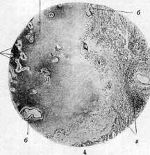
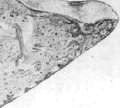
Fig. 1. Chronic endocarditis of the aortic valves and bicuspid valve; parietal endocarditis of the apex of the left ventricle; along the line of closure of the aortic valves—fresh warts. Figure 2. Ulcerative endocarditis of the aortic valves and chordae of the bicuspid valve; ruptures of both; sharp myogenic dilation of the heart (left ventricle). Fig. 3. Multilocular echinococcus of the liver: a—a piece of preserved liver parenchyma; b—thickened capsule; c—necrotized tissue around the parasites; the latter are in the form of small cysts; d—scar layers (cirrhosis); the total mass of the affected tissue resembles fine-pored cheese or stale semi-white bread. Figure 4. The same with slight magnification: a—cysts of the parasite with clearly visible membranes (b); c—necrotized tissue; d—granulation-scar tissue with infiltrates.
and not have such consequences, therefore the concept of endocarditis should be strictly distinguished from the concept of an organic valve defect. It is necessary to distinguish them, however, also because organic valve defects may not be of endocarditic origin, e.g., in congenital defects of the development of the valvular apparatus, in arteriosclerosis, in injuries in the region of the heart, etc. In other words, the summary concept of an organic 'valve defect of the heart' (vitium cordis) requires anatomical and pathogenetic clarification each time. The consequences of endocarditis outside the heart are very significant and in themselves are often the cause of death. First of all, thrombotic deposits on the valves carry danger in the sense of the possible detachment of these deposits and the subsequent development of embolism and infarcts. In this regard, aortic defects present the greatest danger, which is quite understandable, since the intensity of the work of the valves is of great importance here. Embolisms of the spleen and kidneys with ischemic infarcts in them are most frequent. Brain embolisms most often relate to the blockage of either the main trunk of the Sylvian artery (more often the left) or its small intracerebral branches. The consequences of embolism here will be foci of gray, sometimes red softening of one volume or another, sometimes softening covering almost the entire hemisphere. Comparatively rarely, emboli block the trunk and branches of the mesenteric arteries (gangrene of the intestine), the hepatic artery (usually without special consequences in view of the organ's supply also from the portal vein), and the arteries of the extremities. Embolisms of the vessels of the pituitary gland, retina, coronary arteries of the heart, adrenal glands, and other organs are known. Very often in endocarditis, one observes infarcts of the lungs (as a rule, hemorrhagic). Since in the majority of cases sources for such infarcts from the right heart are not found (the frequency of pulmonary infarcts does not at all correspond to the relative rarity of right-sided endocarditis), a position has arisen and strengthened that the main mass of such infarcts is caused not by emboli, but by spontaneous thrombosis of the branches of the pulmonary artery on the basis of marantic circulation in the lesser circulation. Sometimes thrombi from such infarcts continue to the very hilum of the lung. In the majority of cases, pulmonary infarcts arise only in the final period of endocarditis, e.g., in the presence of obvious decompensatory phenomena. Under conditions of sufficient compensation from the right ventricle, even in the presence of undoubted sources for embolism (for example, from the tricuspid valve), pulmonary infarcts usually do not develop, in which the relative perfection of collaterals in the arterial system of the lesser circulation is manifested. The serous covering (pleura, peritoneum) often bears a fibrinous effusion during the development of infarcts. In pulmonary infarcts (they almost as a rule emerge with their base under the pleura), serous-hemorrhagic effusions are not rare. These same infarcts are sometimes complicated by pneumonic processes from the surrounding tissues. Since emboli often contain microorganisms, the fate of the infarcts can undergo corresponding changes, e.g., in the form of suppuration, gangrenous decay. It is necessary to specifically note the possibility of the development of so-called mycotic arterial aneurysms. Such aneurysms are comparatively often found in the brain, in the extremities, and in various internal organs. The mechanism of the development of such aneurysms has not yet been finally clarified. Etiology of endocarditis. In the main mass of cases, we are dealing with an infection, and acute rheumatism dominates over all infections (see below). The pathogenesis of endocarditis is not fully clarified. The purely mechanical idea of direct damage to the endocardium by one virus or toxin or another should be considered abandoned, since the overwhelming number of infectious diseases, including sepsis, despite the existence of bacteremic phenomena, gives a comparatively very limited number of endocarditis cases. In other words, infection in itself is no more than one of the conditions required for the development of endocarditis, and furthermore, non-obligatory conditions, since, as indicated, endocarditis is possible even in non-infectious diseases. There is an attempt to explain the origin of valvular endocarditis by the peculiarities of the anatomical structure of the valves, in the sense of the pre-formation of vessels in them: the latter create the possibility of direct bacterial emboli. In favor of such a theory, such indisputable considerations are cited that embolic endocarditis indeed exists, that the development of vessels in the valves on the basis of past endocarditis can take place, that this creates a prerequisite for new emboli and for the exacerbation of the process, for its course according to the type of recurrent endocarditis, etc. On the other hand, it can be considered proven that fresh embolic endocarditis is still comparatively rare and, moreover, as a rule, it is anatomically peculiar, since pathological phenomena develop in this case at the base of the valves, and not along the line of their closure, which is characteristic of the mass of endocarditis cases. The embolic theory is also contradicted by the fact that with the undoubted existence of vessels in the valves and with the undoubted embolism of these vessels (e.g., in some cases of recurrent ulcerative endocarditis), the anatomical pictures characterizing endocarditis develop not at the sites of embolism, but at a distance, where bacterial masses in the vessels are precisely absent, namely near the edge of the valve. At the present time, the view of endocarditis as a particular, but territorially most important sign of damage to the vascular system as a whole, is gaining the greatest significance. This damage should be imagined as a more or less diffuse mesenchymal reaction of the organism, covering primarily the system of so-called littoral cells of the circulatory system, i.e., endothelial, intimal, and adventitial. The expansion of the anatomical base of this reaction can also cover the endocardium, which in its main mass represents the same intima. At the basis of such a diffuse mesenchymal reaction lie, first of all, intensified processes of sensitization of the organism, its increased sensitivity under the influence of a corresponding (especially repeated) infection or intoxication; perhaps the special reactivity of the indicated system, in particular the endocardium, as an expression of special properties of the genotype, also has significance. The possibility of increasing the adsorption capabilities of the endocardium, its reactivity, has been somewhat clarified at the present time also in an experimental regard, e.g., by introducing various dyes to sensitized animals, by the method of their intensified vaccination, etc. These data (Siegmund, Freifeld, and others), however, cannot yet be considered final. Along with the increase in the sensitivity of the endocardium, specifically from the side of the valvular apparatus, an auxiliary, purely mechanical factor is also put forward—the work of the valves. This work intensifies and complicates the initial phenomena to those degrees which we call endocarditis in the anatomical sense of the word. From the stated point of view, some extracardial processes in endocarditis could also receive a different illumination, such as: arteritis and mycotic aneurysms on their basis, thrombovasculitis in various organs, in particular in the renal glomeruli. However, the possibility of the development of such independent thrombovasculitis in endocarditis does not have to be disputed in general, since cases of pure sclerosis of the valves are observed, without vegetations, but with numerous infarcts, precisely on the basis of vasculitis with thrombus formation. Findings from the internal organs in endocarditis vary significantly depending on the duration of the suffering, the character and place of the lesion of the valves, the causes of death, etc. In particular, the answer to the question is of great importance: did the subject die from endocarditis as an infectious process, or did he die with phenomena of cardiovascular decompensation in connection with a heart defect that arose as a result of this process, at the given moment more or less completed. In the first case, at autopsy, we will obtain a picture of endocarditis against the background of a general infection (a classic representative of such cases is so-called sepsis lenta), and infection and intoxication often turn out to be the cause of death here. In the second case, the pictures of infection as such recede into the background, they may not be noticed at all, and in the center of everything stands heart failure with characteristic edema of the subcutaneous tissue, body cavities, cyanosis of organs, etc. A common cause of death for both cases is often embolism or thrombosis of organs vital for life. It is also natural that between pure endocarditis and possessors of pure heart defects there exist all transitions. On the skin, especially in ulcerative endocarditis, hemorrhagic rashes are often observed; the same on the conjunctivae, especially in so-called sepsis lenta. As indicated, the main mass of endocarditis has as its finale heart defects with more or less long periods of decompensation, which leaves a special imprint on the state of almost all organs. In contrast to septic forms, one never observes flabby, boiled, i.e., degenerated organs here; on the contrary, the majority of the latter are in a state of clear induration and cyanosis. Cyanotic induration of the kidneys and spleen is especially striking. At the basis of such induration lie, mainly, two factors: strong blood congestion and an increase in the general tone of the organ, mainly of its stroma and smooth musculature. It is incorrect to associate cyanotic induration with an increase in the mass of the connective tissue of the organ itself.
It is interesting that an increase in tone and significant induration are usually noted in certain hollow organs, such as the stomach, small and large intestines, and the urinary bladder; at the same time, one's attention is often drawn to a simultaneous decrease in the lumen of these organs, a kind of spastic state. In connection with this, it would be more correct to speak not of cyanotic induration, but of cyanotic hypertonia or cyanotic tension of the organs. Combinations of endocarditis with nephritis are not rare, especially in ulcerative, septic forms; chronic forms of nephritis (of intra- and extracapillary character) with phenomena of secondary contraction are also sometimes noted, and during autopsy, the pictures of cardiac decompensation intertwine with pictures of renal insufficiency, up to the phenomena of uremia. Diffuse forms of nephritis in endocarditis apparently have a common pathogenesis with the latter. There exists, however, an opinion that focal forms of nephritis are connected with multiple bacterial embolisms. Infarcts of the kidneys are a very frequent phenomenon; in comparatively rare cases, one observes massive formation of infarcts with necrosis of the greater part of the kidney. The same effect is sometimes achieved by the occlusion of the main trunk of the renal artery or its main branches. Pathological-anatomical mortality statistics (Moscow, 1923–27) show that endocarditis (and the heart defects based on them) occupy a very large place among other diseases of the circulatory organs (44.3%). In general, among autopsy material, the percentage of endocarditis fluctuates between 4 and 5. Congenital endocarditis represents a rare phenomenon. Usually, they are expressed in sclerotic changes of the valves, and almost exclusively of the bicuspid valve. Warty phenomena are not observed. The difficulty of distinguishing the indicated scleroses from simple defects in the development of the valvular apparatus of the heart is significant. Another question is the layering of typical forms of endocarditis onto undoubted defects in the development of the heart and its valves; such layering is a common phenomenon, e.g., around defects in the ventricular septum, in congenital stenosis of the pulmonary artery, etc.
I. Davydovsky. Etiology. The microbe causing inflammation of the endocardium is more often than others the streptococcus in all its varieties. When culturing blood from patients at the height of the process in various forms of endocarditis according to the Freund-Berger-Heilig method, streptococcus is most often cultured; moreover, regardless of the form of endocarditis, sometimes hemolytic, sometimes non-hemolytic, sometimes greening streptococcus is cultured. More often than other microbes, streptococcus is also found in blood cultures from the heart or emulsion from vegetations on the valves in individuals who died from endocarditis. But at the same time, staphylococcus, pneumococcus, gonococcus, influenza bacillus, tubercle bacilli, treponema, etc., are also sometimes found on the valves. Thus, any microbe, having entered the blood, can cause endocarditis; in practice, however, the causative agent of endocarditis is most often streptococcus, and in acute endocarditis, the hemolytic type of streptococcus is more common, while in chronic, on the contrary, the greening form (Str. viridans), which is an adapted mutational form of streptococcus. In the opinion of some authors (Schottmüller, Greff, Zuckerstein, and others), the cause of rheumatic endocarditis is a specific causative agent of rheumatism, which has not yet been discovered, but not streptococcus. Similar results are given by blood culture in endocarditis developing during rheumatic polyarthritis. According to the research of Heilig (in the clinic of Strazhesko), in 68% of cases of rheumatic endocarditis, streptococcus is also cultured. From the data presented, it is clear that the various clinical forms and the course of endocarditis depend not so much on the type of microbe as on the constitutional and immunobiological properties of the person who has contracted endocarditis and the environment in which the disease proceeds. Thus, the development of one or another form of endocarditis is conditioned and explained by the variability of the interaction between the virulence of the microbes and the protective forces of the organism. The significance of the biological properties of the organism is clearly evident from the fact that individuals with similar constitutional properties, for example, those originating from the same family, often contract endocarditis. For example, Schott, among individuals suffering from heart defects, counted 23.7% belonging to the same families. Endocarditis is observed in various infectious diseases when the causative agent of the infectious disease, settling on the endocardium, causes inflammation or, more often, due to the fact that a secondary septic infection joins, which causes endocarditis. Endocarditis is especially often a complication of various septic processes. Most often, streptococcal angina, streptococcal puerperal sepsis, panaritium, or a purulent wound, as well as chronic streptococcal foci in the tonsils, in the lymph nodes, and in granulomas around the roots of "dead" teeth, etc., cause complications in the form of endocarditis. A predisposing factor for the onset of the endocarditic process is already existing changes on the valves. Clinic of endocarditis. From a clinical point of view, acute, subacute, and chronic endocarditis are distinguished by their course, and by the severity of the clinical picture, symptomatology, prognosis, and outcome - benign (endocarditis benigna), having a tendency to recovery, and malignant (endocarditis maligna), not showing a tendency to recovery and ending in death at one time or another. In the chronic course of endocarditis, one can distinguish a third - "indefinite" form - endocarditis infausta, which is characterized by a moderate severity of the course and the simultaneous presence of symptoms characteristic of the benign and malignant form, with an alternation of periods of remission and periods of exacerbation (e. recurrens) being noted, and for a long time it is difficult to determine in which direction the process will turn, towards recovery or a fatal outcome, in short, where the prognosis appears extremely difficult.
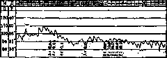
I. Benign Endocarditis. It seems completely impossible among cases of benign endocarditis to distinguish simple, plastic endocarditis (endocarditis simplex) from rheumatic endocarditis. The benign nature of the course of this form depends mainly on the immunobiological properties of the diseased organism and on the local tissue immunity at the site of valve damage and the speed of development of granulomatous tissue. If one takes the point of view that acute rheumatism is not a separate infectious disease, but only a special state of the organism, conditioned by the presence in it of an allergic, resp. hyperergic, reactive capacity acquired by the organism as a result of its sensitization, most often under the influence of streptococcal infection, then it becomes clear that neither from a clinical nor from a pathological-anatomical point of view is it possible to draw a line between these two forms. Both of them end in the vast majority of cases with recovery with wrinkling of the valves and the development of a heart defect. The difference here is quantitative, not qualitative, and during the patient's life, it is elusive. To consider the presence of rheumatic articular and other clinical phenomena as a differential sign between these two forms is not possible, because one often has to observe how, during simple endocarditis, rheumatic symptoms suddenly begin to appear. Some authors (Talalaev, Egorov, Zuckerstein, and others) classify both forms of endocarditis as rheumatism, believing that in reality there is a special form of rheumatism - namely, only rheumatism of the heart without damage to the locomotor apparatus. Endocarditis can be observed at any age, with women suffering from endocarditis approximately twice as often as men. In adults, endocarditis is more common between 15 and 40 years of age; it is rarely observed in children under 5 years of age, but as an exception, it can also be encountered in newborns. Endocarditis also occurs in intrauterine infants (A. Kisel), and in these cases, it is localized most often on the valves of the right heart, which is explained by the fact that bacteria and their toxins from the mother's organism directly enter through the placenta into the right heart of the infant. Benign endocarditis can develop simultaneously with rheumatic polyarthritis, sometimes precede it, or develop during or after various infectious diseases, such as pneumonia, influenza, scarlet fever, gonorrhea, and all kinds of septic processes. Symptomatology. In some cases, endocarditis proceeds without fever, without any symptoms from the heart - there are no changes in rhythm, no subjective sensations from the heart, no murmurs upon auscultation (aphonic form of endocarditis), in others only slight increases in temperature to 37.4-37.6° in the evenings and sensations of slight malaise. Such endocarditis, localizing along the edge of the valves or slightly away from it, can resolve, even without leaving a valve defect, within a few weeks, without being noticed by either the patient or the attending physician. In most cases, benign endocarditis is still accompanied by certain signs, such as pressure in the chest, a feeling of dull pain in the heart region, pain upon pressure on the precordial region (Head's zones), palpitations, and sometimes a sensation of heart irregularities. Among general symptoms, the most important is a moderate increase in temperature, most often of an irregular type (Fig. 1). Often, endocarditis, arising during one or another infection, initially causes only an exacerbation of the fever; Figure 1. Temperature in a favorable form of acute mitral endocarditis.
In other cases, the temperature, which had dropped almost to normal, rises again as if for no reason, and little by little other symptoms of endocarditis begin to appear. Headaches, malaise, pain in the lower back and limbs, and sometimes chills and sweats during the fall in temperature may periodically occur. Local symptoms from the heart are reduced to a change in the quality of the heart sounds and the appearance of murmurs. One of the first signs of endocarditis is the appearing indistinctness, lack of clarity of the corresponding heart sound, and its muffling. Soon, a murmur joins this acoustic phenomenon, progressing in its clarity from day to day. There is an opinion that these symptoms are the result of a myocarditic process occurring simultaneously with endocarditis. In view of the most frequent localization of endocarditis on the mitral valve, this murmur is best heard at the apex or at the edge of the sternum in the III or IV intercostal space on the left, usually clearer in the lying position. Two or three weeks later, an accent of the 2nd sound on the pulmonary artery joins this phenomenon; thus, the melody of mitral insufficiency is formed and revealed. In the case of the formation of a narrowing of the left atrioventricular orifice due to the gluing of the edges of the mitral valve, the 1st sound at the apex gradually acquires greater and greater sonority, and a small, indefinite murmur appears before it, and sometimes simultaneously a protodiastolic flow; thus, the melody of mitral stenosis is gradually formed independently or alongside the symptoms of insufficiency. But the process of its formation proceeds significantly slower than with pure mitral insufficiency. It lasts from 3 to 6 months. With inflammation on the aortic valves, a short systolic murmur usually appears first, to which a diastolic murmur, spreading to the lower third of the sternum, soon joins. Depending on the further direction in the formation of the defect, either the systolic murmur intensifies and becomes noticeably coarser during the formation of valve stenosis, or the diastolic murmur increases and becomes flowing during the development of aortic insufficiency. A number of symptoms that are observed from the heart depend not on the endocarditis itself, but on the lesion of the myocardium and the nervous apparatus of the heart—this concerns the irregularity of the pulse, such as extrasystoles, dropped ventricular systoles, or irregularity of the pulse in the form of arrhythmia during a disorder of conduction along the His bundle, attacks of tachycardia, etc. (see Myocarditis). It should be mentioned that already in this form of endocarditis, lesions of the capillary system, arteritis, periarteritis, and phlebitis are often encountered. Myocarditis to one degree or another always accompanies benign endocarditis. Pericarditis is also often encountered in one form or another (see Pericarditis). Glomerulonephritis is also observed, most often in the form of focal lesions. Then albuminuria, cylindruria, and, most importantly, hematuria of one intensity or another appear. Complications develop on the basis of either allergic (rheumatic) vasculitis and the formation of rheumatic granulomas in the vessel wall or on the basis of embolism (see above). Emboli, entering the terminal arteries and causing successive infarcts, do not produce suppuration despite the presence of virulent bacteria in the emboli, thanks to the presence of good immunity and hyperergic reactivity in the organism. Changes in the blood are reduced to an increase in the globulin fraction, acceleration of the ESR, an increase in the fibrinogen content, moderate leukocytosis with relative lymphocytosis, and also the development of secondary hypochromic anemia. The diagnosis of benign endocarditis has its difficulties. Often, rheumatic acute myocarditis, which can also be accompanied by a systolic murmur at the mitral valve due to dilation of the left heart cavity, as well as all kinds of arteritis, phlebitis, etc., without the involvement of the endocardium, gives cause for erroneous recognition. The presence of bacteria in the blood in case of doubt in the diagnosis always speaks in favor of endocarditis. The course and outcome of benign endocarditis do not lend themselves to calculation for a long time. In the majority of cases, it ends in recovery with the formation of a defect within one period or another. Cases of the transition of benign endocarditis into a malignant form are encountered far from rarely, especially in children. Death can sometimes occur from those complications that are associated with embolisms—e.g., embolism to the brain, embolism to the mesenteric arteries, etc. Thus, even benign endocarditis must still be recognized as a dangerous and serious disease in relation to life and the complete restoration of the organs and systems affected by it. In other cases, benign rheumatic endocarditis acquires a prolonged, sometimes lasting for years, course with periods of lull and exacerbation. In general, it must be said that benign endocarditis as a rule proceeds chronically and as an exception acutely. A characteristic sign of it in the presence of the described changes from the heart is a prolonged, sometimes lasting for months, rise in temperature in the range from 36.5° to 37.5° with individual flare-ups to 38.0–38.7° for two or three days, and sometimes for a longer period. During this period, all phenomena from the heart are exacerbated and the general condition worsens, and often an embolism occurs in some organ. The treatment of benign endocarditis is reduced to the energetic treatment of the underlying suffering. The earlier endocarditis is recognized and the more energetically the underlying disease, most often a septic disease, is treated, the faster the process ends in recovery. Treatment consists of maintaining the patient's strength with good nutrition and creating rest for the entire organism and the diseased heart. The patient must observe strict bed rest and keep a light ice pack or cold water on the heart for most of the day. One can recommend sodium salicylate with urotropin internally (4.0 sodium salicylate + 2.0 urotropin per day), as well as light cardiac agents, such as lily of the valley, valerian. More strongly stimulating cardiac activity agents, such as adonis, digitalis, etc., are permissible only for special indications. The timely intravenous use of silver, gold, and copper preparations may also have significance. The best agent is argoflavin (0.03–0.05 in 10.0 distilled water, collargol in the form of a 2% aqueous solution at 1.0–2.0–3.0, electrargol at 5.0–10.0 intravenously or at 20.0 intramuscularly). These medications are used daily or every other day depending on the effect from them, alternating with the intravenous infusion of 40% urotropin in an amount from 5.0 to 10.0 cm3. In other cases, it is useful to perform a blood transfusion 1 or 2 times in an amount of 250–400 cm3, best of all from a donor immunized with the streptococcus found in the patient. Finally, treatment with polyvalent anti-streptococcal serum sometimes has a good effect. With a sluggish course of chronic benign endocarditis, it is useful to add vaccination to the drug therapy. The removal of a septic focus, if it is discovered, may also have significance. The removal of such a focus in the tonsils or in a dental granuloma, where the streptococcus nests and lives, the removal of a sactosalpinx, a purulent cyst of the vermiform appendix, or the gallbladder can have a favorable influence on the course of endocarditis. Regarding tonsillectomy in endocarditis, there is no unanimous opinion. One should also not forget the climate treatment of chronic benign endocarditis. This treatment raises the strength of the diseased organism, strengthens immunity, and removes the hyperergic reaction in the rheumatic patient, in view of which it contributes to the suspension of the endocarditic process and accelerates the formation of the defect. Climate treatment must be carried out cautiously, in sanatorium conditions, under semi-bed rest, and under strict medical supervision. II. Malignant endocarditis with septic phenomena. A. Acute septic endocarditis (acute ulcerative endocarditis). This form of endocarditis is not a separate special disease form, since it is not caused by special microbes, but it deserves to be described separately due to the severity of clinical manifestations and the severity of the prognosis. Bouillaud already distinguished this form. He wrote: "The phenomena of inflammation here, just as in the previous form (forme simple), constitute an essential element, but they are so changed by the addition of typhoidal phenomena that one must agree that one should not confuse this kind of endocarditis with ordinary inflammatory endocarditis, and to distinguish it, we will give it the name of typhoidal endocarditis." These words of the great French clinician are full of meaning even in our days. This endocarditis differs from the preceding one not in its etiopathogenesis, but only in the severity of clinical manifestations. Together with Peter and Jaccoud, one can admit that malignant endocarditis develops in a poor state of the organism of the sick person, in subjects who are poorly nourished, cachectic, living in poor hygienic conditions, or weakened by previous diseases, in a word, in people with poor immunobiological properties, with a functionally weak reticuloendothelial system that does not provide sufficient resistance to infection. It is possible that the special virulence of the microbe and the number of microbes entering the blood from the site of the primary focus at one time also have significance here.
In this form of endocarditis, microbes are cultured from the blood, even using the Schottmüller method; most often these are various species of streptococcus, although in very rare cases pneumococci, staphylococci, and even gonococci have been detected in cultures. The endocardium can be affected in any sepsis. Very often, the severe picture of general sepsis, accompanied by endocarditis, myocarditis, and pericarditis, masks the picture of endocarditis. This form of septic endocarditis is called secondary endocarditis. In other cases, the only recognizable septic focus is the focus on the endocardium, and then endocarditis is the primary disease—primary septic endocarditis. In these cases, another primary septic focus is either not clinically detectable or does not exist at all, i.e., microbes enter the blood, passing through the mucous membrane without detectable damage, settle on the endocardium, and produce a primary septic focus there. Acute malignant endocarditis is not encountered so very often among cases of endocarditis. Out of 33,539 patients, among whom there were 243 cases of sepsis, Romberg counted only 42 cases of acute septic endocarditis. Out of 1,070 cases of endocarditis observed over 10 years in the clinics of Strazhesko, there were 67 cases of acute septic malignant endocarditis. Among the symptoms of acute malignant endocarditis, subjective cardiac complaints, as with
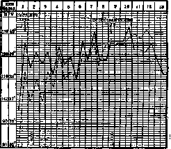
Fig. 2. Typhoid form of acute septic endocarditis. In simple cases, they recede into the background. More pronounced complaints regarding the heart always point to various complications involving the myocardium or pericardium, and in some cases to diseases of the coronary arteries or the nervous system of the heart. Symptoms from the heart itself do not appear so quickly. The disease often proceeds in the form of a severe infection, accompanied by general severe symptoms, often with clear signs of a progressing anemic state. In many cases, only an unexpectedly occurring embolism to the eye, brain, spleen, or kidneys, with insignificant phenomena from the heart in the form of a soft systolic murmur at the mitral valve, forces one to think about the possibility of acute ulcerative endocarditis. The diagnosis is facilitated if a diastolic murmur is heard at the aorta, which is rarely observed in severe forms of anemia. Among the general symptoms, fever deserves attention first of all. Temperature elevations have the character of either a constant high fever or a remittent type of fever, sometimes an irregular fever with individual spikes up to 40° and higher. In some cases, periods of more moderate fever are replaced by periods of intermittent fever with chills and sweats during the fall in temperature (Figs. 2, 3, and 4). The general condition of patients suffers quite severely, often from the very first days of the disease. Great general malaise, severe headaches, exhaustion, sometimes drowsiness and clouding of consciousness with the development of a state resembling typhoid status, distinguish acute septic endocarditis from benign endocarditis. The tendency toward embolisms is also expressed much more strongly here. Often the entire disease proceeds in the form of a severe fever with successive embolisms to the central artery of the retina, then to the brain, then to the spleen, then to the kidney, and finally to the skin with the subsequent formation of large petechial spots and individual hemorrhages. In some cases, larger arteries of the limbs and even the abdominal aorta are blocked by embolism, with subsequent gangrene of the fingers and even entire limbs in some cases. Figure 3. Temperature in the Schottmüller form of endocarditis lenta; Str. viridans in the blood. In other cases, the blockage of arteries or veins arises not on the basis of embolism, but as a result of septic arteritis or phlebitis. The entire vascular system (arteries, veins, capillaries) is necessarily affected in acute malignant endocarditis. Sometimes lesions of the lymphatic system—lymphangitis and lymphadenitis—are also observed. On the basis of lesions of small vessels—septic vasculitis—a true hemorrhagic diathesis develops, manifesting on the skin in the form of a petechial rash of pinpoint and larger diameter, nosebleeds, menorrhagia, and sometimes significant hematuria. In the blood, the percentage of protein content falls, the globulin fraction increases significantly, and the ESR (erythrocyte sedimentation rate) accelerates sharply. At the same time, the percentage of Hb (hemoglobin) content and the number of erythrocytes fall, among which pathological elements appear—microcytes and poikilocytes, even normoblasts. The blood picture resembles that of malignant anemia, but always with a color index less than one (hypochromic anemia), and in most cases with significant neutrophilic leukocytosis (up to 25-40 thousand), with a significant shift to the left and even with the appearance of individual myelocytes. The number of eosinophils is reduced. Large cells belonging to the reticuloendothelial system appear—typical and atypical histiocytes according to Schilling. In some cases, on the contrary, leukopenia is observed. Myocarditis is the most constant companion of malignant endocarditis, either in the form of inflammatory or degenerative myocarditis. It manifests as cardiac dilation, muffled heart sounds, sometimes a gallop rhythm, and, most importantly, tachycardia and all types of arrhythmias, and finally the development of a picture of cardiac decompensation with a drop in diuresis, the appearance of congestive phenomena in organs, and edema, the formation of which is facilitated by the altered composition of the blood and increased permeability of the capillary wall as a result of vasculitis. Figure 4. Acute aortic endocarditis (Str. haemolyticus); recovery with early use of argoflavin. Among other complications, pericarditis is encountered, either dry or exudative, more often with serous or, less frequently, seropurulent exudate; less frequently, pleurisy and septic pneumonia; and finally, pyelitis of embolic origin and enterocolitis, most often of toxic origin. The liver and spleen in most cases appear enlarged due to congestion and septic intoxication, and the spleen, in addition, due to frequent embolisms with the subsequent development of infarcts (white infarct), the formation of which each time causes sudden severe pain in the left hypochondrium, which can last for a fairly long time due to the subsequently joining perisplenitis. The diagnosis of acute malignant endocarditis is initially very difficult. Only the appearance of all kinds of embolisms and increasing heart murmurs in parallel with the weakening of its function determine the final diagnosis. Finding streptococci in the blood supports the diagnosis. Acute ulcerative endocarditis at the beginning of the disease can easily be confused with the cryptogenetic form of sepsis, with typhoid fever, with miliary tuberculosis, with pernicious anemia, with severe malaria, and finally with all kinds of septic processes, such as, for example, angiocholitis, paranephritis, etc. Increasing phenomena from the cardiovascular system and embolic processes decide the matter. The duration of the course is from several days to two to three months. Death is the rule, recovery the exception. Patients die either from some complications, e.g., cerebral hemorrhage, severe pneumonia, or more often from intoxication and exhaustion of strength during phenomena of progressive anemia. Treatment is symptomatic; in most cases, it remains ineffective. Nevertheless, it is necessary to treat, and as energetically and early as possible, according to the same rules as for benign endocarditis. In rare cases, having begun acutely, malignant endocarditis, with the gradual subsiding of septic phenomena, can pass into a chronic form of lingering, creeping malignant endocarditis. Benign endocarditis can also pass into this same form upon worsening of the process; when septic phenomena are masked to a certain extent, then the endocarditis is called indeterminate (endocarditis infausta), and when they are expressed quite clearly, this is the so-called creeping endocarditis with acute septic phenomena according to the nomenclature of Strazhesko, or endocarditis septica lenta according to the name given to this form by H. Schottmüller. The possibility of transitions of one form of endocarditis into another, as well as the finding of the same species of streptococcus in the blood in different forms of endocarditis, testifies to the fact that endocarditis is essentially one, while the course and form of endocarditis depend not on the specificity of the microbe, but on the constantly changing interaction of the immunobiological (protective) properties of the organism and the infecting microbe, which, in the case of a protracted course of endocarditis, adapts to the conditions of life in the organism fighting the infection and gradually mutates. Schottmüller's opinion that creeping endocarditis acquires such a course only due to the specificity of the microbe—namely, the greening streptococcus (Str. viridans)—must be recognized as incorrect after the works of Freund-Berger-Krieger-Loewengardt, Heilig, and others, especially since end. septica lenta is caused not only by the greening streptococcus but also by hemolytic non-greening and non-hemolytic streptococcus. The main characteristic of typical protracted endocarditis is an extremely slow, imperceptible onset, a lingering development of the disease with mild manifestations for a long time, but with a fatal outcome in many cases, sometimes after a whole series of years of the course. Of course, in life, there are many deviations from the indicated picture—the onset can be more stormy, as in acute septic endocarditis, and in the course of endocarditis, periods of remission can be replaced by periods of exacerbation, and the symptomatology can be weakly expressed, etc.—these are atypical cases. Most cases of lingering endocarditis arise on the basis of old changes on the valves. Whether this is the result of an outbreak of the endocarditic process from a smoldering streptococcal focus in verrucous deposits, where, as Reiss, Salus, and Kreitz found, virulent streptococci remain for a long time among thrombotic organized masses, or whether a secondary infection from a smoldering focus elsewhere (in the tonsils, in a tooth granuloma, etc.) takes place here is difficult to say. People of prime age fall ill, with women significantly more often than men (according to Strazhesko's statistics, in a ratio of 3:1). The onset of the disease, even in typical cases, and phenomena from the heart are often unclear and indefinite.
General malaise, resembling a grippal state, pain in the back and limbs, general fatigue, remittent temperature within the range of 37.3–37.5°, rising for a short time, usually during the day, between one and seven o'clock, gradually developing pallor, loss of appetite, and weight loss are the only signs of the onset of the disease. There are no complaints from the heart in non-stigmatized, nervous people. Examination of the heart during this period either yields no indications at all or only a systolic murmur is heard over the bicuspid valve. These symptoms are often taken by both the patient and the physician for symptoms of influenza, the onset of pulmonary tuberculosis, tuberculous bronchoadenitis, latent malaria, or thyrotoxicosis. Patients, feeling generally tolerable, continue their usual life and work until some new phenomenon, most often of an embolic nature, or a significant decline in strength and anemia forces the patient, who often presents no complaints regarding the heart, to turn to a physician again. In this period of the already developed disease, examination of the patient reveals excessive pallor (the skin color resembles coffee with milk) and clear indications from the cardiovascular system. Endocarditis, having begun in most cases on the bicuspid valve, has spread to the aortic valves, and examination reveals the entire symptomatology of aortic insufficiency. Examination of the vascular system reveals scattered capillaritis (Skulsky), arteritis, nodular periarteritis, phlebitis; the Rumpel-Leede sign is always present; petechial hemorrhages in the skin are often noticeable; pallor with a cyanotic tint of the fingers, the terminal phalanges of which thicken and resemble drumsticks. The liver and spleen appear dense and enlarged—a hepatolienal syndrome develops along with anemia, resembling the syndrome in chronic malaria or Banti's disease. Blood appears in the urine, as well as isolated hyaline casts. In the blood, the percentage of protein is lowered, the globulin fraction is increased; the erythrocyte sedimentation rate is accelerated. The number of erythrocytes and hemoglobin drops sharply, misshapen forms appear among the erythrocytes, nucleated forms unequal in size, the number of polychromatophils increases, but the developed severe anemia always remains hypochromic. The number of white elements in most cases fluctuates around the norm, but there are cases with leukocytosis and even hyperleukocytosis (35,000–142,000); sometimes, on the contrary, leukopenia is observed (usually in far-advanced cases). In some cases, there is significant monocytosis (up to 15–20%). Special large epithelioid cells appear in the blood—typical and atypical histiocytes (Schilling, Hess, Bittorf, Tushinsky, and others), indicating, according to Schilling, an intense struggle of the protective reticulo-endothelial apparatus with the infection, and according to Tushinsky—endothelial damage to the vascular wall (endovasculitis). A characteristic phenomenon for this form of endocarditis is periodically recurring embolisms in various organs: in the eye, brain, kidneys, spleen, intestine, bladder, and limbs with corresponding clinical symptomatology. Diagnosis of protracted endocarditis in the initial stages is difficult or even impossible. A detailed analysis of symptoms, characteristic changes in the aorta, hematuria, a dense large spleen, and hypochromic, rather than hyperchromic, anemia without megaloblasts make it possible to distinguish endocarditis from Biermer's anemia. It is more difficult to distinguish protracted endocarditis from malaria, but heart and vascular lesions, drumstick fingers, and repeated embolisms in the absence of plasmodia in the blood always point to endocarditis. The diagnosis is decided by a blood culture, which in almost all cases yields growth, most often of green-producing streptococcus. If a single culture is negative, the blood culture must be repeated using the Freund-Berger-Heilig method, and then the streptococcus is always eventually discovered (Loewenhardt, Heilig). Cases where enterococcus, diplostreptococcus, influenza bacilli, or micrococci were discovered are rare in the literature. The course of protracted endocarditis is long—months and years. Usually, patients die from exhaustion and chronic intoxication in a state of positive anergy: they have developed tissue immunity, but a great sensitivity to bacterial toxins remains, and patients die in a state resembling chronic uremia, however, without major functional disorders of the kidneys. Rarely do patients die from phenomena of circulatory decompensation or from accidental complications, which develop most often on the basis of various embolisms. The prognosis for protracted endocarditis is grave; however, not all patients, as Schottmüller thought, necessarily die. A number of recoveries have been described—everything depends on the form of protracted endocarditis and on how early treatment is started. It is carried out according to the principle already described above for benign and acute malignant endocarditis. Cases of recovery relate to protracted, septic endocarditis of an indefinite form (e. infausta). Cases of protracted endocarditis with acute septic phenomena of the type emphasized by Schottmüller, which represents only the final stage of septic endocarditis and not a separate disease as Schottmüller thinks, do indeed, as a rule, end in death. At autopsy, an ulcerous-verrucous form of endocarditis is found with large vegetations, sclerosing myocarditis, widespread endo-, meso-, and peri-vasculitis with local thrombi, cell proliferation throughout the entire reticulo-endothelial system with a transition to fibrosis and proliferation of connective tissue in parenchymatous organs, the parenchyma of which has undergone all kinds of degeneration and infarction with the development of scar tissue at the site of the infarcts. Capillary vessels in the organs show phenomena of endotheliitis. III. Parietal Endocarditis. Often, alongside the lesion of the valve endocardium, a disease of the endocardium lining the heart cavities is observed. It is perfectly understandable that cases can also be observed where microbes appear only on the endocardium lining the walls of the heart, producing the same inflammatory-ulcerative process with the deposition and organization of thrombotic masses as in valvular endocarditis. The exclusive localization of endocarditis on the parietal endocardium gave Baeumler (Baumler) reason to isolate this endocarditis into a separate clinical form, calling it "parietal." From the clinical side, this form is characterized by the absence of valvular phenomena in the presence of symptoms of myocarditis, proceeding with an increase in temperature and leukocytosis and multiple embolisms. Clinically, the picture very much resembles the picture of the first days after a heart attack during occlusion of the coronary artery system, in which parietal endocarditis with the deposition of thrombotic masses is also often observed. Isolated parietal endocarditis is observed relatively rarely in practice, and during life, it is most often diagnosed as myocarditis, and only an increase in temperature and repeated embolisms give reason to suspect endocarditis. Treatment is according to the general rules for the treatment of acute endocarditis. IV. Traumatic Endocarditis. By this name is understood endocarditis arising after a gunshot or incised wound, as well as after rupture, during a blow or sharp compression of the chest, of the valvular apparatus of the heart and the subsequent settling on the wound surface of the valves of pathogenic microbes circulating in the blood. In essence, therefore, this form is an infectious endocarditis, but with preceding damage to the valves. The clinical picture of the course is the same as in acute or chronic infectious endocarditis, with the only difference that in these cases there are always symptoms from the myocardium, which usually also suffered during the trauma. Treatment is according to the general rules of endocarditis therapy. V. Tuberculous Endocarditis has been studied mainly by the French clinic (Vaquez, Lion, Pic et Morenas, and others). It is encountered most often in children in the rare form of caseous endocarditis, in the form of follicular endocarditis, or vegetating endocarditis. The latter form is the most frequent. At the autopsy of those who died from miliary tuberculosis, neither tubercles nor giant cells are found under the microscope on the valvular apparatus in the latter case, but in the vegetations on the mitral or arterial valves among the fibrinous masses, Koch's bacilli are discovered. However, whether Koch's bacilli are the microbes provoking endocarditis in the presence of bacillemia is a controversial question: it is possible that the tubercle bacillus settles already secondarily at the site of inflammation caused by other microbes. The symptomatology of this endocarditis is extremely indefinite. In the opinion of Vaquez, this endocarditis is most often discovered only at autopsy. It is suspected during life only upon the appearance of cardiac phenomena, murmurs on the valves, or arrhythmia in a tuberculous patient with bacillemia, and also when endocarditis diagnosed in a tuberculous patient takes an atypical course, Koch's bacilli are present in the blood when cultured by the Löwenstein method, and there are no other microbes, for example, in a blood culture by the Freund-Berger method. VI. Syphilitic Endocarditis is known as a disease of the aortic valves, joining a disease of the aortic wall above the attachment of the valves. Syphilitic inflammation of the media, when affecting the vessels nourishing the aortic wall, also captures the valvular apparatus, which gradually shrinks, eventually causing aortic valve insufficiency or, more rarely, aortic stenosis. Aortic insufficiency, according to Schrumpf, is of syphilitic origin in 74.3%, and according to Hubert in 67%.
Simultaneously with this, inflammatory phenomena are sometimes observed also on the mitral valve and on the mural endocardium. This endocarditis is accompanied by retrosternal pain, a feeling of pressure in the sternal region during physical exertion, and subfebrile temperature. Upon objective examination, one finds on the sternum a systolic splitting of the 1st heart sound, an accentuated 2nd sound on the aorta, and alongside it systolic and diastolic murmurs, a slight enlargement of the left ventricle, a high and rapid pulse, and upon X-ray examination, a typical picture of syphilitic aortitis (see). Upon blood examination, reactions typical for syphilis are often discovered. The course depends on early recognition of the nature of the disease and energetic specific treatment. In case of delayed recognition, aneurysms of the initial part of the aorta develop with valvular insufficiency (see Aortic aneurysm), N. Strazhesko.
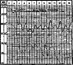
Endocarditis in childhood. Endocarditis in children has great practical and theoretical significance, therefore the diagnostics and clinical picture of endocarditis represent a question of paramount importance for pediatricians. Unfortunately, diagnostic possibilities for endocarditis are still extremely limited. Endocarditis sometimes runs a latent course. With involvement of the endocardium, the myocardium and pericardium are also frequently involved. It seems difficult to differentiate the symptoms that occur with one or the other lesion. Many symptoms observed in endocarditis can also be explained by the infectious disease that is observed in the child simultaneously with the endocarditis. It is understandable, therefore, that in clinics, the consequences of endocarditis—valvular defects—are diagnosed more often, and acute endocarditis significantly less often. Endocarditis rarely develops in the fetus during intrauterine life. Such endocarditis develops especially readily with cardiac malformations. Fetal endocarditis affects not only the right but also the left heart. Cases of congenital endocarditis have been described by a number of authors (Fischer, Weigert, and others). If sometimes congenital endocarditis develops with a congenital heart defect, then, on the other hand, sometimes endocarditis leads to the formation of a congenital heart defect. The etiological moment of congenital endocarditis is intrauterine infection—rheumatic, gonococcal, etc. In early childhood, up to 2-3 years, endocarditis is encountered very rarely. As a rule, they are of a septic character, caused by streptococcus, staphylococcus, and pneumococcus; the course is severe and rapid and usually ends in a fatal outcome. Diagnostics are very difficult and often not established during life. Sometimes there are no symptoms from the heart or they are poorly expressed: a systolic murmur, some enlargement of the heart, a change in the timbre of the first sound. Only pallor, dyspnea, cyanosis, irregular temperature in the absence of data from the lungs and other organs force one to think about heart involvement. Pathoanatomically, one is most often dealing here with ulcerative endocarditis. Treatment must be directed at the primary septic process. In the majority of cases, it is unsuccessful. In children after 5 years, rare cases of septic endocarditis are observed, but most often, one can say, almost exclusively, endocarditis in children after 5 years develops on the basis of rheumatism. Judging by the frequency of valvular defects that developed on the basis of endocarditis in rheumatism, one can say that rheumatic endocarditis (Fig. 5) is encountered not infrequently and affects about 0.5-1% of the entire child population. There is no pathognomonic sign in rheumatic endocarditis. A whole series of symptoms, such as pain in the heart region, a feeling of fear, dyspnea, increased heart activity, temperature elevation, change in heart borders, change in the timbre of the first sound at the apex of the heart, a systolic murmur in the same place, with positive anamnestic data about previously occurring attacks of rheumatism, will speak for heart involvement. Further observation and the gradual drawing out of features characteristic of endocarditis—involvement of individual parts of the heart, typical murmurs, etc.—makes it possible to determine endocarditis involvement. Not infrequently, general symptoms, subjective sensations, and functional changes are either absent or are so insignificant that endocarditis passes unnoticed if one does not pedantically examine the heart in rheumatism every day. Findlay, on the basis of a study of 701 cases of rheumatism over a number of years, considers that in the arthritic form of rheumatism, heart involvement is observed in 75.1%, and in chorea—in 53.6%, and in the presence of both arthritis and chorea in one and the same patient—in 69.2%. The frequency of rheumatism according to data from the Central Institute for the Protection of Health of Children and Adolescents in the city of Moscow among school-age children reaches 2%. Endocardial involvement, clinically determinable, is encountered in rheumatism, if the child was observed for a long time, in 90%. According to data from the pathoanatomical material of the Model Children's Hospital and the therapeutic clinic of the Central Institute for the Protection of Health of Children and Adolescents.
Figure 5. Temperature in so-called rheumatic endocarditis.
Most often the bicuspid valve is affected (in 55 cases out of 69); the second place is occupied by the aortic valves (38 cases out of 69); the third—the tricuspid valve (29 out of 69); most rarely the valves of the pulmonary artery are affected (1 out of 69). The bicuspid valves are affected first in time; the aortic valves are affected more often with a duration of rheumatism of several years. If endocarditis developed only in the form of warty deposits along the edge of the valve, then, since the closure of the valves occurs not linearly, but along a plane, there may be no sound phenomena (murmurs); if, however, murmurs have developed, they may disappear upon the regression of the warty deposits. Upon the shrinkage of a diffusely affected valve, restoration of the valve is impossible, just as is the disappearance of existing sound data. With manifestations of rheumatism in the form of chorea, endocardial involvement is encountered more rarely and runs a more benign course. Involvement of the endocardium of the papillary muscles and tendons is sometimes possible to assume; mural endocardial involvement is not diagnosed. A rapid fatal outcome in rheumatic endocarditis is not observed so often (according to Talalaev—in 4-6%) and depends on total heart involvement, multiple valve involvement, involvement of the conduction pathways, or coronary vessels, as a result of which the nutrition of the heart muscle is disrupted. The course of rheumatic endocarditis depends, apparently, mainly on the constitutional properties of the child. With a constitutional predisposition, already the first attack of rheumatism in the majority of cases gives heart involvement. With repeated attacks, heart involvement intensifies and sometimes leads to a fatal outcome. In other cases, the course acquires a favorable character: endocarditis passes into a valvular defect, with which the child can live for many years. Treatment in the absence of compensation disorder reduces to the prescription of rest in the acute period, cold on the heart region, sedatives, and proper nutrition: small frequent portions of tasty, varied food with moderate restriction of fluids. The primary disease—rheumatism—is treated. In some cases, for reasons not yet closer known to us, the course of endocarditis is peculiar. An aortic defect is observed more often with enlargement of the spleen and glomerulonephritis, with prolonged elevated temperature, with phenomena of anemia, and sometimes with embolisms. The course is persistent, progressive; children more often perish. Such a symptom complex bears the name endocarditis lenta (Fig. 3). According to data from pathologists (Talalaev), primary forms of endocarditis lenta are observed in 20%, the remaining 80% are made up of rheumatic verrucous endocarditis. The prognosis in endocarditis lenta is most often unfavorable. The duration of life is about 1-1 1/2 years. Treatment reduces to raising the patient's strength, the use of cardiac agents, and symptomatic treatment. Of great clinical interest and great diagnostic difficulties are cases of recurrences of rheumatism, when one has to decide the question of whether there are old changes in the heart or, in addition, there are also new changes. The clinic teaches that rheumatism is prone to recurrences, and any cardiac pathology especially readily settles on an already altered soil. On pathoanatomical material of the Model Children's Hospital (Moscow), the development of fresh endocarditic deposits on the site of old ones was noted in 15%. Exacerbation of the rheumatic process and the variability of data from the heart indicate the possibility of the addition of new endocarditic changes. N. Omshovsky.
Related articles
Mentioned in
Cite this page
“Endocarditis.” Soviet Medical Encyclopedia. English translation of Bolshaya Meditsinskaya Entsiklopediya, 1st ed. (Moscow, 1928–1936), ed. N. A. Semashko. https://sovietmedicalencyclopedia.pages.dev/article/endocarditis/