Myelitis
Historical document, translated for reference. It reflects medical knowledge of the 1920s–30s and is not medical advice.
Summary
Myelitis refers to inflammatory diseases of the spinal cord. The article traces the historical evolution of understanding myelitis from the early 19th century through various classifications by prominent physicians, including Leonhardi, Olivier, Charcot, Leyden, and others, who differentiated various forms and causes of spinal cord inflammation.
Encyclopedia article (1928–1936)
MYELITIS (from Greek myelos - spinal cord), inflammatory diseases of the spinal cord. At different times and up to the present, various pathological processes in the spinal cord were described under the name M. Until the beginning of the 19th century, diseases of the spinal cord and its membranes were designated as spinitis, rhachialgitis, notomyelitis. Leonhardi was the first to introduce the designation M. for them. In 1821, Olivier gave the first detailed description of the macroscopic picture of M. and differentiated it from inflammation of the meninges. He also included softening of the spinal cord under M., placing it in parallel with encephalitis, i.e., softening of the brain. 'Chronic M.', taught Olivier, 'lead to sclerosis'. Charcot and Dujardin-Baumetz (1873) distinguished M. with softening from M. without such. In 1874, Westphal was the first to describe acute disseminated M. Leyden (1874-76) included traumatic myelomalacia, compressive M., spontaneous acute softening of the spinal cord, acute bulbar M., acute M. without softening (including acute spinal paralysis of adults, Landry's paralysis, paralysis from cooling, acute ataxia), abscess of the spinal cord, and acute myelomeningitis under acute M. Leyden included tabes dorsalis, multiple sclerosis, and combined sclerosis under chronic M. Erb (1878) tried to separate inflammatory softening of the spinal cord from vascular and compressive M. He attributed multiple sclerosis, especially its spinal form, to chronic disseminated M. To the present day, the controversy over the relationship between acute forms of multiple sclerosis and disseminated myelitis has not been resolved. Tietzen (1886) expressed the idea that M. is not an inflammatory process, but only a degenerative one, depending on embolism and thrombosis. After Oppenheim (1891), who significantly narrowed the concept of M., it had to be expanded again when it became clear that even acute poliomyelitis was considered a form of M.

Fig. 1. Sagittal section of the brain of a fetus 1 - corpus callosum; 2 - hemispheres of the cerebellum; 3 - thalamus; 4 - pons; 5 - medulla oblongata; 6 - cerebellum; 7 - white matter of the cerebellum; 8 - cerebral hemispheres; 9 - anterior central gyrus; 10 - optic thalamus; 11 - corpus striatum; 12 - globus pallidus; P - pons Varoli; Ik
t
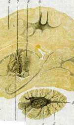
Fig. 2. Sagittal section of the brain of a newborn 1 - pons Varoli; 2 - thalamus; 3 - cerebellum; 4 - white matter of the cerebellum; 5 - cerebral hemispheres; 6 - anterior central gyrus; 7 - optic thalamus; 8 - medulla oblongata; 9 - nucleus tegmentalis;
Figure 3. Sagittal section of the brain of a 9-year-old child: 1 - hemispheres of the cerebellum; 2 - white matter of the cerebellum; 3 - area of the brain; 4 - medulla oblongata; 5 - central sulcus; 6 - thalamus opticus; 7 - internal capsule; 8 - lenticular nucleus. (The dark color is the staining of the nerve tissue.) constitutes a typical inflammation of the spinal cord. If poliomyelitis is still described separately from other M., then exclusively for historical reasons. Also, Landry's paralysis, which is closely related to poliomyelitis, belongs to the group of acute M. Chronic forms of M. also include spinal processes developing on the basis of anemias and cachexia. These include funicular M., or so-called combined sclerosis (see). The discussion about inflammation (Aschoff, Marchand, Rossle and others.) has also extended to the problem of inflammation in the central nervous system. The role of neuroglia (see) in inflammatory processes became the starting point in the classification of myelitis and encephalitis. An attempt was made to oppose infections that mainly affect the ectodermal tissue of the nervous system, nerve cells, fibers, and glia, to those that cause a reaction mainly from the mesenchymal elements of the nervous system (meninges and vessels). Levaditi distinguished neurotropic ectodermoses under the name of poliomyelitis, epidemic encephalitis, rabies, and herpes. To these forms, caused by Levaditi by a filterable ultravisible virus, some authors also include the so-called disease of Born, i.e., encephalomyelitis in horses, as well as M. and encephalitis after measles, whooping cough and other infectious diseases, which supposedly activate the virus present in the body or alter the body's own allergy. These also include those myeloencephalites that have recently been observed after vaccination. The recently mentioned 'neurotropic' infections are opposed to M. and encephalites in which a mesenchymal reaction is observed, namely M. and encephalites in typhoid, purulent diseases, meningococcal diseases, from chronic diseases - in tbc and syphilis. Spielmeyer rightly points out to the artificiality of such classifications based on histological differences. The same ectodermoses in their different stages cause reactions sometimes from the mesenchyme, sometimes from the glia. Besides the factor of time, the local factor is also important for the nature of the reaction: different tissue reactions near the site of infection and at a distance from it. Rossle's experimental research on inflammation showed how as the organism becomes more complex, the nature of inflammatory reactions also becomes more complex. In the most differentiated organ, the central nervous system, glia, originating from ectoderm, acquired the functions of stroma, interstitium, which are characteristic of mesenchyme everywhere and participate to a large extent in inflammatory processes in the central nervous system, acquiring some other qualities. But this does not completely 'remove' the function of mesenchymal elements of the central nervous system, but only modifies it. From this breach of the 'law of the mesenchymal nature of interstitium' it is clear how artificial are the constructions that do not take into account the complexity of phenomena in M. and encephalomyelitis, but single out only one side of it. But the main disadvantage of classifications based only on the histological picture is the ignoring of the fact that M. and encephalomyelitis are clinical-anatomical phenomena in which all elements of the nervous system and the whole organism participate both morphologically and functionally. In the further presentation, M. will imply clinical pictures of the disease of the spinal cord, which are based on its inflammatory reaction to various, mainly external, harmful influences. - To M. should be attributed diseases of the spinal cord in connection with acute and chronic infection, intoxication, trauma, when the latter causes secondary, or 'symptomatic' (according to Spielmeyer) inflammation of the spinal cord. For the same reason, it is difficult to single out compressive M. and some forms of myelomalacia (softening of the spinal cord) from this group, which depend on vascular lesions and also cause inflammatory reactions. Often the involvement of the meninges of the spinal cord - meningomyelitis; very often the brain is also affected at the same time - myeloencephalitis, or encephalomyelitis. The simultaneous involvement of all these elements gives a picture of meningomyeloencephalitis. - Depending on the spread of the process, focal M. and diffuse, or disseminated, are distinguished. Focal M. can occupy the entire cross-section of the spinal cord (myelitis transversa). Disseminated myelitis sometimes may be limited only to the gray matter (see Poliomyelitis) or only to certain systems of white matter (see Combined sclerosis). Etiology. Acute infectious M. and myeloencephalites have been described after influenza, measles, whooping cough, scarlet fever, chickenpox and smallpox, typhoid, dysentery, diphtheria, mumps, gonorrhea, malaria, erysipelas, cholera, pneumonia, appendicitis, trichinosis, rheumatism. Infectious M. also include epidemic diseases of the central nervous system: Heine-Medina disease (see), epidemic, or lethargic, encephalitis, or, better, encephalomyelitis, disease of Born in horses, rabies. Purulent M., up to the formation of abscesses, can develop as a result of suppuration located anywhere in the body: in lung abscess, liver, in parametritis, cystitis, phlegmons, in purulent inflammation of the paranasal sinuses, in meningococcal meningitis. Chronic infections - syphilis and tbc - cause both acute and chronic M., often with significant involvement of the meninges - meningomyelitis (meningomyelitis, or myelo-meningitis), sometimes giving a picture of compressive M. Multiple sclerosis also belongs to chronic infectious M., although it sometimes has an acute onset. - Toxic M. and encephalomyelites develop after acute or chronic poisonings with CO, illuminating gas, carbon disulfide, chloroform, nitrobenzene, in autointoxications, in eclampsia of pregnant women, in chronic nephritis, in cachexias from malignant tumors. This should also include M. developing after tetanus, diphtheria, poisoning with fish and sausage poison (see Botulism), after rabies vaccinations. (On the connection of funicular M. with malignant anemia, helminthic diseases, etc. - see Combined sclerosis. - On the toxic disease of the brain after alcohol - see Polioencephalitis.) M. after trauma are caused by penetrating wounds or strong contusions of the spine (concussive M.), when the disintegrated area of the spinal cord provides a ground for the development of bacteria present in the body, but sometimes develop without infection (so-called symptomatic M.). - Decompression M. in caisson disease (see Decompression diseases, Caisson work) is caused by gas embolism of the capillaries of the spinal cord. Occasionally decompression M. is observed in ascents to high mountains or in aviators, more often in divers, in workers in caissons under pressure up to 4 atm. with too rapid exit from the caissons. In the etiology of compressive M., the greatest role is played by tub. or other diseases of the spine (see Spondylitis) and meninges (see Meningitis), hemorrhages into the meninges (see Haematorrhachis), as well as extramedullary tumors. (On the causative agent, statistics and epidemiology of epidemic forms of M. encephalomyelitis - see Heine-Medina disease, Encephalitis.) As for the frequency of individual forms of M. and meningomyelites, recently M. after various infectious diseases (measles, whooping cough, etc.) as well as after rabies vaccinations are often observed. Even more common are syphilitic and tuberculous myelites, which often run under the guise of compressive myelitis. Pathological anatomy. Pathological changes in M. cannot always be detected macroscopically. But sometimes, especially in acute cases, it is easy to note hyperemia, general increase in volume, softer consistency of the spinal cord. When the spinal cord is cut, its white matter protrudes. With a knife, a soft mushy mass is scraped off. When the entire cross-section is affected, the characteristic butterfly figure disappears. The boundaries between gray and white matter are smoothed out. In hemorrhagic M. (m. haemorrhagica), the red color of the cut is striking, in fatty degeneration - yellow staining. On stained preparations, even without the help of a microscope, many additional details are found. Thus, dilated small vessels and capillaries are more prominent. Along their path, sometimes visible to the naked eye, there are infiltrates. In purulent M., sometimes small abscesses are visible to the naked eye. - Chronic M. are already macroscopically characterized by greater densification, 'sclerosis' of the spinal cord (m. sclerotica). In the most severe degree, such a process is observed in multiple sclerosis. In long-lasting M., ascending and descending degenerations develop in the long pathways above and below the inflammatory focus. Macroscopically on stained preparations, this is sometimes detected as 'areas of rarefaction' [see separate table (pp. 71-72), figure 5].
The most characteristic is the microscopic picture of M., with varying degrees of involvement of different elements (neuroglia, mesenchyme) in the inflammatory reaction, which determines the various histological pictures of M. In acute non-purulent M., severe changes are sometimes observed in the ganglion cells of the nervous parenchyma, with loss of processes and disintegration of the nucleus (m. parenchymatosa). The glia at this time sometimes also does not proliferate, but disintegrates or undergoes ameboid degeneration. Sometimes (in sepsis, after typhus) swelling of ganglion cells occurs with increased activity and proliferation of glial elements, which, along with leukocytes, actively participates in neuronophagy. Glial cells, especially Hortega cells, form nodules with the character of granulomas (see Neuroglia). In other cases, infection particularly affects the brain vessels, thrombi and hemorrhages occur (m. haemorrhagica, s. apoplectica), leading to disintegration of nervous tissue (m. interstitialis). Finally, in third cases, the entire usual inflammatory complex comes to the fore, with the participation of the entire vascular-connective tissue apparatus of the spinal cord, along with the parenchyma. Alongside degenerative changes, vascular and proliferative-exudative processes, mesenchymal, leukocytic and lymphocytic infiltrations, and reactive proliferations of glial tissue appear. In the initial stages of acute non-purulent M., polymorphonuclear cells predominate microscopically, which are later replaced by lymphocytes and plasma cells. In protracted processes, the lymphocytic reaction is almost always prominent. In the microscopic picture of most cases reaching autopsy, degeneration and softening (myelomalacia) predominate. The axis cylinders swell, myelin disintegrates, forming spheres and products of fatty breakdown. Nerve cells die and undergo neuronophagy. Depending on the stage, the outcome of acute non-purulent M. microscopically presents either a picture of 'fields of rarefaction' ('Luckenfeld') in the form of a wide-meshed tissue, or a picture of foci of softening with myelin disintegration, detritus, and granular cells, or a picture of scar-forming proliferating glia. In a large number of cases, pathological changes in M. are limited to vascular reaction without significant participation of the parenchyma and even the glia. In acute purulent M., enormous amounts of purulent cells are visible microscopically, filling not only the mesenchymal tissue, especially the vessel walls, but also the nervous parenchyma, sometimes with the formation of abscesses of various sizes in it. In chronic M., microscopy reveals proliferation of connective tissue of glial cells and fibers, and also proliferation of vessels and thickening of the membranes (m. sclerotica). Syphilitic M. usually begins with the membranes and presents a picture of specific meningomyelitis with thickening of the membranes. In the newly formed tissues, gummas sometimes develop. Myelitic phenomena, often more pronounced in the posterior columns, consist either of mild changes in the periphery of the spinal cord or more often of wedge-shaped ingrowths of granulation tissue into the spinal cord, accompanied by death of nervous tissue and inflammatory phenomena. The participation of arteries and veins is significant, the walls of which are thickened to complete obliteration. Softening of tissue is also associated with impaired circulation, sometimes accompanied by hemorrhages. Sometimes syphilis causes disseminated (m. disseminata) and diffuse M. (m. diffusa). It is often impossible to differentiate it pathologically, especially when it runs under the picture of acute M. It is not easy to distinguish it from tuberculous M., all the more so since spirochetes are not found so often. Tuberculous M., as an exception, is a direct continuation of the process from the membranes. In such cases, typical tubercles, changes in vessels, and degeneration of cells and fibers develop in the spinal cord. Sometimes tuberculosis develops along the vessels, especially along a. sulci, causing obliterating arteritis and softening of the brain substance. In toxic M. and encephalomyelitis, degenerative changes predominate, but in cases of acute poisoning, the vascular reaction is often very significant. In compressive M., the spinal cord is rarely subjected to direct pressure from the diseased vertebra or thickened membranes or tumor. Fungous masses in tuberculous spondylitis, by pressing on the dura mater, cause a chronic inflammatory process in the form of peripachymeningitis. The membranes thicken 3-4 times. Compression of blood and lymphatic vessels leads to edema of the spinal cord, to degenerative, and subsequently to true inflammatory phenomena in the form of transverse M. In early stages, changes are limited to swelling of the axis cylinders of the spinal cord columns. Here the veins are often very dilated, while the arteries, on the contrary, are very narrow. Granular cells appear. The glia is initially little involved, but in other cases it undergoes ameboid degeneration. Ganglion cells undergo various metamorphoses. A characteristic feature is significant dilation of the central canal of the spinal cord. In cases of very significant compression, sharp changes in the configuration of the spinal cord occur. In the areas above and below, secondary degenerations are observed. In cases where recovery occurs after severe compression phenomena, the loss of function of the spinal cord is explained exclusively by edema. Decompression M. is characterized by small necrobiotic foci due to air embolism of small spinal arteries, especially in the posterior and lateral columns. The pathogenesis of M. has not yet been fully clarified. Experimental introduction of living bacteria and toxins into the spinal cord in rare cases caused myelitic changes (experimental myelitis). Experiments were made with cultures of roseus (Babinski, Charrin), streptococci (Widal, Besançon), streptococcal toxins (Hoten), erysipelas (Roger), typhus, colon bacillus, etc. Bacteria quickly disappeared from the spinal cord, the changes proved to be of various characters, and sometimes amounted to zero. Obviously other factors also play a role. Among them, changes in circulation are in the first place. (Research by Ricker indicated the exceptional role of disorders of vascular innervation.) Mention can also be made of experiments with the introduction of lycopodium, dyes, air into the vessels. These experiments sometimes caused softening, hemorrhages, abscesses, sometimes the slightest changes. Finally, mention should be made of experiments with the introduction of liquid wax (Kahler), blood (Blumenfeldt, Favorsky), small pieces of silver (Shcherbak and Rosenbach), laminaria into the vertebral canal with the aim of causing compressive M. The results corresponded approximately to those observed in humans, both from the clinical and pathological-anatomical sides. Recent works (Levaditi, Doerr, Steiner, Pette, etc.) with the introduction of herpes, poliomyelitis, rabies virus, etc., deserve attention. These experiments showed that the virus spreads along the nerve tracts through lymphatic spaces to the spinal cord and here predominantly affects the gray matter. In the pathogenesis of M. in humans, the transmission of bacteria or toxins via lymphatic pathways or the bloodstream plays a role. Moments leading to disruption of the blood-brain barrier are of great importance in this (see Barrier function). Especially in syphilis and tuberculosis, vascular changes and the resulting disintegration of tissues play no small role in the pathogenesis of M. A number of M., especially 'primary' ones, such as poliomyelitis, measles M., M. after whooping cough, vaccination, etc., must be explained by the specific affinity of nervous tissue. Intoxication M., including auto-intoxication (in diabetes, malignant anemia, etc.), rather have the character of myelodegenerations or myelomalacias. The same applies to compressive and traumatic M. In decompression M. (caisson disease), the physical factor, not chemical. Despite obtaining M. in animal experiments after cooling (with carbon dioxide), 'cold M.' should still be greatly doubted. The course of the disease and symptomatology. If the causative agent of an acute infectious disease affects the central nervous system, then in exceptional cases the matter is limited to one M., but for the most part, encephalomyelitis is found in the clinical picture. It is especially common in children. Adults over 40 years rarely get sick. The female sex is somewhat more predisposed, especially persons of weak constitution. Most often the disease in the form of disseminated encephalomyelitis begins already in the period of recovery from the main disease (influenza, measles, etc.), rarely a week after the fall in temperature, even more rarely during the febrile period. In some cases, it is not possible to establish a preceding infectious disease. After 1-2 prodromal days, characterized by lassitude, dizziness, headache, pain in the sacrum and bones, drowsiness sets in, often reaching coma. Often the accompanying serous meningitis of encephalomyelitis causes nuchal rigidity and opisthotonus. In children more often than in adults, vomiting, convulsions, and mental, especially delirious, states are observed.
Sometimes true psychoses are observed, especially during influenza (Postovsky, Kleist). The temperature is not always high. In severe cases, especially those with a fatal outcome, it reaches 40-41°C, and in rare cases it is completely absent. The pulse is initially slowed, later becoming rapid. However, the opposite is also observed. Breathing is often rapid and shallow; there is leukocytosis in the blood, and no significant deviations are found in the cerebrospinal fluid. Often, especially in severe cases with significant involvement of the spinal cord, there is urinary and fecal incontinence. Focal symptoms are extremely diverse. With localization of foci in the cortex or subcortex, in the cerebellum, in the brain or spinal trunk, monoplegia, hemiplegia, paraplegia, aphasia, hemianopia, paralysis of eye muscles, ataxia, and bulbar phenomena may occur. Some diseases described as acute ataxia, acute bulbar paralysis, etc., belong here. In some cases, these phenomena appear from the very beginning of the disease, sometimes they develop gradually. Jacksonian-type epileptic seizures or generalized seizures are particularly common in children. Sometimes the condition is limited only to local clonic seizures, while in other cases M. takes the clinical form of Kozhevnikov epilepsy (see). Epileptic seizures persist throughout life. Of the cranial nerves, in addition to frequent lesions of the oculomotor, abducens, and facial nerves, special mention should be made of optic neuritis, which sometimes leads to complete blindness but in mild cases can result in complete recovery. Such forms are sometimes described as optico-myelitis or neuro-myelitis optica. Stagnation of the optic disc is very rarely observed. Sometimes the auditory nerve is also involved in the process. With predominant involvement of the brainstem and cerebellum, bulbar symptoms (m. bulbaris) (see Bulbar paralysis) and cerebellar phenomena appear. With predominant involvement of the spinal cord, paraplegia, ataxia, sensory disorders, pelvic organ disorders, and bedsores are prominent. Tendon reflexes are usually increased; rarely, with lesions of reflex centers, they are absent. Pathological reflexes are common. Later, with incomplete recovery, spastic states and contractures almost always develop. As for the course, in rare cases it is fulminating (m. foudroyant). This also includes cases described as Landry paralysis. In such cases, symptoms sometimes begin in the lower limbs, then the upper limbs become involved, and finally the bulbar nerves—ascending myelitis (myelitis ascendens). The disease often drags on; sometimes fluctuations and even remissions are observed. The clinical picture of toxic myelitis and encephalomyelitis completely resembles the infectious myelitis and encephalomyelitis just described. The so-called Born disease, which occurs in horses, belongs to the epidemic forms of disseminated myeloencephalitis. It was first described in the 1890s in the Saxon town of Born, and since then it has been observed in various countries of Europe and America. It appears at the end of spring and reaches its peak in summer; in winter it completely disappears. Agricultural horses are predominantly affected. The bacteriology of the disease has not been clarified. It is assumed that it is based on a filterable, ultravisible virus. It has been possible to transmit it to guinea pigs, rabbits, rats, chickens, monkeys, and sheep. The epidemic myeloencephalitis in sheep is apparently identical with Born disease. The pathological anatomy amounts to a picture of disseminated encephalomyelitis; particularly characteristic are lymphocytic infiltrates and acidophilic round and diplococcal bodies included in cell nuclei. The clinical picture is variable, consisting of states of excitement and more often of depressions, forced movements, disorders of equilibrium, twitchings, and later—paralysis of cranial and spinal nerves. Temperature is subfebrile. The disease lasts 1-2-3, rarely 4-6 weeks. Mortality is 80-90%. Both infectious and toxic myelitis, in addition to the disseminated forms described above, can sometimes also be focal. In these cases, myelitis most often proceeds as acute transverse myelitis. Paralysis develops rapidly, has the character of spastic paraplegia, and with high cervical myelitis affects all four limbs. Only the lower limbs are affected with thoracic or upper lumbar myelitis. With localization in the cervical enlargement, flaccid paralysis may develop in the upper limbs. With lumbo-sacral myelitis, paralysis of the lower limbs may have a flaccid character. Sometimes flaccid paralysis also develops with localization in the thoracic or cervical part if the pathological process has involved the entire cross-section of the spinal cord (Bastian's rule). In such cases, the "mass reflex" of Head and Riddoch is sometimes observed: when any part below the lesion is irritated, different reflex automatisms are simultaneously brought into motion, such as defecation, urination, sweating, etc. Disorders of sensitivity are rarely absent, although they are not always sharply expressed. The boundary of thermal anesthesia is usually higher than the boundary of tactile anesthesia. Contractures of the lower limbs sometimes deviate from the usual extensor type and take on a flexor character. Pelvic disorders are common, especially with localization in the lumbo-sacral part of the spinal cord. Residual urine and catheterization of the bladder often lead to cystitis. The function of the rectum is also disturbed. Trophic disorders manifest as bedsores on the sacrum, heels, greater trochanters, and ankles. In the cerebrospinal fluid, positive globulin reactions are sometimes observed. With specific etiology (syphilis, tuberculosis), the fluid may reveal characteristic signs of these forms. If the patient does not die from cystitis or bedsores, paraplegias usually remain.
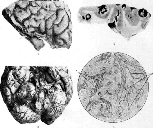
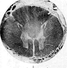

1 h 1 fp >-^;1 Figure 1. Cysticercus on the surface of the brain. Figure 2. Cysticercus in a cross-section of the cerebral cortex. Figure 3. Tumor of the right cerebellar-pontine angle. Figure 4. Sputum in gangrene: 1 - elastic fibers of Koppen-Jones; 2 - bundles of elastic fibers. Figure 5. Myelitic focus in the posterior columns of the spinal cord. Figure 6. Longitudinal section through the root of the bulb. In the middle passes a column of large cells, dividing the section into symmetrical halves. Figure 7. Oblique section of the proximal end of the root, indicating the affected side. or Paraparesis. The course of cervical myelitis is the worst. Clinically, very close to acute transverse M. is compressive M. Processes in the spine (see.. Spondylitis) and in the membranes (see Pachymeningitis, meningitis), whether tuberculosis, syphilis, tumors, hemorrhages, first of all usually give symptoms from the roots, especially posterior, in the form of girdle "neuralgic" pains, sometimes accompanied by girdle-shaped herpes. With cervical localization, the pains radiate to the back of the head, shoulders and upper extremities. Compression of the thoracic roots causes persistent intercostal pains. Lumbo-sacral localization often gives severe pains in the area of the sciatic nerve, often on both sides. At the beginning of the disease in the absence of other symptoms, the diagnosis of intercostal neuralgia, sciatica, etc., is sometimes made for a long time. With increasing pressure on the membranes, these symptoms are joined by the above-described picture of transverse myelitis. Extremely characteristic for compression of the spinal cord is the compressive syndrome of the cerebrospinal fluid. It consists in an increase in the amount of protein with a slight pleocytosis (cytoalbumin dissociation or Nonne's symptom). Sometimes in this case a greenish coloration (xanthochromia) and rapid clotting of the released fluid is observed due to the increased content of fibrin, exuding from the blood due to stagnation (Froin's coagulation syndrome). In the case of spondylitis, the picture is supplemented by changes from the spine. Not always curvature of the spine (even the most significant) causes compression of the spinal cord. And, conversely, even in the absence of clear changes on the x-ray in tuberculous spondylitis, it can give phenomena of severe compressive M. Compression of the spinal cord can in this case proceed in two completely different forms. In the initial stage of spondylitis soon after the appearance of the first root symptoms, paraplegia can develop lightning-fast, within a few days or weeks. In such cases, it is caused by edema of the spinal cord, sometimes due to the formation of an extradural abscess. If the phenomena of compressive M. develop slowly and in later stages of spondylitis in the presence of obvious bone changes, then the mechanism of compressive myelitis is completely different. It is caused by a slowly developing chronic pachymeningitis, which, as it were, "protects" the spinal cord from the purulent process, in turn leads to its severe compression, mainly by gradually disrupting blood and lymph circulation. 'Correspondingly' to this, the prognosis in both cases is different. In the first case, the edema can completely pass with immobilization of the spine (within 1-1½ years). In the second case, the prognosis of compressive M. is completely hopeless. The nature of compression can sometimes be established by a test with lipiodol or iodipin, which is introduced into the dura mater sac by means of occipital puncture. Lipiodol detained at the place of compression is detected by means of x-ray film. - Compressive M. due to syphilitic pachymeningitis usually develops in the cervical part, causing first pains in the back of the head and upper extremities, and later flaccid paralysis of the muscles of the shoulder girdle and upper extremities. Gradually the entire picture of cervical M. is added. - hemorrhages into the membranes, causing a picture of compressive myelitis (haematorrhachis, see.), are detected by the presence of blood in the cerebrospinal punctate and the acute onset after trauma. For extramedullary tumor or limited serous meningitis (serous cyst), slow onset, Brown-Séquard type of paralysis; paresthesias, often observed in extramedullary tumor, begin from the distal ends and only later pass to the proximal parts. This depends on the fact that with compression, the paths going from the distal parts of the extremities and lying on the periphery of the spinal cord are involved in the process earlier (law of E. Flatau on the eccentric location of the long paths of the spinal cord). Diagnosis. If M. or more often myeloencephalitis develop during or soon after general infectious diseases, especially in children, the diagnosis presents no difficulties. It must always be kept in mind in such cases when a child, who has just had an infection, develops symptoms from the central nervous system. It can be confused with epidemic meningitis, all the more so that encephalomyelitis is often accompanied by serous meningitis. On the contrary, and in epidemic meningitis sometimes (and far from rarely) the disease is complicated by encephalomyelitis. The most valuable differential diagnostic signs are the changes in the cerebrospinal fluid (see Meningitis). It is especially difficult to differentiate acute disseminated encephalomyelitis or M. from acute forms of multiple sclerosis, if it is not possible to establish a preceding infection. Sometimes the question is finally resolved only on the basis of the epikrisis and the subsequent remissions, characteristic of multiple sclerosis. However, disseminated encephalomyelitis in rare cases gives remissions. For disseminated encephalomyelitis will be spoken of preceding febrile diseases, especially whooping cough, measles, smallpox, etc. But multiple sclerosis can also flare up after the mentioned diseases. Epidemic encephalomyelitis differs from disseminated mainly in that with it the foci are very rarely located in the white matter. Therefore, pyramidal symptoms, spastic paralyses with Babinski's reflexes and others always speak rather for disseminated encephalomyelitis. Heine-Medin disease can also give a picture of disseminated... encephalomyelitis, but with it ataxias, 1 sensory disorders, pelvic disorders, bedsores, pathological reflexes are rare. In polyneuritis the nerve trunks are painful, there are no pelvic disorders. The diagnosis of transverse M. was formerly made very widely. But this clinical form, as our knowledge grew, gradually differentiated into the most various diseases: multiple sclerosis, brain tumor, syphilis, spondylitis, etc. And if therefore formerly transverse M. occupied one of the most honorable places both in the nervous departments and in the literature, now it is diagnosed comparatively rarely. As already indicated, it is most often accompanied by symptoms from other parts of the spinal cord and even the brain. Therefore, in the presence of transverse lesion of the spinal cord, one more often has to think of multiple sclerosis, sometimes long-lasting under the guise of transverse M. The same applies to intramedullary tumor. Anamnesis (transferred infections), course, examination of the cerebrospinal fluid help in the correct diagnosis. It is much easier to make the diagnosis of compressive M. due to extramedullary tumor, spondylitis, hemorrhage, syphilitic or other meningitis. Anamnesis, x-ray of the spine, examination of the cerebrospinal fluid, sequence of development of symptoms, serological studies decide the question of the nature of compression. The prognosis of M. and encephalomyelitis is always serious, especially when the brain stem is involved. Bedsores, cystitis should be regarded as prognostically unfavorable complications, often leading to death. Encephalomyelitis, especially in children, usually give recovery with defects in the form of paralyses, sometimes associated with forced movements, athetosis, or in the form of epilepsy, often with intellectual disorders. However, there are M. and encephalomyelitis with complete recovery. This especially applies to those mild epidemics that have been observed in recent years. - Treatment of infectious M. and encephalomyelitis consists of rest, bed regime, struggle for cleanliness, prevention of bedsores, cystitis, strengthening of the heart. Urotropin, collargol, iodine and mercury preparations should be used. Frequent lumbar punctures are useful, especially in cases of symptoms from the optic nerves. Complications (cystitis and bedsores) are treated on the usual basis. The paralyses remaining after the acute period are subject to treatment by massage, baths, mechanotherapy, electricity. Luetic M. require specific treatment. Treatment of compressive M. consists in treating the main cause that caused the compression. On the treatment of epidemic encephalitis - see Encephalitis. - Prevention of infectious M. consists in the struggle with infections in general, in improving the way of life, etc. Rest and sufficient rest are necessary during and after infectious diseases. It is important to remove from the body all foci of infection, to which belong chronic purulent diseases of the tonsils, carious teeth, periodontitis, etc.
Related articles
Mentioned in
Cite this page
“Myelitis.” Soviet Medical Encyclopedia. English translation of Bolshaya Meditsinskaya Entsiklopediya, 1st ed. (Moscow, 1928–1936), ed. N. A. Semashko. https://sovietmedicalencyclopedia.pages.dev/article/myelitis/