Tabes Dorsalis
Historical document, translated for reference. It reflects medical knowledge of the 1920s–30s and is not medical advice.
Summary
Tabes dorsalis is a chronic syphilitic disease of the nervous system that primarily affects the posterior columns and roots of the spinal cord. The article details its historical recognition, clinical features, pathology, and the ongoing debate about its syphilitic etiology in the 1930s medical understanding.
Encyclopedia article (1928–1936)
TABES DORSALIS (spinal atrophy, tabes dorsalis, progressive locomotor ataxia, ataxie locomotrice progressive), a chronic syphilitic disease of the nervous system that primarily affects the posterior columns and posterior roots of the spinal cord, but also spares neither the brain nor the peripheral nerves. Among organic nervous diseases that most severely affect the spinal cord, T. d. occupies a relatively significant place. Thus, in a number of statistics, it in frequency among organic diseases of the spinal cord is surpassed only by multiple sclerosis. Due to the large number of bright and unique symptoms, T. d. was rightly called a clinical giant by Schaffer. On the other hand, it is not easy to isolate from the entire mixed array of symptoms those that are characteristic specifically of T. d. and not of syphilitic damage to the central nervous system in general. It is no less difficult to precisely determine the time when, against the general background of neurosyphilis, the clinical picture of spinal atrophy develops. In any case, T. d., like its relative progressive paralysis, which until recently was considered not syphilitic but metasyphilitic disease, in its phenomenology, course, and especially in prognosis and relation to specific treatment presents a very characteristic picture that justifies isolating this form from other syphilitic diseases of the central nervous system. History. The beginnings of the identification of the clinical form of T. d. date to the second half of the 19th century. The leading symptom that prompted the description of an independent clinical form was ataxia, characterizing far advanced cases. The disease, especially in France (Charcot, Fournier) and in England, was described on the basis of this main symptom as progressive locomotor ataxia. Undoubtedly, at that time other types of ataxia, for example Friedreich's disease, were also often included in this group. But gradually around the central symptom of T. d. - ataxia - new symptoms began to group themselves, the discovery of which constitutes historical milestones in the evolution of neuropathology in general and the doctrine of T. d. in particular. These include the discovery of the Argyll-Robertson symptom (pupillary rigidity; see Argyll-Robertson symptom), Westphal's discovery of loss of knee reflexes, and finally the discovery of the Romberg symptom (see Romberg symptom). This classical triad for a long time was considered a mandatory prerequisite for the diagnosis of T. d., precisely at a time when the disease was still viewed as a sum of symptoms. However, the significant polymorphism of T. d. did not escape the attention of authors. At present, it is diagnosed completely independently of the presence of the mentioned symptoms. A huge role in the history of T. d. was played by the discovery of the importance of degeneration of the posterior columns of the spinal cord. It was already seen by Olivier (1837), Cruveilhier, however, the connection of the gray degeneration of the posterior columns, as well as of the posterior roots, with spinal atrophy was established by Leyden, as well as by Bourdon and Luys. No less important in the history of the doctrine of T. d. is the question of its etiology. Its syphilitic etiology was by no means unanimously recognized by authors. A fierce struggle 'for' and 'against' the significance of syphilis in the origin of T. d. was waged. Among the supporters of syphilitic etiology were Fournier, Gowers, Erb. In the camp of opponents were such major authorities as Leyden, K. Westphal. The main argumentation of the latter authors included the indication that in a large number of tabetics there were no indications of syphilitic infection either in the anamnesis or in the status. But thanks to modern biological research methods, including that of cerebrospinal fluid, the number of cases of T. d. where there are no indications of syphilitic infection is constantly decreasing. At present, there is no doubt that the main cause of T. d. is syphilitic infection. Former authors, in particular Leyden, attributed the main role to cold influences, cooling, overexertion, injuries. There is no doubt that the factors just listed sometimes play a certain role in the occurrence of the tabetic symptom complex, but their significance is secondary. They are only additional, activating factors which, without a previous syphilitic infection, are unable to cause T. d. Without syphilis there is no spinal atrophy. However, as already mentioned above, spinal atrophy, like progressive paralysis, differs from a syphilitic disease not only in its histological picture but also in that it does not respond to ordinary specific treatment. Therefore, supporters of the syphilitic etiology of T. d., such as Fournier, Erb, and others, believed that T. d., although it depends on syphilis, is not a syphilitic disease but a meta- or parasiphilitic disease (see Metasyphilis). However, since the discovery of the pale spirochete in the brain of tabetics and progressive paralytics, the view of T. d. as a syphilitic disease has firmly established itself in science. The difficulty in detecting the pale spirochete in the spinal cord, which plays an etiological role in the origin of T. d., is explained by Levaditi and others by the special biological properties of spirochetes. They take in the body and in particular in the nervous parenchyma the form of granules, sometimes barely visible in the ultramicroscope and in this state exist for a long time in the body. The peculiar course of T. d. and its clinical features depend on the fact that in this form of neurolues the spirochete affects mainly or primarily the nervous substance itself - fibers and cells - and to a lesser extent the vessels and meninges. Thus, the spirochete, having penetrated the brain tissue through the blood-brain barrier, is 'beyond the reach' of those immune bodies in the blood that do not pass through the barrier. Medicinal substances, such as mercury, neosalvarsan, bismuth, under normal conditions also do not pass through the barrier. This explains the relative refractoriness of tabes to specific treatment. Pathology and pathological anatomy. The most characteristic changes are found in the spinal cord, where even macroscopically on a fresh preparation the gray color of the posterior columns, sharply contrasting with the rest of the white mass on a cross-section, catches the eye. The changes are most pronounced in the lumbar part. In the cervical part of the spinal cord, the process is usually limited to the posterior columns. The posterior roots are thinned and colored grayish. Sometimes the same color is found in the optic nerves. The meninges of the spinal cord are often cloudy, especially in the area of the posterior columns. Microscopically, one can find disintegration in the area of the posterior roots, always beginning at their point of entry into the spinal cord and then extending to the segments of the roots outside the spinal cord. Some authors especially emphasize that for T. d. it is characteristic precisely the intramedullary beginning of the disease of the posterior roots, in contrast to their secondary degeneration due to damage to their extramedullary segments in diseases of the meninges, compression by tumors, etc. Spielmeyer showed by experiments on dogs that infection of dogs with certain strains of Trypanosoma nagani can cause fresh degenerations of the intramedullary segments of the posterior roots, as well as in the optic nerve. At the same time, both the meninges and the spinal ganglia remained normal. Thus, by experimental trypanosomal T. d., the primary nature of selective lesions of the posterior roots was proven. The outcome of the lesion is the complete destruction of the myelin sheaths and axis cylinders. Nerve elements are replaced by glial scars. The posterior horn, namely the zone of Lissauer, and the columns of Clarke are almost always involved. The ependymal cells of the central canal often proliferate. Pathological changes are also found in the spinal root of the trigeminal nerve, the nucleus of the vagus nerve, and sometimes in other nuclei of cranial nerves. In a number of cases, specific changes in the vessels are found in the spinal as well as in the brain, but to a lesser extent than in the mesenchymal forms of neurolues. Some authors consider tabetic arthropathies not as trophic disorders but as a syphilitic process, based on the fact that in them they find infiltrates of plasma cells, endarteritis and endophlebitis. Some authors, unlike the above view of T. d. as a primary selective disease of ectodermal elements of the nervous system, consider the starting point of degeneration of the posterior roots and posterior columns to be the extramedullary segment of the posterior root. Thus, Richter found in the t. i. place of Nageotte, where the spinal nerve passes through the arachnoid and dura mater, syphilitic granulation tissue, which is caused by spirochetes settled in the lymphatic spaces and connective tissue sheaths. The granulation tissue penetrates into the nerve bundles, causing in them local foci of disintegration. Other authors attribute the decisive importance in the degeneration of the posterior roots to the place of Obersteiner-Redlich, where the root nerve enters the spinal cord.
However, at the present time, it should be acknowledged that T. d. represents an independent primary syphilitic disease, predominantly of the nervous parenchyma, of ectodermal elements, caused by the penetration of the spirochete through the hemato-encephalic barrier. Onset and incubation period. On average, the nervous symptoms of T. d. appear 10-15 years after the primary lesion. However, there are also shorter intervals, in rare cases 3-5 years. Intervals of 20-25 years and even more have been described. There are undoubtedly cases of T. d. after congenital syphilis. In juvenile T. d., the disease sometimes develops, although rarely, in the first years of life. Some authors believe that specific therapy for syphilis lengthens the incubation period, however, this point of view is shared by far from all authors. Regarding the frequency of the disease among syphilitics, the figures for T. d. diverge. Thus, Mattauschek found 2.37% tabetics among 4,134 syphilitics, while Hoche (Nothnagel) stated that out of 100 syphilitics, 90 avoid progressive paralysis and tabes. Clinical picture. Among the early symptoms of tabes dorsalis are various kinds of paresthesias and pains in different parts of the body. They are predominantly localized in the extremities and have a distinctly radicular character. On the upper extremities, they prefer the ulnar side, corresponding to the Cvm and D: roots. On the lower extremities, they most often correspond to the S1 root. The paresthesias are characterized by a sensation of crawling ants, numbness, and a dull feeling. The patient feels as if they have thick rubber soles or are walking on a carpet. On the trunk, a belt-like or girdling sensation is observed. Patients complain of a tightening around the trunk at the level of the subcostal line, sometimes higher or lower. The back and lateral parts are often free at the same time. At this time, the extremely characteristic shooting or lancing pains of T. d. also appear. At different points, most often in the lower extremities, the patient experiences excruciating, piercing, instantaneous pains that appear in attacks and move from one place to another, often corresponding to the distribution of some nerve branch. These pains intensify depending on the weather. A drop in barometric pressure, snow, and rain have an intensifying effect on them. The belt-like sensation is also often accompanied by severe pains. Even at this time, it is possible to detect objective sensory disturbances with careful examination. The favorite areas are the nipple areas, i.e., the distribution of Dv-D1 roots (Hitzig's zone). Often, the left mammary anesthesia is more pronounced. It affects all types of sensitivity, but often irritation with cold, on the contrary, causes an extremely unpleasant, burning sensation with significant radiation. Such hyperpathia to cold is most often observed on the skin of the abdomen and on the lateral parts of the trunk. Pain sensitivity is often disturbed in a peculiar way. Sometimes painful irritation is perceived not at its point of application, but at a distance, often in a symmetrical place on the opposite side (see Allodynia). This is explained by disease of the posterior horn. A prick is sometimes perceived with an especially unpleasant sensory tone, not as sharp, but as burning or as ice, it is poorly localized, radiates, i.e., has all the properties of hyperpathia. This phenomenon depends on the lesion of the posterior columns, which phylogenetically conduct the more epicritic sensitivity. Due to its destruction, the phylogenetically oldest protopathic sensitivity, possessing the properties outlined above, sharply emerges. It also explains the characteristic paresthesias mentioned above. Acroparesthesias (see) are in most cases also explained by lesions of the posterior columns. A very characteristic disturbance is pain sensitivity. One prick with a pin is perceived in such cases twice: first as a touch, and then only as a prick. Pressure on nerve trunks is not painful - this is the basis of Bernatsky's symptom (see Bernatsky's symptom). Similar to it is Abadie's symptom (see Abadie's symptom). Sometimes superficial pain sensitivity also disappears. Due to severe radicular pains, a picture of anaesthesia dolorosa (see) arises. Deep sensitivity is particularly affected. This is most noticeably detected on the toes of the lower extremities. Due to the loss of deep sensitivity stands a very bright symptom of tabes dorsalis - ataxia, which develops already in a later stage of the disease. In more severe cases, it is noticeable even when the patient is sitting and even lying down. In the finger-nose test or when attempting to place the heel on the other knee, the patient misses. Ataxia is particularly sharply evident when the patient stands with feet close together and eyes closed (see Romberg's symptom). Ataxia is even more noticeable when walking. The peculiarities of gait are determined not only by ataxia but also by a sharp decrease in tone, which usually accompanies the loss of deep sensitivity. Due to this, the tabetic walks widely spaced, throwing out the legs, stamping the heels, lifting them higher than normal and excessively extending at the knees. At the same time, he carefully watches his legs with his eyes. As soon as the eyes are closed, ataxia increases so much that the patient even falls. It deserves attention that the ataxia of a tabetic increases not only when closing the eyes but also when looking up at the ceiling or when tilting the face upward. Perhaps, in this case, according to the law of the neck tonic reflexes of Magnus and de Kleijn, the tone of the lower extremities decreases. Finally, there is another way to 'sensitize' Romberg's symptom. The patient is made to squat and then quickly straighten up. At this time, ataxia is clearly evident. Another method of sensitizing Romberg's symptom is to make the patient, while standing, not bring one foot to the other, but place one in front of the other. Usually, there are no paralyses in tabes, however, it is sometimes complicated by paralysis of peripheral nerves, most often the peroneal. Tendon reflexes disappear early. This especially applies to the knee reflex and the Achilles tendon reflex. But on the upper extremities, tendon reflexes also disappear. Also, plantar and abdominal reflexes are often absent. With the loss of deep sensitivity, partly with a lesion of the peripheral motor neuron, is associated tabetic hypotonia. It reaches particularly large dimensions in the lower extremities. Both the active movements of patients and, especially, the passive movements in volume in all joints significantly exceed the norm, which gives them a clownish character. The patient is able to place his feet behind his head. When standing, hypotonia of the knee flexors is manifested in excessive knee extension (genu recurvatum). This creates conditions for traumatization of the ligamentous and the entire joint apparatus and intensification of the arthropathic process, one of the most characteristic trophic disorders in T. d. Among the most characteristic symptoms of tabes dorsalis are pupillary symptoms. Argyll Robertson's symptom (see Argyll Robertson symptom) should still be counted among the most typical signs of T. d. It is most often bilateral; according to Frey's observations in 850 cases, it is on both sides in 58.82% of all cases of tabes dorsalis, and unilateral in 11.76%. In other words, in more than 70% of cases of T. d., this symptom is present. According to Frey, other pupillary disorders in T. d. are distributed as follows: irregularly shaped pupils - 21.76%, miosis - 65.06%, mydriasis - 19.64%, anisocoria - 52.35%, absolute immobility of pupils - 21.18%. Normal reaction was found in 4.7% in the same material of Frey. Among the relatively early and frequent symptoms are also paralyses of the ocular nerves, most often the oculomotor, less often, but still in 59 cases out of 850 (Frey), and the abducens. The participation of the optic nerve, much less often the auditory nerve, is very tragic. Often being an early symptom of the disease, optic atrophy (see Optic nerve) leads to blindness on average in 9-10 months, more often in 2-3 years. In more advanced cases, optic atrophy occurs less frequently. Frey observed it in 240 cases out of 850 tabetic patients. It deserves attention that in almost all patients in whom the optic nerves are affected still in the preataxic stage, ataxia does not develop. Among the main symptoms of T. d. should also be included disorders of the bladder and sexual activity. Most often, difficult urination, retention, less often incontinence, absence of urges are observed. The fading of sexual functions is expressed in decreased libido, erection, orgasm, and defects in ejaculation. The trophic disorders in T. d. are very peculiar. In 5-10% of all cases of tabes dorsalis, such disorders of bones and joints are observed. Most often, tabetic arthropathies are observed in the knee joint (Fig. 1), then in the ankle and hip, in the elbow. Osteoarthropathies of the spine are also encountered. Often, arthropathy appears acutely in connection with trauma, sometimes insignificant.
Undoubtedly, in such cases, there were previously latent changes in the joint. In the joint, a serous or bloody fluid appears. Upon examination, fluctuation is detected. The most characteristic feature is the absence of pain, redness, and elevation of skin temperature. The fluid may be absorbed, or the condition may progress to destructive changes in the articular surfaces, cartilage, and epiphyses. Along with resorption processes, hypertrophic changes also occur in the form of exostoses and proliferation of synovial villi. The result is the characteristic osteoarthropathies in T. d. (osteoarthropathia hypertrophica) (Fig. 2). The joints are disfigured, and dislocations and subluxations appear, giving the tabetic arthropathy its characteristic appearance. In addition to the knee, such disfigurement is also observed in the foot (pied bot tabetique). Trophic changes in the bone can lead to spontaneous pathological fractures as a result of a slight impact, and sometimes even simply from muscular tension. At the same time, due to the absence of pain, patients continue to walk; because of insufficient immobilization, the ends of the bones rub against each other, which delays the healing process, which may not occur at all
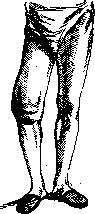
Fig. 1.
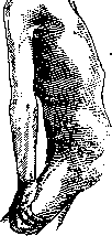
bone serous or bloody, Upon examination, fluctuation is detected. The most characteristic feature is the absence of pain, redness, and elevation of skin temperature. The fluid may be absorbed, or the condition may progress to destructive changes in the articular surfaces, cartilage, and epiphyses. Along with resorption processes, hypertrophic changes also occur in the form of exostoses and proliferation of synovial villi. The result is the characteristic osteoarthropathies in Tabes Dorsalis (osteoarthropathia hypertrophica) (Fig. 2). The joints are disfigured, and dislocations and subluxations appear, giving the tabetic arthropathy its characteristic appearance. In addition to the knee, such disfigurement is also observed in the foot (pied bot tabetique). Trophic changes in the bone can lead to spontaneous pathological fractures as a result of a slight impact, and sometimes even simply from muscular tension. At the same time, due to the absence of pain, patients continue to walk; because of insufficient immobilization, the ends of the bones rub against each other, which delays the healing process. Fig. 2.
which may not
occur. Trophic disorders of the skin and subcutaneous tissue in tabes dorsalis are not so frequent. Spontaneous gangrene (mal perforant du pied) may develop over the metatarsophalangeal joint of the big toe or little toe of the lower extremities or on the heel. Cases of callus formation on the dorsum of the foot, on the palm of the hand, and on the ala nasi have been described. The callus turns into a blister and abscess, the epidermis is shed, and a deep ulcer forms, which may extend to the bones and joints. Particularly remarkable is the painlessness of this process. Also encountered are painless loss of teeth and defects in nail growth, graying of hair, sometimes its segmental loss, bruises, and pigmentary atrophy. In some cases, Dupuytren's contractures as well as Raynaud's disease can be classified as tabetic trophic disorders. It is remarkable that most tabetics are characterized by significant emaciation. It remains unresolved whether this depends on some correlation of T. d. with an asthenic constitution or whether the emaciation is connected with general trophic anomalies or with crises. Among the most tormenting symptoms of T. d. are visceral crises (see Crises), accompanied by severe pains. Most frequently, approximately in 8% of all cases, gastric crises occur. They manifest as attacks of vomiting or severe spasms below the navel. Often they are initially interpreted as a primary stomach disease. During crises, hypersalivation and increased acidity of gastric juice are usually observed. Sometimes patients complain of heartburn, belching, and regurgitation. With very prolonged gastric crises leading to severe exhaustion, there may also be a danger to the patient's life. X-rays in tabetic gastric crises sometimes reveal an hourglass-shaped spasm. Intestinal crises manifest as diarrhea and spasmodic contractions of the intestines; laryngeal crises as spasmodic cough and difficult breathing; respiratory, cardiac and vascular, bladder, uterine, vaginal, and temperature crises also occur in T. d. Finally, the limb crises described by Förster should also be mentioned, which manifest as attacks of lancinating pains accompanied by muscle cramps or other involuntary muscle movements. Finally, it should be pointed out that tabetic symptoms from the nervous system are often combined with syphilitic changes in the heart and aorta. In contrast to this, severe tertiary symptoms from the skin and bones are extremely rarely observed in T. d. In the cerebrospinal fluid of tabetics, characteristic changes are found. Its appearance is clear, and the pressure is moderately high. The number of cellular elements is increased 90 times, but rarely reaches 200 cells per 1 mm3. The main mass of them are small lymphocytes. Globulin reactions are almost always positive (phase I according to Nonne-Appelt, Pandy, Weichbrodt). Colloidal reactions with gold or mastic show changes in the 2nd-4th test tubes. The Wassermann reaction is less pronounced. With 0.2 amounts of cerebrospinal fluid, it is sharply positive in 20%, with larger doses—approximately in 95%. In the blood, the RW is positive in 60-70%. The Sachs-Georgi reaction is positive somewhat less frequently. (For the combination of T. d. with general paresis—see Taboparalysis.) The course of T. d. is very diverse in different cases. Sometimes, besides pupillary symptoms and absence of tendon reflexes, mild sensory disturbances in patients for many years nothing else is noted. They do not lose their ability to work. The latter is significantly affected when ataxia and shooting, lancinating pains are added. In particularly severe cases, hypotonia becomes so serious that patients lose the ability to walk—the so-called paralytic stage. In this stage, the disorder of pelvic organs also intensifies, often ending in inflammation of the bladder, urosepsis, bedsores, etc. Some authors distinguish, besides the usual, typical lower T. d. with predominant involvement of the lower extremities, also an upper, or cervical, as well as T. d. of the conus. A unique course is taken by the form of T. d. in which atrophy of the optic nerve develops first. In these cases, ataxia usually does not develop. More rarely is the combination of T. d. with spinal atrophies due to lesions of the anterior horns encountered. According to some authors, amyotrophies occur in 4-8% of all cases. In women, T. d. often runs more favorably than in men. In a large number of cases (approximately in one third) T. d. runs favorably. After the appearance of the first symptoms, sometimes 18-20 years pass before any more serious symptoms appear, such as urinary incontinence. Deterioration often occurs after an infectious disease, trauma, etc. Some cases that initially showed a tendency to progression later take a favorable course when patients are placed under conditions of rest and bed rest. In less than a quarter of all cases, an unfavorable course is observed, when after the first symptoms, severe ataxia appears after only 4-5 years, soon depriving the patient of the ability to walk. In late stages, in severe cases, a severe decline in nutrition develops relatively early. This marantic form is especially common with crises, especially gastric ones, however it cannot be entirely explained by the patient's starvation. It is based on a particular disorder of the central vegetative apparatus. The prognosis in T. d., as can be seen from the description of its course, is very varied. There are completely favorable forms where the disease for many years manifests with only one or two symptoms, not depriving the patient of their ability to work. Such stationary cases are not uncommon. The ataxia that appears may stop and even completely disappear. In other cases, ataxia, which most often leads to disability, develops only after 15-20 years. Only in a small percentage of cases does ataxia lead to disability after only 5-6 years. In general, it should be noted that the phenomena of irritation, such as pains and crises, more likely pass, although they often show great persistence. A worse prognosis is given by symptoms of loss, but they too sometimes show an intermittent course. Under the influence of persistent treatment, symptoms such as ataxia, bladder disorders, sexual weakness, etc., can improve. Usually it can be said that the long incubation period between syphilitic infection and the first appearance of tabetic symptoms gives a right to a better prognosis both in terms of course and in terms of the appearance of certain symptoms. Infectious diseases that join in, such as influenza, typhoid, pneumonia, significantly darken the prognosis. The prognosis should also take into account the condition of the heart and large vessels, as well as the danger of drug addiction, which results from the use of narcotic drugs for shooting pains. The diagnosis of T. d. usually does not present difficulties when the classic triad is present: Argyll-Robertson symptom, absence of knee reflexes, ataxia; but also shooting pains, bladder disorders, typical sensory losses, tabetic atrophy of the optic nerves, arthropathy, pathological reflexes are sufficiently bright symptoms helping recognition. Differentiation is sometimes necessary with pseudotabes (see Polyneuritis). In the latter, pupillary symptoms and bladder disorders are usually absent. Nevertheless, peripheral neuritis is not always easy to differentiate from T. d. due to the pains, sensory disturbances, and absence of tendon reflexes that occur with it. An essential sign that allows differentiation of T. d. from polyneuritis is the Abadie symptom. Pressure on the nerves in polyneuritis is usually extremely painful, in T. d., on the contrary, completely painless. From diseases of the central nervous system, one must sometimes think of Friedreich's ataxia (see Ataxia), as well as funicular myelitis, combined system diseases of the spinal cord, developing with certain intoxications or pernicious anemia. As already mentioned above, it is sometimes difficult to recognize the tabetic nature of visceral crises, which can be confused with a primary disease of the internal organ. Thus, gastric crises sometimes for a long time give reason for a diagnosis of perforating gastric ulcer. The pupillary symptoms, almost always observed with crises, and the absence of tendon reflexes make it possible to make the correct diagnosis, provided one remembers the possibility of a tabetic basis for internal symptoms. Treatment. The recent nihilism regarding specific treatment of T. d. must be considered obsolete. Since the syphilitic nature of T. d. was proven, and it was clarified that by destroying the blood-brain barrier one can achieve penetration of salvarsan, mercury, and bismuth preparations into the central nervous system, specific treatment should be systematically applied to every tabetic. When prescribing specific treatment, one must take into account the courses of treatment previously conducted.
With insufficient previous treatment, it is necessary to conduct such treatment in the form of mercury rubs and simultaneously or after their completion, to use intravenous administration of neosalvarsan. The total amount of neosalvarsan administered reaches 4-5 g. If the treatment is successful, it should be repeated in a year or even earlier. Sometimes at the beginning of treatment, a certain worsening of the condition occurs, which, according to Steiner, is rather a favorable prognostic symptom. Some specialists, for example Dreyfus, prescribe significantly more intensive courses of neosalvarsan, for example six courses of 6-8 weeks each with a subsequent break of 8-12 weeks. He starts with 0.15 neosalvarsan two to three times a week and goes up to 0.45. Also French authors recommend courses of approximately the following type: first series of injections-0.3; 0.6; 0.9; 0.9; a four-week break. Second series-0.6; 0.9; 0.9; a four-week break. Third series-0.9; 0.9; 0.9. Stationary, as well as incipient forms of T. d. should not be subjected to specific treatment. Sometimes it is useful to combine intravenous administration of neosalvarsan with endolumbar. Sodium iodide is given either orally or preferably in a 10% solution intravenously at 5-10 cc3. T. d. and Basedow's disease are contraindications for the use of iodine. General strengthening treatment is of enormous importance. One should strive for weight gain. Protein and vaccine therapy are also recommended, but on the condition that the body's reaction is not too severe. Treatment with malaria for T. d. does not give the same effect as in general paralysis, however in some cases combined treatment with malaria, neosalvarsan and bismuth gives good results. It is also worth trying the treatment of T. d. with the introduction of sodoku, typhoid fever, which gives a certain effect in the treatment of general paralysis. Palliative treatment should mainly be directed against shooting pains. Morphine and its derivatives should be avoided due to the danger of morphinism. It is better to resort to pyramidone, antipyrine. Intravenous administration of dextrose is also useful. Dattner explains this by the fact that all measures that have an alkalizing effect on tissues lead to a reduction in pain. Wagner-Jauregg proposed insulin therapy for tabetic pains, based on the fact that a sugar-rich diet causes pain. Insulin is successfully used in crises. In view of the fact that the disturbance of acid-base balance, according to Dattner, lies at the basis of gastric crises and in turn is caused by local vagotonia due to disease of the spinal nerve roots, mainly Dv-x, the fight against gastric crises is recommended to be carried out by desensitization with the help of the smallest doses of alttuberculin. Physiotherapy in the form of half-baths with douches and rubdowns, electrization and massage is of great importance. Treatment of ataxia is carried out according to the Frenkel method, i.e. through systematic exercises, with the participation of vision acting vicariously. It is especially useful in the early stages of ataxia and should be carried out under strict control of the general condition and especially cardiac activity. The most difficult tasks face the physician in the treatment of tabetic crises. Regulation of regimen and diet should be foremost here; in the most common gastric crises, one must beware of narcotic agents. Spinal anesthesia, paravertebral or epidural injections of novocaine with adrenaline also help little. Foerster's operation of section of the posterior roots is only advisable if the gastric crises depend on sympathetic defects in the area of the thoracic roots (Dvn--Dx or Dv--Dxn). If the crises have a pronounced vagal character in the form of vomiting, then subdiaphragmatic section of the vagus nerve is more indicated. In view of the complexity and incomplete effectiveness of the surgical interventions just mentioned, they are now used much less frequently. The most common means giving the best result is physiotherapy in the form of half-baths, electrization, wrapping. Care of the skin, of the bladder to prevent bedsores, trophic disorders, and urosepsis play an important role in the treatment of a tabetic patient. Prevention. The most essential measure for the prevention of T. d. is the prevention of syphilis. Therefore, raising the cultural level, sanitary measures, and the fight against household syphilis are the most effective preventive measures against T. d. When specific infection has occurred, one must beware of all factors that weaken the nervous system: alcohol, cooling, especially of the feet, dietary errors, excessive transitions, physical and mental fatigue. Reasonably conducted specific treatment under the control of cerebrospinal fluid is the most important preventive factor.
M. Krol.
Related articles
Mentioned in
Cite this page
“Tabes Dorsalis.” Soviet Medical Encyclopedia. English translation of Bolshaya Meditsinskaya Entsiklopediya, 1st ed. (Moscow, 1928–1936), ed. N. A. Semashko. https://sovietmedicalencyclopedia.pages.dev/article/tabes-dorsalis/