Neuroglia
Historical document, translated for reference. It reflects medical knowledge of the 1920s–30s and is not medical advice.
Summary
This article from the 1928–1936 Great Medical Encyclopedia discusses the structure, classification, development, and cytoarchitectonics of neuroglia in the central nervous system, detailing the debate between cellular and reticular theories, as well as the types of glial cells such as astrocytes, oligodendrocytes, and microglia.
Encyclopedia article (1928–1936)
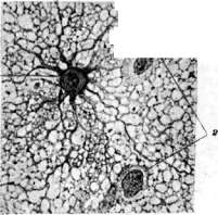
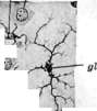
NEUROGLIA (neuroglia) (from the Greek glia - glue), glia, the interstitial tissue of the brain, has a common ectodermal origin with the nerve parenchyma. The primary epithelial cells of the embryonic neural tube differentiate in two directions: neuroblasts arising from them develop into nerve cells with fibers, while spongioblasts give rise to the ependyma and glia. Spongioblasts initially form a single cellular syncytium, from which various glial cells originate. Ependymal cells retain the shape of epithelium in the mature brain and are equipped in some places with cilia. General structure of neuroglia. In mature glial tissue, one can distinguish 1) a structureless ground substance, 2) cells, and 3) fibers. The first was attached great importance by older authors, hence the very name of glia. As for the ratio of other elements - cells and fibers - two theories still struggle over the question of the general structure of glia. According to one (Cajal and his school), glia consists of separate cells whose ramifications form a fibrous plexus that gives only the impression of a network. According to the other (Held and the German school), glial cells are not isolated, but are partly connected by their processes to each other, and partly woven into a true three-dimensional protoplasmic network (glio-reticulum), filling all spaces between nerve cells, fibers, vessels, ependyma, and the pia mater of the brain (see separate plate, Fig. 1). On the outer (pial) surface of the brain and around vessels, this unified protoplasmic network of glia thickens to form marginal protoplasmic membranes (membrana gliae superficialis et perivascularis). The latter closely adjoin connective tissue elements, leaving no "epycerebral" or "perivascular" lymphatic spaces. Processes of glial astrocytes approach and fuse with the terminal membranes. Cells of the neuroglia are diverse in shape and function. In older authors, cells rich in protoplasm were designated by the general name of Deiters' cells. Held singled out under the name of marginal glia those of them that participate with their processes in the formation of the brain periphery; he classified the rest as main glia. But this primary classification by Held proved insufficient. The works of Cajal, Rio-Hortega, and others, carried out using new methods of glial reproduction, provided new principles for dividing glial cells. According to Cajal, there are two types of process-bearing glial cells (astrocytes) and one so-called "third element," or non-process glia (adendroglia). Among the former, he distinguishes 1) protoplasmic astrocytes and 2) fibrous, or fiber-forming astrocytes. Protoplasmic astrocytes have a relatively large light nucleus, round, oval, or slightly indented on the side in shape. Protoplasmic astrocytes are scattered in moderate numbers in the gray matter of the brain and have no tendency to connect their processes with vessels or nerve cells. They are encountered in even smaller numbers in the white matter of the brain. Fibrous astrocytes are also large stellate cells; some of their processes, sometimes very long ones, are directed toward vessels and the outer surface of the brain. Weigert fibers, or glial fibrils, run in the marginal, concave border of their protoplasmic bodies and in their processes. Fibrous astrocytes are encountered in huge numbers in the white matter, as well as in the thalamus, pallidum, and anterior horns of the spinal cord. They are almost absent in the cortex. Cajal's third element consists of small protoplasmic cells which he thought were non-process cells. In reality, these cells also have processes, but less long and less numerous (see separate plate, figure 2). Hortega divided this category of cells into two subspecies, differing from each other not only morphologically, but, as he thinks, genetically as well. These subspecies are sparsely branched cells (oligodendroglia) and more abundantly branched ones, which have been given the name of microglia or mesoglia, as well as "Hortega cells." Sparingly branched cells, when reproduced by Hortega's method, have the following appearance: around a light round nucleus, there is an uneven rim of protoplasm, from which sparsely branched short processes depart with small swellings along their course. Hortega cells usually have an elongated nucleus, at the ends of which the protoplasm is significantly elongated; branching processes extend from the protoplasm (see separate plate, Fig. 3). Sparingly branched cells are close to astrocytes in their origin. In their position, these cells are satellites of nerve cells, fibers, and vessels; in the white matter, their arrangement in rows along the fibers is characteristic. Microglia, according to Hortega and Cajal, is of mesodermal origin and appears in the brain along with the development of vessels. These are elements having various inclusions within their bodies in the form of pigment, lipoids, and so on. Their predominant place of distribution is the cerebral cortex. Often they embrace nerve cells with their body and processes or lie longitudinally along the apical process of pyramidal cells, as well as near vessels. In smaller numbers, they are admixed with other glial cells in the white matter of the brain. Mesodermal origin and their isolation from the glio-reticulum, however, are disputed by German authors. There are also glial cells that occupy an intermediate place between ependymal cells and proper neuroglia, namely the epithelial cells of Golgi and Fafianas' cells in the cerebellum and subependymal accumulations in the midbrain, medulla oblongata, and spinal cord. Golgi's epithelial cells are particularly close to ependymal cells; these are the cells of the molecular layer of the cerebellum whose radial processes are known as Bergmann fibers. The bodies of these cells lie in several rows between Purkinje cells, and the processes (one or several), reaching the pial surface, form cone-shaped expansions; the latter, fusing, form the membrana gliae superficialis in the cerebellum. There are also Fafianas cells in the cerebellum, related simultaneously to both ependymal and epithelial cells. They are located in the same place as the epithelial ones, but are concentrated exclusively in the deep layer of the stratum molecularis; their processes (one, two, or several), which are also radial, are equipped with short rounded side branches and terminate in the molecular layer without reaching the periphery. Finally, subependymal accumulations of glial cells in the midbrain, medulla oblongata, and spinal cord, sometimes designated as gliomatous, are arranged in groups and represent ependymal cells that have detached from the surface and been drawn inward. Gliosomes. Using special methods (Altmann's acid fuchsin, etc.), special organoid granules of round, oval, or rod-like shape - gliosomes - are revealed in glial cells. They correspond, on the one hand, to Altmann's granularity, and on the other hand to mitochondria. They are distributed not only in the cell body, but also in the protoplasmic network of the glia, which is why they sometimes appear scattered in the interstitial structureless substance. In addition to small granules brightly stained with acid fuchsin, larger granules with a light center and also large ones poorly stained with fuchsin are distinguished. It is believed that these are various stages of the transformation of fuchsinophilic granules into secretion (see separate plate, Fig. 4). Neuroglial fibers. A distinction must be made between the protoplasmic network of the glia and the so-called Weigert fibers, or glial fibrils, embedded in it. The latter are independent elements, distinct from protoplasm, and represent smooth, cylindrical, straight or curved fibrils that do not anastomose with each other and do not form networks. Running in the body and processes of glial cells and in the protoplasmic network, they often turn out to belong not to one, but to several cellular elements. Cemented by protoplasm, however, they form reticulated membranous formations in places (for example, around large myelin fibers). Weigert's initial assumption about their complete isolation from cells turned out to be erroneous, although it cannot be completely denied in certain pathological cases. The development of glial fibrils occurs in the protoplasm of cells. Glioarchitectonics. We have spoken of various glial cells. Their morphological difference corresponds to a difference in functions, and in connection with this, their distribution is completely typical for individual parts of the brain. The distribution of Weigert fibers in the brain is similarly typical. For example, in the cerebral cortex, they are located in a thin layer in the subpial marginal zone and are almost absent in other layers of the cortex; furthermore, they are almost absent in the neostriatum, but a significant amount is present in the pallidum and thalamus, and so on. Thus, each department of the brain in the sense of the distribution of glial cells and fibers has its own definite physiognomy, its own glioarchitectonics. Function of glia. Not only cells, but also fibers of the neuroglia are distributed in the brain non-randomly. Very fine patterns of glioarchitectonics, striking in the unusual regularity of fiber distribution, are largely explained mechanically
ogl da da-

-&'
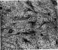
Figure 1. Protoplasmic network of the neuroglia (glia-reticulum), into which an astrocyte (g) weaves with its processes; н - nerve cells. Figure 2. Glial cells of the cerebral cortex (after Hortega): odg - oligodendroglia; mgl - microglia or mesoglia (Hortega cells); gl - astrocyte nucleus; nc - nerve cell; в - blood vessel; adnt and endt - adventitial and endothelial cells. Figure 3. Glial cells of the white matter of the brain (after Hortega): 1 (B) - astrocytes; 2 (A) - oligodendroglia; 3 (C) - Hortega cells. Figure 4. Fibrous astrocytes of the white matter of the spinal cord. Weigert fibers, or gliofibrils. Round bodies - transversely sectioned myelin fibers; in the middle of them - axon. Figure 5. Regressive forms of glia: 1 - swollen fibrous astrocyte, amoeboid transformation; 2 - amoeboid-altered oligodendroglia, methyl blue-eosinophil granularity in the cells; 3 - blood vessel; 4 - axon; 5 - myelin sheath. Figure 6. Floor of the urinary bladder from within: 1 - fossa retroureterica; 2 - area interureterica; 3 - orificium urethrae int.; 4 - uvula vesicae; 5 - ligamentum vesicae (Lieutaudi); 6 - orific. ureteris et plica ureterica.
To the article Ureter, Neuroglia. Along with vessels and membranes, the glia primarily performs the role of an interstitial supporting tissue. Its fibrous layers around the vessels serve here as if to protect the parenchyma from the mechanical pressure of the vessels during their pulsation. But this is not all. Proceeding from the conception of the glia as a continuous protoplasmic syncytium filling the entire space between vessels and nerve cells, we must imagine that through this glio-reticulum, nutritive material is conducted from some elements to others. A close symbiosis is thus realized between the glial cells and the nerve cells (P. Schiefferdecker). A fine chemical interaction between them can also be imagined as taking place in another respect. Gliosomes were mentioned above. A number of authors (Nageotte and others) prove that they produce a special secretion and that the glia is thus, as it were, a gland of internal secretion. In a purely humoral way, it thereby enters into relationships both with the nervous parenchyma and possibly also with certain distant organs. However, even if the glia is not a true gland, it nevertheless exhibits a definite internal secretory function of great importance in metabolism. The physico-chemical work of the glia is manifested particularly brightly in pathological cases, specifically during the breakdown of nervous tissue, when the glial tissue perceives various decay products from the lymph, processes them, and transports them toward the vessels. Sometimes its cells even dissociate from the glio-reticulum, acting in the role of true phagocytes. Of no small importance is the fact that the glia forms, as it were, an additional barrier to the reticulo-endothelial system of the brain vessels. In this respect, it is not only a mechanical obstacle to various bodies coming from the vessels, but can enter into chemical combat with various poisons and neutralize them (Lugaro). Microglial cells (Hortega) are of special importance in this regard. They play an exceptional role in the adsorption of substances that penetrate into the brain from the blood vessels and via the cerebrospinal fluid (Beletsky and Harkavi). In addition, they participate in immuno-biological processes, in particular exhibiting phagocytosis especially in cerebral spirochetoses, such as, for example, relapsing fever, progressive paralysis (Beletsky and Umanskaya). Nor should it be forgotten that already in the normal state the glia performs the role of a lymph-carrying tissue. At the same time, one must always keep in mind two pathways for the conduction of various soluble and insoluble bodies: one pathway is the protoplasm of the glial network itself. Golgi had already concluded from the fact of the fusion of thickened processes of glial cells with brain vessels that nutritive substances are absorbed from the blood bed through these processes and further transmitted to nerve cells. The passage of various substances from vessels to nerve cells and vice versa has been further proved by a series of experiments. But a lymph current is also possible along the surface of glial elements, in particular in the loops of its network. Much has been written about the direction of this current and the existence of special lymphatic spaces in the brain. One can think that special glial cells of the oligodendrocyte type of the white matter of the brain lie in small space-sinuses, facilitating the movement of tissue fluid among nerve fibers (drainage cells - Snesarev). One thing is unquestionable: that no pericellular, perivascular (His), and epioerebral spaces exist. Neither are there Arnold's and Kronthal's vessels. It has been proved that many substances (in particular toxins) spread along nerve trunks from the periphery of the body, enter the subarachnoid space, and penetrate into the brain via roots and root filaments (Rakhmanov). Substances injected into the ventricles of the brain likewise rapidly pass into the nervous tissue (Stern), heading through the ependymal barrier to the intracerebral vessels. In conclusion, one can mention the ideas—having only historical interest—on the role of glial cell processes as insulators of the nerve current and even on their vasomotor significance. The pathology of the glia can manifest itself, on the one hand, in anomalies of growth, and on the other, in diverse specific reactions to mechanical, physical, and chemical irritants. The latter can occur both primarily and secondarily (following the destruction of the nervous parenchyma). Growth anomalies of the glia also occur under the influence of irritants, but an embryonic predisposition is always additionally assumed. Gliomas stand in the first place here (see Glioma). Anomalous development of the glia can also manifest itself in the formation of monstrously large, protoplasm-rich glial cells, for example in tuberous sclerosis and in diseases of the Wilson group—pseudosclerosis. Specific fibrous formations in the form of curved, interweaving bundles of fibers developing during disease processes of early childhood also bear the traits of anomalous development. Progressive reactions of the glia. Reacting to various stimuli, the glia can produce, on the one hand, hyperplastic forms (cell multiplication), and on the other, hypertrophic forms (increase in volume). Multiplication can affect all types of glial cells mentioned above and proceeds by both mitotic and amitotic pathways. Hypertrophy can also undergo any type of glial cell; in this case, protoplasmic increase may predominate in some cases, and the development of fibers in others, or they are combined with each other. The protoplasmic reaction can be expressed either in simple swelling of the glial cells or in the fusion of individual cells and the formation of plasmatic structures (Gliarasen). Sometimes hypertrophy occurs not only in the cells, but also in the protoplasmic network. Hypertrophied astrocytes, becoming rounded, take on the appearance of "fattened cells" (gemastete Zellen). But hypertrophy can also affect pauci-branched glia and Hortega cells. Due to the multiplication and hypertrophy of individual cells, complex figures of nodules, rosettes, bush-like proliferation of the glia, etc., are sometimes obtained. The fibrous reaction of the glia proceeds mainly at the expense of fibrous astrocytes as well. But purely protoplasmic astrocytes and pauci-branched glia can also undergo fibrous transformation. The formation of fibers by Hortega cells is doubtful. The fibrous reaction occurs both primarily and secondarily, i.e., by way of replacing the perishing parenchyma. Gliosis, or local proliferation of the glia, leads to the formation of glial scars, or gliosclerosis. Gliosis is called isomorphic if it develops according to the type of glial tissue of a given brain area; otherwise, it is called anisomorphic or atypical. If the proliferation of fibers runs along the edge of the pial surface of the brain, one speaks of marginal gliosis; if cells and fibers proliferate around vessels, this is perivascular gliosis. A completely peculiar form of glial reaction is obtained in those cases when the physico-chemical activity of glial cells increases. Young glial cells appearing in the order of regeneration and specifically anaplasia and having a rich mitochondrial apparatus are particularly active in this respect. In addition, some glial cells can completely dissociate from the general cell syncytium and exhibit their vital activity in the form of independent cells; at the same time, they also act in the role of gliophages. The nucleus of these cells is usually disproportionately small compared to the large rounded body. Reticularity and various inclusions (lipoids, pigments, etc.) are visible in their protoplasm. These are lattice cells and granular corpuscles. Any glial cell can become granular, but a significant number of them are given by Hortega cells. Granular corpuscles found in the young, maturing brain are ascribed participation in the formation of myelin of nerve fibers (see separate table, fig. 5). Regressive reactions of the glia. The glia can undergo both simple atrophy, degeneration, and necrosis. Already in the normal mature brain in the outer layers of the cortex, one can encounter sclerotic, atrophied glial cells. They can be met in large numbers in old age. Degenerative reactions to various harmful influences can be relatively diverse. Their usual forms are: swelling, vacuolization, loss of gliofibrils, degenerative obesity, etc. A large number of fuchsinophilic grains and a special pathological granularity—methyl blue granularity (named after the dye with which it was first discovered)—can appear in swollen cells. Basophilic granularity accumulating usually simultaneously with it in the interstitial substance bears the name of "fibrinoid." Sometimes the interstitial pathological granularity has the appearance of irregular, angular-shaped bodies arranged in a mosaic (Füllkörperchen of German authors). Swollen degenerative cells of the glia often show signs of incipient dying (necrobiosis): pyknosis, hyperchromatosis of nuclei, short swollen processes. Such cells are called amoeboid.
Related articles
Mentioned in
- Agenesia of the Central Nervous System
- Alizarin Dyes
- Alzheimer's Stains
- Amyloid Bodies
- Astrocytes
- Astrocytoma
- Brain (551 General diagnostics of brain diseases)
- Brain (cerebrum, a comprehensive term for the entire)
- Fatty Necroses
- Ganglioneuroma
- Glioma
- Granular Bodies
- Hamartoma, Hamartia, Hamartoblastoma
- Lobar Sclerosis
- Macrophages
- Mann Method
- Myelitis
- Nervous Tissue
- Neurinoma
- Neurogenic
Cite this page
“Neuroglia.” Soviet Medical Encyclopedia. English translation of Bolshaya Meditsinskaya Entsiklopediya, 1st ed. (Moscow, 1928–1936), ed. N. A. Semashko. https://sovietmedicalencyclopedia.pages.dev/article/neuroglia/