Nerve Cells
Historical document, translated for reference. It reflects medical knowledge of the 1920s–30s and is not medical advice.
Summary
Nerve cells are the basic elements of nervous tissue, discovered by Ehrenberg in 1833 and further studied by Purkinje, Remak, and others. This article details their morphology, classification, structure, and various staining techniques developed to study them.
Encyclopedia article (1928–1936)
NERVE CELLS, the basic elements of nervous tissue. Discovered by N. k. Ehrenberg and first described by him in 1833. More detailed data about N. k. with indication of their form and the existence of the axon-cylinder process, as well as the topography of N. cells, are found in the works of Purkinje (1838). Remak (R. Re^ tak) more fully described the origin of nerve fibers and recognized the cells of sympathetic ganglia as elements of the nervous system (1837, 1854). The works of Schwann, defining more precisely the concept of the cell in general, also gave a more definite description of N. k. with its processes and nerve fibers. Hannover (1840) first began to study the structure of the nervous system on fixed and hardened preparations of the brain, which made it possible to more accurately establish the relationship of individual elements and areas of the brain. Deiters (1865) and Gerlach (1871) significantly expanded the understanding of the forms of N. k. and their processes thanks to the latter's proposed technique of staining animal tissues with carmine. However, ordinary histological methods did not make it possible to stain N. k. completely, i.e., N. k. with all their b. c. numerous processes. Therefore, the actual study of the morphology of N. k. begins with the discovery of special methods for staining nervous tissue. The main methods that created chapters in the doctrine of N. k. are the following: the silver impregnation method of Golgi (Golgi, 1883), which for the first time showed in detail the external appearance of N. k. with all processes; the method of vital staining of N. k. and fibers with methylene blue by Ehrlich (Ehrlich, 1885-1894), developed by Aronson (N. Aronson), Arnstein, as well as A. Dogel, Smirnov, and Timofeev; the staining method with basic aniline dyes of Nissl (Nissl, 1894); various methods of silver impregnation of nervous tissue and special staining of neurofibrils: Apathy (Apathy, 1877), Bethe (Bethe, 1898), Simarro (Simarro, 1900), Ramon y Cajal (R. y Cajal, 1903), Bielschowsky (Bielschowsky, 1903), Donaggio (Donaggio, 1904), A. V. Rakhmanov (1905); other methods of combined staining with various aniline dyes, revealing the fine structure of the nucleus and plasma, in particular the method of Altmann, various methods of staining lipoids, etc. N. k., originating from one common source—the outer embryonic leaflet, ectoderm (the question of the origin of part of the N. k. of the sympathetic nervous system is disputed, which some authors attribute to mesenchyme), in the further development of the organism, differentiate in higher animals into elements that differ both in function and in external appearance. The external appearance and internal structure of N. k. are extremely diverse depending on their functions and location in different parts of the nervous system. The connection of certain structural features of N. k. (form, character of processes, Nissl substance, etc.) with certain physiological features has been far from fully revealed to the present time, especially in relation to the brain. At the same time, it is impossible to establish complete morphological similarity of nerve cells that are close in function, although some common features are still noted, for example, N. k. of the motor area of the cerebral cortex and N. k. of the anterior horns of the spinal cord are isochromatic (see below). Cells of the vegetative centers of the medulla oblongata have a special characteristic arrangement of Nissl blocks—near the nucleus smaller, toward the periphery larger, etc.—By function, one can distinguish N. k. belonging to motor, sensory, and vegetative centers of the nervous system; cells of peripheral sensory receptor apparatuses can be placed in a separate group: visual, auditory, and olfactory, of which the cells of the retina are called sensory nerve cells, and the cells of the latter two apparatuses are simply called sensory cells.—By location, N. k. are distinguished in the central nervous system: N. k. of the cerebral cortex, N. k. of subcortical ganglia, N. k. of the cerebellum, N. k. of the medulla oblongata, N. k. of the spinal cord. In each part of the brain, N. k. are divided even more in detail; thus, in the spinal cord, N. k. of the anterior and posterior horns, whose axon-cylinder processes pass into the roots, are called root cells (cellulae radiculares); N. k. that give processes into the lateral or anterior columns of the spinal cord are called column cells (cellulae funiculares); N. k. that give processes into the anterior commissure are called commissural cells (cellulae commissurales). (On the division of cells by location in other parts of the brain, see the description of the corresponding parts—Brain, Basal ganglia.) Outside the brain, N. k. are located in spinal ganglia, in nodes of the sympathetic nerve trunk, in paravertebral ganglia, and in peripheral plexuses of the autonomic nervous system. The diversity of the morphological character of N. k. is manifested in their very different size, different shape, great diversity in the number, development, and place of origin of processes, and in some peculiarities of structure, in particular the so-called Nissl granularity. The size of N. k. in humans varies from 5-7 μ (the so-called grain cells of the cerebellar cortex) to 120-150 μ (Betz cells, N. k. of the anterior horns of the spinal cord; in mollusks, N. k. reach 300-500 μ). There are reasons to believe that the size of N. k. is to some degree proportional to the length and number of branches of the axon-cylinder process and the number of elements connected with it (Cajal).—The shape of the body of N. k. is very diverse; N. k. can be polyhedral, round, pear-shaped, spindle-shaped, and pyramidal. One of the main distinguishing features of N. k. are the processes. Already at an early stage of embryogenesis, the cells from which N. k. develop (neuroblasts) form processes; initially there is one, then two, and they do not yet differ from each other either morphologically or functionally; it is impossible to determine in which direction nerve irritation spreads in them, there is as yet no so-called "dynamic polarization" of the processes. In the further development, the processes of most cells as a rule differentiate, and one of them—the axon-cylinder process, or neurite, or axon—acquires the property of centrifugal conductivity of excitation (from the cell), while other protoplasmic processes, or dendrites—have centripetal conductivity (to the cell). However, even in the adult organism, as an exception, there are N. k. in which it is impossible to distinguish among the processes the axon; these are some N. k. of sympathetic nodes (the so-called type II cells of Dogel), horizontal cells of the outer layer of the cerebral cortex (Cajal cells); to them can also be attributed the "amacrine cells", or "anaxones", described by Cajal in the retina. The axon, or neurite, usually begins from the body of the cell with a small cone; more rarely it originates from one of the dendrites. The neurite is usually thinner than the dendrites, has smooth even contours, its length can be very significant, especially when it leaves the central nervous system and in the form of the axial cylinder of a peripheral nerve fiber reaches the periphery of the animal's body. The neurite usually does not divide into large branches until its terminal apparatus, but along its path, thin branches, collaterals, sometimes branch off. The method of termination of neurites is extremely diverse (see Nerve endings).—The fine structure of dendrites resembles the structure of the nerve cell body (contain Nissl blocks), which is why dendrites are sometimes called protoplasmic processes. A characteristic feature of most dendrites is their branching. Some dendrites begin to branch immediately after leaving the body of the N. k. In the arrangement of dendrites and their branches in some cases, no special regularities can be noted, but in other cases, dendrites are oriented in space with a clearly expressed regularity. This includes, for example, Purkinje cells in the cerebellum, whose dendrites and their branches are located in one plane (see Cerebellum). In recent years, Figure 1. Severe disease of a nerve cell. Staining according to Nissl. (From Jacob.) Figure 2. Sclerosis of a nerve cell. Staining according to Nissl. (From Jacob.) Figure 3. Karyochrome nerve cells of the cerebral cortex. Staining according to Nissl. (From Jacob.) Figure 4. Cytochrome nerve cell of the granular layer of the cortex. Staining according to Nissl. (From Jacob.) Figure 5. Acute disease of a nerve cell. Staining according to Nissl. (From Spilmeyer.) Figure 6 and 12. Comparative position of the Schwann nucleus and the appearance of myelin in thick and thin myelinated fibers. Staining with hematoxylin-Sudan. (According to Rakhmanov.) Figure 7. Segmental neuritis. The node of Ranvier (2) is visible, on one side of which the nerve fiber is normal (3), on the other side—in the initial stage of disintegration (4). Staining with hematoxylin-Sudan. (According to Rakhmanov.) Figure 8. Normal polyhedral nerve cell from the spinal cord. Somatochrome nerve cell. A "crystalloid" is visible in the nucleus. Staining according to Nissl. (From Spilmeyer.) Figure 9. "Incrustation of the Golgi network". Staining according to Nissl. (From Spilmeyer.) Figure 10. Normal pyramidal cell (Betz cell). Somatochrome nerve cell. Pigment deposition is visible. Staining according to Nissl. (From Belyanovsky.) Figure 11. Schwann cell with inclusions of β-granules. Staining with thionin.
(According to Rakhmanov.) Figure 13 - General view of secondary degeneration of a nerve trunk 4 weeks after injury. Remnants of myelin are stained dark, fat - red. Longitudinal section. Stained with hematoxylin-eosin. (From Spilmeier.) Figure 14. Fat pigment in a pyramidal nerve cell. Stained with eosin-red. (From Spilmeier.) Figure 15. Chromophilic nerve cell of the spinal ganglion. Pigment, capsule cells are visible. Stained according to Nissl. (From Spilmeier.) Figure 16. Nerve cell of the anterior horns of the spinal cord on the 5th day after injury of the axis cylinder. Chromatolysis. Stained according to Nissl. (From Spilmeier.) Figure 17. Fatty degeneration of a nerve cell. Stained with eosin-red. (From Spilmeier.) (For illustrations in the articles Nerve fibers, Nerve cells.) -z* Oh!

/ . :-. k £4f^^«s .^..л *-" ** чг> :.% To the article Nerve cells. Nerve fibers. 62fr special plate-like expansions of dendrites ("lamellae") of N. k. of the vegetative system, increasing many times the total surface of N. k. (Lavrentiev, Ph. Stohr junior). The question of the endings of dendrites cannot be considered resolved. For some N. k. anastomoses between dendrites of neighboring N. k. are described (in N. k. of the retina, in type I cells of Dogel of the vegetative nervous system). In this case N. cells can be interpreted as areas of syncytium. In a number of other cases free endings of dendrites are also described. - When staining preparations according to Golgi and sometimes according to Ehrlich, dendrites of some N. k. (Purkinje cells in the cerebellum, pyramids of the brain, some cells of the striated body and Ammon's horn) appear densely covered with short thorn-like processes (moniliform processes, proc. spinosi or moniliformes). Some authors (Kölliker, S. Mayer) consider them an artificial product of processing, others (Held, Cajal) attribute to them the significance of establishing contact with other cells and their processes. The physiological significance of the neurite, cell body, and dendrites is far from sufficiently clarified. It is usually believed that the neurite conducts excitation away from the cell (centrifugally). However, under physiological experimental conditions the neurite conducts excitation equally in both directions (Kuhne). The possibility of such conduction in normal conditions is not excluded (especially in the so-called axon reflexes, see Vegetative nervous system). The cell body is usually attributed with the ability to change the nature of excitation, its strength and rhythm, or to give rise to excitation. To what extent these processes depend on the body of N. k. and to what extent on "synapses", i.e., points of articulation of the cell body with the endings of another neuron (see Neuron theory, Synapsis), remains insufficiently clarified. Dendrites with their branching can increase the surface of N. k. many times. Therefore, a number of authors attribute to dendrites a trophic, nutritive significance in the sense of exchange of N. k. with surrounding tissue fluid. It is believed that dendrites conduct excitation to the body of N. k. (centripetally). This is evidenced by the existence of very long dendrites (see below sensory N. k.), which end on the periphery with sensory nerve endings. But short dendrites also obviously possess the property of centripetal conduction. The connections of N. k. with other neurons are of great importance here. The endings of these neurons are located not only on the bodies of nerve cells but very often extend to dendrites as well, and nerve fibers (the so-called pericellular apparatuses) surround the dendrites in the form of spirals or creep along them ("climbing fibers"). In some cases, branches of dendrites connect with appropriate endings of other neurons in certain areas, and the entire formation takes the form of a tangle or plexus (olfactory bulb, mossy fibers of the cerebellum). In the area of the autonomous nervous system, plexuses of dendrites with special expansions are described, in which dendrites of many N. k. participate. Endings of central neurons approach these plexuses. Such formations received the name "receptive plates" (de Castro, Lavrentiev). However, there are data that suggest the possibility of reverse, centrifugal conduction along at least some dendrites. This includes cases of the so-called antidromic action (see Vegetative nervous system, vasoconstrictor nerves). - According to the external form of cells and the number of their processes, they can be divided into N. k. with one process - unipolar, with two processes - bipolar, and with many processes - multipolar. The general appearance of N. k. is best studied by the Golgi, Cajal and other impregnation methods. Unipolar N. k. in adult vertebrates are very rare; they are described only in the nucleus of the descending motor root of the trigeminal nerve, while in some invertebrate animals they are the predominant type of N. k. False unipolar N. k. are the N. k. of the spinal ganglia. Their unipolarity is the result of displacement during development of two processes of an initially bipolar cell and their subsequent fusion; the process obtained in this way in the adult cell divides into two branches, one of which, going to the spinal cord, is a neurite, while the other, going to the periphery, is a dendrite, but, unlike ordinary dendrites, is very long and covered with a myelin sheath (see Nerve fibers). - Bipolar N. k. are found almost exclusively in peripheral sensory organs; they include olfactory cells, N. k. of the spiral ganglion of the cochlea and vestibular ganglion, and bipolar N. k. of the retina. There are also elongated N. k., which have one branching dendrite at each pole, while their neurite departs from the dendrite. Cajal and Retzius described in the molecular layer of the cortex of young animals "horizontal" N. k. of this type, from which an axon departs from each dendrite and which are thus, as an exception, pluriaxonal. Cells of the same type, but with one axon, are N. k. in tectum opticum, the so-called "staff" cells (Bischofsstabzellen). - Most nerve cells are multipolar and have one neurite. According to the properties of the latter, these N. k. can be divided into two types: 1) cells with long processes - type Deiters, or Golgi type I and 2) Golgi type II cells. Some authors also distinguish type III - cells with a neurite that most often divides in a T-shape into two large independent long branches. To the first type belong, for example, motor cells of the spinal cord, pyramidal cells [see separate table (art. 623-624), figure 10], Purkinje cells; an example of type II can be N. k. of the granular layer of the cerebellum; type III can be represented by granule cells of the cerebellar cortex, some cells of the columns of the spinal cord [see separate "27 table (art. 623-624), fig. 8]. There are also transitional forms between N. k. with "long and short neurites"; to such "belong Martinotti cells in the cerebral cortex, whose axon, going to the surface of the gyrus, at a relatively short distance from the cell body, breaks up into several branches. To this same transitional group can also be attributed basket cells of the cerebellum. According to the nature of dendrites, one can distinguish (Cajal): 1) N. k. of star-shaped form; processes depart in all directions (e.g. motor cells of the anterior horns). 2) N. k. with a main, or stem, protoplasmic process; one of the processes stands out as a powerful trunk and gives abundant lateral branches (for example pyramidal nerve cells) (fig. 1). 3) Tree-like N. k. with opposite polar dendrites; the body of tree-like cells is spindle-shaped, gives dendrites in two directions, sometimes processes from one end resemble tree roots, the other end resembles a branching trunk (for example pyramidal cells of Ammon's horn). 4) N. k. with monopolar dendrites; from one pole of the cell body depart one or several trunks, breaking up into many branches; the nerve process departs from the other pole of the cell (e.g. Purkinje cells, figure 2). - Müller divides N. k. of the vegetative system into star-shaped cells with long dendrites (Sternzellen) and crown-shaped cells (Kronenzellen) with short ones. A special external form is presented by some cells of the spinal ganglia, belonging to false unipolar cells. Their process often bends and twists, bending around the cell body or forming a spiral inside the capsule characteristic of these N. k.; sometimes it begins with many separate legs from the cell body. The body of such N. k. forms special "thick processes ending in swellings, or it appears with loop-like openings at the edges - "end cells". The fine structure of N. k. In the basically undifferentiated substance of protoplasm (neuroplasm, hyaloplasm, cytoplasm) there are structures generally characteristic of the cell: chondrioma, intracellular
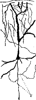
Figure 1. Pyramidal
cell of the large brain with cytoplasmic processes (C). the Golgi apparatus, Holmgren's trophospongium (see Cell), and structures characteristic only for N. k.: neurofibrils and chromophilic substance, as well as
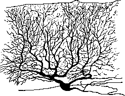
Figure 2. Purkinje cell (according to Kölliker).
some special inclusions. - N e v r o - f i b r i l s (Apathy, 1895, 1898) become visible only with special treatments (see above). They represent thin, clearly defined threads that are found both in the body of N. k. and in its processes, forming according to some authors (Ramón y Cajal, Held, Retzius, M. Heidenhain, Deineka, Rahmanov) a true network, and according to others (Apathy, Bethe, Nissl) only a plexus of free threads; some authors recognize in N. k. along with a true fibrillar network the existence of long independent fibrils passing from one process of N. k. to another (Donaggio, Bel'shovskiy^ (fig. 3). - The appearance of N. k. with stained neurofibrils has the same relationship to the appearance with Nissl staining as a photographic negative has to a positive. In places where there are large accumulations of chromatic substances, caps on nuclei, and clumps during the division of dendrites, there are no neurofibrils or very few. In the processes, neurofibrils are arranged more parallel to each other; entering the cell body, they diverge in different directions, twisting and branching and connecting with each other in one way or another, forming a network with loops of various sizes and shapes. In some cells, neurofibrils are arranged in bundles, which form larger loops, in others a fine-grained homogeneous network is obtained. The diversity of the reticular structure is very significant. At the point of departure of the neurite, neurofibrils collect into a bundle, closely adjacent to each other, so that they often become indistinguishable. Cajal attempted to group N. k. according to the nature of the neuro
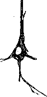
Figure 3. Neurofibrils in a pyramidal nerve cell (from Jacob). "29 fibrillar network and divides them into the following types: 1) plexiform type - fibrils are collected into bundles forming a larger-meshed network (e.g., large N. cells of the spinal cord and medulla oblongata, pyramids). 2) Wide-meshed flat network (e.g., N. cells of spinal ganglia, medium and small cells of the medulla oblongata, retinal cells). 3) Only perinuclear network (small grain cells, basket cells). There are also transitional types; for example, Purkinje cells and pyramidal cells of Ammon's horn represent transitional forms between the 1st and 2nd groups. The most diverse views exist regarding the physiological significance of neurofibrils. Some researchers attributed special significance to neurofibrils in the processes of excitation and conduction in the nervous system. Bethe attempted to prove that neurofibrils are the only conducting elements in the nervous system. Most researchers believed that in the processes of conduction of excitation along a nerve, both neurofibrils and neuroplasm participate equally. This view was particularly convenient for physiological theories of conduction [ionic theory of excitation (see) and conduction; see Nervous system, physiology]. Finally, some authors attributed to neurofibrils the significance of skeletal, supporting formations (Goldschmidt, Koltsov). At present, these disputes have largely lost their acuteness, since the very fact of the existence of neurofibrils during life is being questioned. Neurofibrils in the nervous system of vertebrates are found only on fixed and stained preparations. Observations on living N. cells and nerve fibers in tissue cultures outside the organism (Lewis, Levi, Burrows), as well as observations on freshly cut nerves (Peterfi, Renyi, Auerbach), gave exclusively negative results: neurofibrils were not observed in any case; they become noticeable only at the moment of death of the nerve fiber. Particularly demonstrative are the experiments of Peterfi, performed with the aid of a micromanipulator and filmed with a microcinematograph. The axis cylinders of a living nerve appear optically empty under the microscope. When the nerve fiber is irritated with a microelectrode, thin threads-neurofibrils-instantly become visible. The extraordinary constancy in the arrangement of neurofibrils and their appearance, detected by the most diverse methods of fixation and staining, lead Peterfi to nevertheless assume in N. cells 'something,' some 'latent structure,' which at certain physiological moments, as well as upon fixation of the nerve, 'manifests' in the form of neurofibrils. Other authors tend to consider neurofibrils simply as an artificial formation, an artifact. The fact that neurofibrils are not visible in the living N. cell is not yet direct proof of their absence, since too small a difference in the refractive indices of neurofibrils and neuroplasma can make them inaccessible for microscopic observation. Experiments by Zhivago showed that a number of objects, the structure of which is not resolved by ordinary methods of microscopic observation, can be revealed by means of special photomicrography with enhancement. Thus, the data of Peterfi's experiments can be interpreted as an enlargement, thickening of pre-existing neurofibrils. The existence of neurofibrils during life is also evidenced by the discovery of neurofibrils in living N. cells in invertebrates (Boltzer, Renyi). Thus, the question of the existence of neurofibrils during life remains unresolved. However, the constancy with which they are found in preparations has great significance for neurology and in particular for neuropathology. Changes in neurofibrils in a number of diseases of the nervous system are quite regular and have very great significance. 'Chromophilic substance,' 'chromoidal' substance, 'basophilic' substance, 'Nissl granules,' 'tigroid' substance, 'tigroid' (Lenhossek) represents a granular substance detected in fixed preparations of nervous tissue in greater or lesser quantity and well stained by basic aniline dyes. The property of this substance to take up these dyes, in particular methylene blue, was discovered by Nissl (1884) and had outstanding significance in the study of N. cells and their pathology. Numerous studies carried out by Nissl's method have given a large number of descriptions of N. cells and have led to attempts to classify N. cells according to the amount, type, and arrangement of chromophilic substance, which has generated quite a large terminology. First of all, Nissl distinguishes by the relative size of the nucleus and the cell body, visible when stained by this method. A. Somatochromic N. cells-large and medium-sized cells in which the cell body is visible from all sides of the nucleus when stained with basic dyes. The majority of N. cells belong to this group; Nissl distinguishes in them according to the arrangement of chromophilic substance: 1) tigrochromic N. cells, the body of which is uniformly filled with granules arranged in fairly regular rows; they are also visible in dendrites; at the beginning of the axon there are few granules (such are, for example, the large N. cells of the motor area of the cortex, N. cells of the anterior horns of the spinal cord); 2) argyrochromic N. cells; in them the granules have a reticular arrangement; mixed-type forms are not uncommon; Nissl calls them 3) argyrotigrochromic (for example, Purkinje cells); 4) N. cells with irregularly scattered granules throughout the body are called gryochromic (for example, cells of Clark's columns, some cells of thalami, many cells of spinal ganglia) [see separate table (p. 623-624), Fig. 15]. Cells rich in chromophilic substance, not in the form of granules, but in a finely dispersed state (as, for example, many N. cells of vegetative centers and nodes) are sometimes called pheochromic. - B. Caryochromic N. cells [see separate table (p. 623-624), Fig. 3] 632" Nissl calls those in which the colored part of the cell body is not visible from all sides of the nucleus: the latter lies on one side as if free-an apparent phenomenon, depending on the fact that the cytoplasm is not stained (for example, N. cells of the outer granular layer of the cerebral cortex).-C. In cytchromic N. cells [see separate table (p. 623-624), Fig. 4] the nucleus is small, surrounded by a thin layer of plasma, better visible from one edge of the nucleus, which gives a resemblance to a lymphocyte (for example, so-called grain cells of the granular layer of the cerebellum, cells of fascia dentata).-According to the density of chromophilic substance, Nissl distinguishes among somatochromic N. cells pynomorphic, in which chromophilic substance fills all the plasma without gaps, and pynomorphic N. cells, in which much unstained cytoplasm is visible, and parapynomorphic N. cells-transitional forms between the named ones.-When stained by Nissl's method, a collection of substance closely adjacent to the nuclear membrane and resembling chromophilic granules is visible near the nucleus of the N. cell, the so-called cap (Kernkappe); it is given (Spatz) special significance, attributing participation in exchange processes and considering that its composition also differs from chromophilic substance.-The chemical composition of chromophilic substance has not been precisely established; the main constituent part is recognized by many authors as nucleoproteins (Geld), related to the chromatin of the nucleus (M. Heidenhain, Scott). It is interesting that N. cells with a high content of chromophilic substance have a pale, chromatin-poor nucleus, and vice versa. - The question of whether Nissl granules exist during life or not has a large literature. Photographs taken with ultraviolet rays (Shter Jr., Weimann) showed them on unstained preparations. Just as in the study of neurofibrils (see above), chromophilic substance is not found in the living cell (tissue cultures). Marinesco, using an ultramicroscope, saw in unfixed N. cells a fine granularity, which upon the action of fixing, resp. coagulating, substances enlarged and precipitated in the form of granules similar to Nissl's. One must think that chromophilic substance is a product of protein coagulation, obtained upon fixation of tissue. Essentially important is the fact that with a certain technique (see Nissl's method) a certain picture is always obtained, which makes it possible to compare the appearance of N. cells under various normal and pathological conditions and thus to judge the normal or pathological state of nervous tissue. On the basis of this, Nissl created his doctrine of equivalent pictures (Aquivalentbilder), considering chromophilic substance as a completely constant sign-equivalent of a certain state of the protoplasm of the N. cell. The study of these equivalent pictures has given very much for pathological histology of the nervous system. The nucleus of the N. cell generally differs in a small content of chromatin, and the amount of the latter is inversely proportional to the size of the cell. According to Ramon y Cajal, three types of nuclei can be distinguished: 1) in the nucleus basophilic substance in the form of grains collected in fairly thick crossbars forming a network; nucleoli are only suggested-small cytchromic cells; 2) the linin network is delicate; on it several accumulations of chromatin in the form of small nucleoli-cells of medium size; 3) one large nucleoliu darkly staining, and a very delicate, barely noticeable linin network-large somatochromic cells."
In the nucleolus of N. k. one can often see a strongly light-refracting formation, so-called crystalloid. In the central nervous system, as a rule, all N. k. are mononuclear; multinuclear cells are found mainly in the N. k. of sympathetic ganglia; as an exception, binuclear cells are found in pallidum. - Centrosome. in N. k. has been described in N. k. of both central and peripheral (vegetative) nervous systems (Rio-Hortega). Centrioles look like short sticks. - Nerve cells do not have a true cell membrane and in the central nervous system are surrounded by supporting tissue-neuroglia. Nerve cells of peripheral ganglia are surrounded by a thin connective tissue capsule. Between the connective tissue capsule and the body of these cells there are spindle-shaped or star-shaped cells with short processes ('amphicytes' - Langossek, 'satellites' - Kakhal). Satellites are equated by many authors with neuroglia. In diseases of the vegetative nervous system, the number of satellites can significantly increase. At the same time, phenomena of melting of the protoplasm of N. k. occur. Satellites are undoubtedly connected with Schwann cells of the nerve fiber. Of the inclusions in N. k., the frequently occurring pigment deserves special attention. One can distinguish two types of pigment, differing both morphologically and chemically. 1. Finely granular yellowish pigment, so-called lipochrome, which easily takes up stains that color lipoids (sudan, scarlet, osmic acid). It is found in humans in most N. k. after 30 years of age, and with old age its quantity increases; cells accumulating pigment are called lipophilic (Obersteiner) in contrast to lipophobic, in which even in old age there is almost none (e.g. Purkinje cells). 2. A coarser brown-black pigment of the melanin type is found in N. k. of certain parts of the nervous system: subst. nigra, locus coeruleus, nucL dorsalis n. vagi, N. k. of spinal ganglia and sympathetic ganglia. In addition, in N. k. one can sometimes find neutral fat and other lipoids. --Microreaction for the presence of oxidases made it possible to detect oxidases, mainly 'labile' ones, in the protoplasm of nerve cells and in terminal nerve apparatuses. A very strong reaction to oxidases is given by the peripheral segment of a severed nerve at the time of formation of Büngner's bands. The method of detecting oxidases can, according to Belanovsky, give very important results in pathological histology of the nervous system. - Spatz found in non-pigment-containing N. k. of globi pallidi and subst. nigrae a granularity that gives a reaction to iron (Berlin blue, Turnbull's blue). - The 'pericellular network of Golgi' is a network detectable by this author's method, which covers the entire body of N. k. with its processes (except for the axon) with uniform rounded loops. Golgi himself compares it with neurokeratin (see Nerve fibers). Bete attributes great functional importance to it, considering it an apparatus that sums up irritations and regulates the conductivity of N. k.; Apathy, Donaggio, Held consider it a gliosis formation. Age-related changes of N. k. It has been established that the growth and differentiation of N. k. do not end at the time of birth (mammals). N. k. of newborns have a more primitive character. This concerns above all the processes of N. k. For example, Purkinje cells of the cerebellum at birth have a small number of dendrites and their branches (in puppies 2-3 branches), dendrites are disoriented, and only later abundantly growing dendrites and their branches orient in one plane. Age-related changes of N. k. in ganglia of the vegetative nervous system are particularly demonstrative. Sympathetic N. k. of the heart, border trunks, solar plexus, bladder, vagina of newborns have an embryonic character (type of neuroblasts) and a small number of processes; in the post-embryonic period there is further growth of dendrites, continuing according to de Castro until 21 years. If one takes into account the role of dendrites in terms of increasing the surface of N. k. for perceiving irritations coming from other neurons, then the enormous importance of post-embryonic differentiation of N. k. for the development of the entire nervous system and its functions becomes clear. In elderly individuals (human) excessive growth of processes of N. k. is found. In these cases, N. k. of the vegetative nervous system are entwined with an unusually dense ball of processes. This excessive growth can in some cases be found also in young individuals, but then it is pathological and, as will be indicated below, manifests itself with particular force in a number of lesions of the central and peripheral nervous systems. The doctrine of the interrelationship of N. k. with each other, from its inception to the present time, has focused on the question of whether there is a continuous connection (Kontinuitat) or only contact (Kontiguitat) between N. k. His (W. His, 1886-89) was the first to speak out for the principle of transmission of irritation in the nervous system without continuity of substance; anatomically he represented it this way: that in the development of the nervous system from the embryonic cell of the neuroblast, processes-axial cylinders and dendritic ones grow, which, elongating, pave their way between other elements and end with one or another terminal apparatus. Thus, anatomically independent units are created. In its finished form, this idea was expressed by Waldeyer (1891) with the position that 'the nervous system consists of units (neurons) having neither anatomical nor genetic connection between them', and that 'in the nervous system there is no other element, carrier of nervous function, except the neuron, and the entire function of the nervous system is performed by the sum of neurons'. Thus, in the further development of this position, 'neuron' became not only an anatomical and genetic unit, but also functional and trophic. The word 'neuron' in many authors became synonymous with N. k. This view received special reinforcement with the development of Golgi's method, which nowhere, except in sympathetic plexuses, showed a direct transition of processes of one N. k. into another (Cajal, Forel, Kölliker, Retzius). However, working by other methods, Dogiel, Bielschowsky in the retina, and Schultze and Ster in Purkinje cells saw the fusion of branchings of processes of N. k. The connection of N. k. with each other is carried out according to the neuron theory by touching, by contact of the terminal apparatus of one N. k.-in the form of buttons or loops-with the body or dendrites of another cell, by intertwining of terminal fibers of one N. k. around the body of another, forming sometimes a basket-like structure, by intertwining of branchings of the axon of one N. k. with the dendrite of another. Apathy, Bete, Nissl put forward another theory. According to their views, neurofibrils form a continuous network of anastomosing fibers, which can gain great independence and become independent of the cells, forming in the central nervous system a dense felt-like intercellular plexus, which is called the elementary diffuse network or 'neuropile'. Intracellular neurofibrils are connected with the neuropile by bundles of fibrils, and from them also arise the fibrils forming the axon. Thus, neurofibrils, without breaking anywhere, go from center to periphery, where again they form continuous networks. N. k. are included in the path of fibrils. Nissl in addition recognizes the existence in the gray matter of the brain of a special 'gray mass' (Rindengrau), filling the spaces between fibers and cells, having the closest connection with neurofibrils and playing an important role in the function of the nervous system. Special stands the doctrine of Held, who on the basis of his numerous researches, as well as the latest researches of other authors, puts forward different views. He points out that in the terminal apparatuses the fusion of the plasma of the latter with the plasma of the innervated element takes place, and neurofibrils also enter into direct connection with the plasma or with the fibrillar apparatus of the innervated cell. Genetically he represents it this way: neuroblasts are units that give rise to fibrils-fibrillogenic units. In their development, the plasma of these cells determines the paths of further connections of individual elements with each other-unidirectional and multidirectional-by the enthytial path; at the same time neurofibrils spread in this plasma and penetrate into another, even non-nerve cell, entering into the closest connection with the plasma of these cells. If this is a nerve cell, a connection of fibrils is obtained, in which one can no longer distinguish the system of neurofibrils of this cell from another. Thus, the fibrillar structure of the adult N. k. is not the product of one neuroblast, but a combination of several systems, and the cell is not an isolated bounded unit in the sense of Waldeyer's neuron, but a part of a complex indivisible system of neurentitium, into which can enter both N. k. of the brain and spinal cord, and N. k. of the intervertebral ganglion and peripheral cells of another kind, e.g. muscle cells, in the plasma of which the fibrillar system forms the so-called terminal plexus. The doctrine of the neuron is currently the most widespread, but at the same time the idea of the need to recognize a closer connection between elements is strengthening. - In pathological processes in the central nervous system, both the external appearance of N. k. and their fine structure can change.
The size of nerve cells can vary in one direction or another. Nerve cells of enormous size and abnormal shape are found in the cerebral cortex in tuberous sclerosis. Among other general changes in nerve cells, the appearance of binucleate cells deserves attention, especially among Purkinje cells in certain diseases, for example, in early dementia (dementia praecox). The size, shape, and number of nerve cell processes undergo significant changes in a number of pathological conditions. In some cases, the surface of the nerve cell becomes uneven, as if gnawed, and satellites lie tightly in the resulting depressions. The phenomenon of 'complex dendrites' arises. In other cases, new processes begin to form, and new processes can arise in nerve cells that normally have only one process (pseudo-unipolar cells of the intervertebral ganglia). The formation of new processes is often accompanied by the appearance of spherical or pear-shaped swellings at their ends. These thickenings can reach a giant size. This phenomenon, first described in detail by Cajal, was named by him 'Kugelphänomen'. This phenomenon sometimes occurs in normal and young individuals, but in pathological cases it can be extraordinarily strong, affecting almost all nerve cells of the affected nerve node. Excessive growth of processes and the appearance of spherical swellings are a reaction of the cell body to inadequate stimuli and in some cases can be compared to the phenomenon of excessive growth of nerve fibers on the periphery (regeneration, neuromas). The described changes in processes (pseudodendrites, excessive growth, 'Kugelphänomen') are found mainly in sensory ganglia (intervertebral ganglia, ganglia of cranial nerves, ganglia of the autonomic nervous system); they are found in tabes, progressive paralysis, in alcoholic neuritis, ganglioneuritis, operative damage to nerve trunks, tuberculosis, metastases of cancer, in cardiosclerosis. Pathological changes in the fine structure of nerve cells have been studied mainly by Nissl's method by comparison with the appearance of a normal nerve cell with this staining (equivalent picture). Valuable data are also provided by methods of staining neurofibrils, as well as staining that reveals various inclusions in nerve cells (lipoids, etc.).- The nucleus in a nerve cell may be displaced to its periphery, but this can sometimes be an apparent phenomenon due to the section passing through the nerve cell. More significant is the change in its size, especially its reduction. In this case, its contours may lose their regularity; it becomes uniformly darkly stained, nucleoli disappear; such changes in the nucleus usually occur when the entire nerve cell shrinks. In other cases, the nucleus decreases, more or less maintaining its structure. The nuclear capsule, when the nucleus decreases, either forms numerous folds on its surface or lags behind it. Changes in the nucleolus, when it looks like a mulberry berry, are a sign of severe damage to the cell. Sometimes changes in the nucleus of the karyorrhexis type occur, for example in so-called 'severe diseases of the nerve cell' (see below).'- Chromophilic substance reacts vividly to various states of the nerve cell; in this case, the granules seem to be scattered-'chromolysis', 'chromatolysis', 'tigrolysis' [see separate table (pp. 623-624), figure 16]. This process develops either from the center of the cell around the nucleus or from its periphery. In other cases, the granules seem to fuse into a darkly staining mass, usually unevenly filling the nerve cell body, and the entire cell strongly shrinks; the extreme degree of this process is called sclerosis of the nerve cell [see separate table (pp. 623-624), figure 2]. In this case, the pyramidal cells have a corkscrew-shaped upper process. When the disintegration reaches a high degree, a fine granularity is visible in the plasma, which takes on a special ring-like structure; later the plasma can become homogeneous [see separate table (pp. 119-120), figs. 1-4]; it melts away and only a 'shadow' remains of the nerve cell. Such pallor of the nerve cell can in some cases (local ischemia) occur suddenly and very quickly. - In the marginal disintegration of the nerve cell, voids, vacuoles, can form in its body, in which gliosis elements-neronophagy-are often visible. This phenomenon is considered as an expression of gliocytes capturing the products of disintegration of the nerve cell. This picture is distinguished from pseudoneronophagy, when there is a proliferation of satellites around the nerve cell, but the integrity of the nerve cell is not violated. In individual areas of the cerebral cortex, a large number of satellites are found around normal nerve cells. In some cases of severe damage to nerve cells, e.g., in ischemic foci, in addition to changes in the nucleus and plasma, a special picture is observed, which Nissl called 'inlaying of the Golgi network' [see separate table (pp. 623-624), figure 9]; in preparations treated by Nissl's method, darkly stained granules, sticks, threads are visible, which seem to cover the processes, and sometimes the cell body. In the deposits on the cell, lime can often be detected [see separate table (pp. 607-608), fig. 4]. Changes in the neurofibrillar structure are always observed in parallel with changes in the plasma and chromophilic substance, and to a certain extent the above-mentioned relationship between them is preserved. When the chromophilic substance is scattered, the fibers mostly form a fine network. When there is a complete disintegration of the chromophilic substance and deep changes in the nerve cell, detectable by Nissl's method, the fibrillar structure usually also disappears.-Changes in the neurofibrillar apparatus in animals during winter hibernation and similar pictures of the fibrillar structure of nerve cells in humans in rabies deserve special mention. Here the neurofibrils appear thickened and in smaller numbers than in normal, which is explained by their fusion. Special are the changes in neurofibrils in Alzheimer's disease (see below).-Pathological changes in inclusions of nerve cells mainly concern an increase in the amount of lipoid substances. Two main types of fatty degeneration of nerve cells can be distinguished: 1) gradual excessive accumulation of fatty substances, apparently mainly of the lipofuscin type, in those places of the nerve cell where they are deposited even in normal, 'simple fatty degeneration' according to A. Jakob (A. Jakob), 'pigmentary atrophy' according to Spielmeyer [see separate table (pp. 623-624), figure 14]. This form of fatty degeneration is often combined with shrinking of the nerve cell and with sclerosis. Gliocytes in this case also often contain fat droplets. This type of fatty degeneration is characteristic of senile changes in general, senile dementia, and arteriosclerosis. 2) Diffuse deposition of fatty substances throughout the body of the nerve cell and even in the processes, occurring in nerve cells that usually do not contain lipoids. It develops mostly rapidly and is accompanied by degenerative changes in the nerve cell. This is 'degenerative fatty degeneration' of the nerve cell according to A. Jakob, or 'fatty degeneration of the nerve cell' according to Spielmeyer [see separate table (pp. 623-624), fig. 17]. In these cases, the lipoids do not have the character of lipofuscins. It occurs in acute and chronic poisonings, intoxications, and infectious diseases. Fatty degenerations of nerve cells are studied with fat-staining dyes on preparations not treated with alcohol (Sudan, scarlet, Nile blue, etc.). Pathological changes in nerve cells specifically characteristic of certain diseases are still very little described, and in most cases one can only speak of a certain typical picture of changes in nerve cells. The most widely used classification of changes in nerve cells is that given by Nissl. 'Primary irritation of the nerve cell' and 'retrograde degeneration' (secondary, or consecutive atrophy) is characterized by swelling of the nerve cell, central chromatolysis, eccentric position of the nucleus. This state of the nerve cell then either passes into restoration of the normal picture or leads to the death of the cell: complete chromatolysis, up to complete disappearance of chromophilic substance, sharp changes in the nucleus, sometimes formation of vacuoles in the cell and in the processes-phenomena of neronophagy. These pictures are observed mainly in traumatic damage to peripheral nerve fibers in the corresponding nerve cells and in general in injuries to the central nervous system. 'Acute disease' of the nerve cell [see separate table (pp. 623-624), fig. 5] according to Nissl is expressed in swelling of the hyaloplasm, accompanied by better staining of it with basic dyes, which makes the cell with all its processes stand out well, then in the dusty disintegration of chromophilic substance, swelling of the nucleus with more intense staining of its structure and sometimes with a change in the shape of the nucleolus. The fibrillar structure is usually preserved. Such a picture is observed in general severe diseases (infections, intoxications), in severe forms of epilepsy, in catatonia. It apparently develops rapidly.-'Severe disease' of the nerve cell [see separate table (pp. 623-624), figure 1] according to Nissl is characterized by the following phenomena: the nucleus is diffusely darkly stained, shrinks, and then disintegrates; the cell plasma-at first diffusely darkly stains, and then-melts. Chromophilic substance disintegrates into granules, sometimes forms rings and then disappears. The remains of the disintegrated cell substance form dark clumps and grains, located around and in place of the disintegrating nerve cell. Fibers also disintegrate.
Such changes are found in various severe diseases, especially those accompanied by 'brain edema'—in severe and acute forms of progressive paralysis, in acute delirious states, etc.—'Edema' of nerve cells ('hydrocephalus' of nerve cells Nissl) is observed in disorders of cerebral circulation, in brain edema. The nucleus of the nerve cell is irregular in shape, shrunken. Instead of the proper arrangement of chromatophilic substance, there are separate lumps and grains, and later the plasma first swells and then decreases in volume and stains poorly, forming 'cell shadows.'—A special picture is presented by nerve cells in myoclonic epilepsy of Lafora and Westphal. In the body of the cell, round, fairly large formations are found, which in appearance and in relation to dyes are analogous to amyloid bodies. The cells do not die immediately in this disease; in their presence, neurofibrils are still visible. Also, characteristic changes in nerve cells have been described in amaurotic familial idiocy (see) and in rabies (see).
Related articles
Mentioned in
- Afferent
- Alzheimer's Disease
- Amaurotic Family Idiocy
- ARCHITECTONICS OF THE CEREBRAL CORTEX
- Axon
- Axoplasm
- Benda
- Bethe
- Bidder's Nodes
- Bipolar
- Brain (551 General diagnostics of brain diseases)
- Brain (cerebrum, a comprehensive term for the entire)
- Burns
- Chromaffin System
- Chromatin
- Degeneration (a870)
- Dystopia
- Encephalometry
- Ependyma
- Extrapyramidal System
Cite this page
“Nerve Cells.” Soviet Medical Encyclopedia. English translation of Bolshaya Meditsinskaya Entsiklopediya, 1st ed. (Moscow, 1928–1936), ed. N. A. Semashko. https://sovietmedicalencyclopedia.pages.dev/article/nerve-cells/