NERVE ENDINGS
Historical document, translated for reference. It reflects medical knowledge of the 1920s–30s and is not medical advice.
Summary
Nerve endings are the connection points between nerve fibers and various body tissues. This article discusses their classification, structure, and function in the context of 1930s medical understanding.
Encyclopedia article (1928–1936)
NERVE ENDINGS, places of connection between nerve fibers with elements of various body tissues. Formations connecting nerve cells with each other, so-called pericellular apparatuses, can also be classified as N. e. (see Neuron theory). Physiologically, N. e. represent that portion of the nerve and innervated tissue where excitation passes from the nerve fiber to the innervated tissue—effector N. e. (motor, secretory, trophic) or where excitation passes from the tissue to the nerve fiber—sensory N. e. The doctrine of the structure and function of N. e. as a chapter in the doctrine of nervous tissue has reflected all currents of scientific thought concerning the cell theory, and in particular the theory of neurons, and criticism of these theories. The concept of cells as elements or even elementary organisms, the sum of which gives tissue, required a sharp distinction between cells as well as between individual tissues. Therefore, connections between tissues were conceived only as the contact of some elements with others. In relation to nervous tissue, its connection with others was described as contact (apposition). At the same time, a crude analogy with electrical installations was often used. Kühne (Kiihne) was the first to clearly show that N. e. in muscle fibers do not fit the concept of contact, since the branching of nerve fibers directly penetrates the protoplasm of the muscle fiber. Through the work of Apathy, Kolmer, Heringa, and others, it was shown that such close relationships of nerve fibers to innervated tissues can also be demonstrated in a number of other N. e. (epithelium, epithelium of sensory organs, connective tissue). Thus it was clarified that N. e. represent an organic whole that cannot be reduced to the sum: nerve plus muscle or nerve plus epithelium either morphologically or physiologically. This qualitative specificity of nerve endings was later established by methods of physiology (reciprocal conductivity, "receptive substance" Langley) and methods of morphology. With ordinary methods of staining histological preparations, N. e. are mostly not detected. N. e. were discovered and described in detail only with the appearance of special methods for staining nervous tissue (methylene blue, silver and gold impregnation of Golgi, Cajal, and Bielschowsky). N. e. have some common structural features. Approaching the N. e., the nerve fiber usually loses its myelin sheath and for some distance becomes non-myelinated. Schwann's and Henle's sheaths often merge with the capsules of N. e. or the organ's sheaths. The existence of so-called bare axons in the area of N. e. is currently disputed by many. In the area of nerve endings of the autonomic nervous system, the presence of elements of Schwann's syncytium, having the character of multipolar cells (so-called "interstitial" cells), is characteristic. The axon forming part of the N. e. often branches. The number of branches can be very large. A characteristic feature of many N. e. is the dilation of neurofibrils (see Nerve fibers) and the resulting expansions or swellings of the nerve fiber (so-called varicosities). Sometimes these expansions can reach very large sizes and in such cases are described as plates (sensory N. e.). Many N. e. are supplied with special cells with which nerve fibers enter into the closest connection (so-called sensory cells, tactile cells, etc.). The origin of these cells is not sufficiently clarified. Classification of N. e. Physiologically, N. e. are usually divided into effector and sensory. Morphologically, it is not always possible to determine the classification of N. e. into these two categories, especially in relation to N. e. located in connective tissue. The classification of N. e. in relation to special types of sensitivity (painful, tactile, thermal) is not currently established, and if the pathways for conducting different types of sensitivity in the central nervous system are known, then with respect to the periphery we currently have extremely meager data. Effector N. e. These include motor endings on smooth and striated muscles and secretory endings on cells of external and internal secretion glands. The nerve endings of the autonomic nervous system to which a trophic function is attributed should also be included in this category. Motor N. e. in voluntary musculature. Motor, or motor endings in striated muscle of mammals have a complex structure [see separate table (pp. 607-608), fig. 5]. The nerve fiber approaching the muscle fiber loses its myelin sheath. The Schwann sheath merges with the sarcolemma of the muscle fiber and the axon enters the sarcoplasm of the muscle fiber. At this place the sarcoplasm may have an outgrowth in the form of a more or less noticeable tubercle (highly developed in some insects and birds). Entering the sarcoplasm, the axon divides into branches, the number of which can vary greatly in different muscles and in different animals. The branches of the axon often end in plate-like expansions with a network-like or fan-like arrangement of neurofibrils (fig. 1). Around the branching of the axon there is a collection of nuclei. According to Tello, they originate from the ordinary nuclei of the muscle fiber. The sarcoplasm in the area of N. e. has a fine-grained character and contains large rod-shaped chondrioconts. The entire formation, including the axon, sarcoplasm and nuclei, has received the name motor plate. This complex is also called the "sole." Bouquet (Boeke), using the latest technique for detecting neurofibrils, described a delicate network penetrating the sarcoplasm of the motor plate; this network on the one hand is connected with the neurofibrils of the axon and on the other approaches the myofibrils closely. It received the name "periterminal network." Its existence on fixed and stained preparations is now confirmed by most researchers. Its physiological significance remains unclear. In some cases during the formation of the motor plate
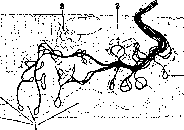
Fig. 1. Motor plate on striated muscle: 1-nuclei; 2-terminal branches; 3-periterminal network.
one of the branches of the axial cylinder emerges from the limits of the plaque and goes to another muscle fiber, where it forms a second-order motor plaque. Such branches have been named ultra-terminal fibers. From the second-order motor plaques, a branch can emerge and form a third-order motor plaque, and so on. In some cases, the number of ultra-terminal fibers is so large that the entire muscle appears to be penetrated by a continuous network of nerve fibers, with motor plaques inserted into this network at certain points (figure 2). Such relationships have been found in amphibians and reptiles (mainly in the tongue) by Stefanelli, Ruffini, and Lavrentiev. The physiological significance of this phenomenon has not been clarified. According to Stefanelli's hypothesis, this arrangement facilitates maximum synchronicity of contraction of all muscle fibers of the organ. In addition to spinal nerve fibers forming the motor plaque of a muscle fiber, thin, mostly unmyelinated nerve fibers also enter the same plaque; these were first observed in amphibians and reptiles by Bremer and Gemelli and were described in detail for mammals by Bouquet. Through a series of experiments, Bouquet established that these fibers belong to the autonomic nervous system. Physiologically, these data were confirmed by Orbeli and his students.-Nerve endings in striated muscles of amphibians are constructed more primitively. The 'sole' usually is absent, and the branches of the axial cylinder lie in the form of long threads along the length of the muscle fiber. In some muscles, mainly in amphibians and reptiles, nerve endings have the appearance of bushes or clusters. Such endings were first described by Chiriev and received the name cluster endings (terminaisons en grappe). Polysegmental nerve endings of striated voluntary muscles. For some striated muscles of mammals, the presence of several motor plaques on the same muscle fiber is characteristic. Agduhr experimentally proved that in such cases the muscle fiber is innervated simultaneously from several segments of the spinal cord; for example, the m. flexor digitorum sublimis of the cat is innervated from three cervical segments, and the muscle fibers here have three plaques, each belonging to a neighboring segment. Polysegmental innervation also occurs in lower vertebrates. Physiologically, it was discovered in the frog by Beritoff and de Boer, and recently confirmed morphologically by Lavrentiev. The physiological significance of this phenomenon remains unclarified.-Motor nerve endings on smooth muscles have the appearance of very dense plexuses of thin unmyelinated nerve fibers; the terminal branches of these fibers are extremely thin and apparently form a terminal network. Varicose thickenings may be present on the fibers. Free button-like terminal apparatuses are possible, which may penetrate inside the cells. It should be noted that on smooth muscles, nerve endings are located rarely, and not every fiber is supplied with them. Trophic nerve endings. A number of physiological observations showing the influence of the nervous system on tissue metabolism have led to the assumption of the existence of special trophic nerve fibers and their endings. Research concerning sympathetic innervation of striated muscles has shown that sympathetic nerve endings in muscles have significance mainly as an apparatus influencing the metabolism of muscle tissue (Orbeli).-The question of the influence of the nervous system on tissue metabolism is connected with the innervation not only of muscle tissue and glandular organs, but also with the innervation of so-called passive tissues, to which are epithelium and a group of connective tissue substances (connective tissue, cartilage, bone). Until the very last time, the innervation of 'passive' tissues in the effector sense was denied. The existence of undoubtedly effector nerve endings on chromatophores (cells of pigmented connective tissue) of some lower vertebrates was considered an exception. In 1930-31, works appeared establishing effector innervation of connective tissue elements. Indications of the possibility of such innervation were given in the work of Akkeringa. In 1931, de Castro showed that osteoblasts, participating in the formation of perichondral and endochondral bone, are supplied with special nerve endings. These endings have the appearance of small loops located in the immediate vicinity of the osteoblast nucleus. It is highly probable that these nerve endings lie inside the protoplasm of the osteoblasts. The same author
/
one of the branches of the axial cylinder emerges from the limits of the plaque and goes to another muscle fiber, where it forms a second-order motor plaque. Such branches have been named ultra-terminal fibers. From the second-order motor plaques, a branch can emerge and form a third-order motor plaque, and so on. In some cases, the number of ultra-terminal fibers is so large that the entire muscle appears to be penetrated by a continuous network of nerve fibers, with motor plaques inserted into this network at certain points (figure 2). Such relationships have been found in amphibians and reptiles (mainly in the tongue) by Stefanelli, Ruffini, and Lavrentiev. The physiological significance of this phenomenon has not been clarified. According to Stefanelli's hypothesis, this arrangement facilitates maximum synchronicity of contraction of all muscle fibers of the organ. In addition to spinal nerve fibers forming the motor plaque of a muscle fiber, thin, mostly unmyelinated nerve fibers also enter the same plaque; these were first observed in amphibians and reptiles by Bremer and Gemelli and were described in detail for mammals by Bouquet. Through a series of experiments, Bouquet established that these fibers belong to the autonomic nervous system. Physiologically, these data were confirmed by Orbeli and his students.-Nerve endings in striated muscles of amphibians are constructed more primitively. The 'sole' usually is absent, and the branches of the axial cylinder lie in the form of long threads along the length of the muscle fiber. In some muscles, mainly in amphibians and reptiles, nerve endings have the appearance of bushes or clusters. Such endings were first described by Chiriev and received the name cluster endings (terminaisons en grappe). Polysegmental nerve endings of striated voluntary muscles. For some striated muscles of mammals, the presence of several motor plaques on the same muscle fiber is characteristic. Agduhr experimentally proved that in such cases the muscle fiber is innervated simultaneously from several segments of the spinal cord; for example, the m. flexor digitorum sublimis of the cat is innervated from three cervical segments, and the muscle fibers here have three plaques, each belonging to a neighboring segment. Polysegmental innervation also occurs in lower vertebrates. Physiologically, it was discovered in the frog by Beritoff and de Boer, and recently confirmed morphologically by Lavrentiev. The physiological significance of this phenomenon remains unclarified.-Motor nerve endings on smooth muscles have the appearance of very dense plexuses of thin unmyelinated nerve fibers; the terminal branches of these fibers are extremely thin and apparently form a terminal network. Varicose thickenings may be present on the fibers. Free button-like terminal apparatuses are possible, which may penetrate inside the cells. It should be noted that on smooth muscles, nerve endings are located rarely, and not every fiber is supplied with them. Trophic nerve endings. A number of physiological observations showing the influence of the nervous system on tissue metabolism have led to the assumption of the existence of special trophic nerve fibers and their endings. Research concerning sympathetic innervation of striated muscles has shown that sympathetic nerve endings in muscles have significance mainly as an apparatus influencing the metabolism of muscle tissue (Orbeli).-The question of the influence of the nervous system on tissue metabolism is connected with the innervation not only of muscle tissue and glandular organs, but also with the innervation of so-called passive tissues, to which are epithelium and a group of connective tissue substances (connective tissue, cartilage, bone). Until the very last time, the innervation of 'passive' tissues in the effector sense was denied. The existence of undoubtedly effector nerve endings on chromatophores (cells of pigmented connective tissue) of some lower vertebrates was considered an exception. In 1930-31, works appeared establishing effector innervation of connective tissue elements. Indications of the possibility of such innervation were given in the work of Akkeringa. In 1931, de Castro showed that osteoblasts, participating in the formation of perichondral and endochondral bone, are supplied with special nerve endings. These endings have the appearance of small loops located in the immediate vicinity of the osteoblast nucleus. It is highly probable that these nerve endings lie inside the protoplasm of the osteoblasts. The same author
/
one of the branches of the axial cylinder emerges from the limits of the plaque and goes to another muscle fiber, where it forms a second-order motor plaque. Such branches have been named ultra-terminal fibers. From the second-order motor plaques, a branch can emerge and form a third-order motor plaque, and so on. In some cases, the number of ultra-terminal fibers is so large that the entire muscle appears to be penetrated by a continuous network of nerve fibers, with motor plaques inserted into this network at certain points (figure 2). Such relationships have been found in amphibians and reptiles (mainly in the tongue) by Stefanelli, Ruffini, and Lavrentiev. The physiological significance of this phenomenon has not been clarified. According to Stefanelli's hypothesis, this arrangement facilitates maximum synchronicity of contraction of all muscle fibers of the organ. In addition to spinal nerve fibers forming the motor plaque of a muscle fiber, thin, mostly unmyelinated nerve fibers also enter the same plaque; these were first observed in amphibians and reptiles by Bremer and Gemelli and were described in detail for mammals by Bouquet. Through a series of experiments, Bouquet established that these fibers belong to the autonomic nervous system. Physiologically, these data were confirmed by Orbeli and his students.-Nerve endings in striated muscles of amphibians are constructed more primitively. The 'sole' usually is absent, and the branches of the axial cylinder lie in the form of long threads along the length of the muscle fiber. In some muscles, mainly in amphibians and reptiles, nerve endings have the appearance of bushes or clusters. Such endings were first described by Chiriev and received the name cluster endings (terminaisons en grappe). Polysegmental nerve endings of striated voluntary muscles. For some striated muscles of mammals, the presence of several motor plaques on the same muscle fiber is characteristic. Agduhr experimentally proved that in such cases the muscle fiber is innervated simultaneously from several segments of the spinal cord; for example, the m. flexor digitorum sublimis of the cat is innervated from three cervical segments, and the muscle fibers here have three plaques, each belonging to a neighboring segment. Polysegmental innervation also occurs in lower vertebrates. Physiologically, it was discovered in the frog by Beritoff and de Boer, and recently confirmed morphologically by Lavrentiev. The physiological significance of this phenomenon remains unclarified.-Motor nerve endings on smooth muscles have the appearance of very dense plexuses of thin unmyelinated nerve fibers; the terminal branches of these fibers are extremely thin and apparently form a terminal network. Varicose thickenings may be present on the fibers. Free button-like terminal apparatuses are possible, which may penetrate inside the cells. It should be noted that on smooth muscles, nerve endings are located rarely, and not every fiber is supplied with them. Trophic nerve endings. A number of physiological observations showing the influence of the nervous system on tissue metabolism have led to the assumption of the existence of special trophic nerve fibers and their endings. Research concerning sympathetic innervation of striated muscles has shown that sympathetic nerve endings in muscles have significance mainly as an apparatus influencing the metabolism of muscle tissue (Orbeli).-The question of the influence of the nervous system on tissue metabolism is connected with the innervation not only of muscle tissue and glandular organs, but also with the innervation of so-called passive tissues, to which are epithelium and a group of connective tissue substances (connective tissue, cartilage, bone). Until the very last time, the innervation of 'passive' tissues in the effector sense was denied. The existence of undoubtedly effector nerve endings on chromatophores (cells of pigmented connective tissue) of some lower vertebrates was considered an exception. In 1930-31, works appeared establishing effector innervation of connective tissue elements. Indications of the possibility of such innervation were given in the work of Akkeringa. In 1931, de Castro showed that osteoblasts, participating in the formation of perichondral and endochondral bone, are supplied with special nerve endings. These endings have the appearance of small loops located in the immediate vicinity of the osteoblast nucleus. It is highly probable that these nerve endings lie inside the protoplasm of the osteoblasts. The same author
/
one of the branches of the axial cylinder emerges from the limits of the plaque and goes to another muscle fiber, where it forms a second-order motor plaque. Such branches have been named ultra-terminal fibers. From the second-order motor plaques, a branch can emerge and form a third-order motor plaque, and so on. In some cases, the number of ultra-terminal fibers is so large that the entire muscle appears to be penetrated by a continuous network of nerve fibers, with motor plaques inserted into this network at certain points (figure 2). Such relationships have been found in amphibians and reptiles (mainly in the tongue) by Stefanelli, Ruffini, and Lavrentiev. The physiological significance of this phenomenon has not been clarified. According to Stefanelli's hypothesis, this arrangement facilitates maximum synchronicity of contraction of all muscle fibers of the organ. In addition to spinal nerve fibers forming the motor plaque of a muscle fiber, thin, mostly unmyelinated nerve fibers also enter the same plaque; these were first observed in amphibians and reptiles by Bremer and Gemelli and were described in detail for mammals by Bouquet. Through a series of experiments, Bouquet established that these fibers belong to the autonomic nervous system. Physiologically, these data were confirmed by Orbeli and his students.-Nerve endings in striated muscles of amphibians are constructed more primitively. The 'sole' usually is absent, and the branches of the axial cylinder lie in the form of long threads along the length of the muscle fiber. In some muscles, mainly in amphibians and reptiles, nerve endings have the appearance of bushes or clusters. Such endings were first described by Chiriev and received the name cluster endings (terminaisons en grappe). Polysegmental nerve endings of striated voluntary muscles. For some striated muscles of mammals, the presence of several motor plaques on the same muscle fiber is characteristic. Agduhr experimentally proved that in such cases the muscle fiber is innervated simultaneously from several segments of the spinal cord; for example, the m. flexor digitorum sublimis of the cat is innervated from three cervical segments, and the muscle fibers here have three plaques, each belonging to a neighboring segment. Polysegmental innervation also occurs in lower vertebrates. Physiologically, it was discovered in the frog by Beritoff and de Boer, and recently confirmed morphologically by Lavrentiev. The physiological significance of this phenomenon remains unclarified.-Motor nerve endings on smooth muscles have the appearance of very dense plexuses of thin unmyelinated nerve fibers; the terminal branches of these fibers are extremely thin and apparently form a terminal network. Varicose thickenings may be present on the fibers. Free button-like terminal apparatuses are possible, which may penetrate inside the cells. It should be noted that on smooth muscles, nerve endings are located rarely, and not every fiber is supplied with them. Trophic nerve endings. A number of physiological observations showing the influence of the nervous system on tissue metabolism have led to the assumption of the existence of special trophic nerve fibers and their endings. Research concerning sympathetic innervation of striated muscles has shown that sympathetic nerve endings in muscles have significance mainly as an apparatus influencing the metabolism of muscle tissue (Orbeli).-The question of the influence of the nervous system on tissue metabolism is connected with the innervation not only of muscle tissue and glandular organs, but also with the innervation of so-called passive tissues, to which are epithelium and a group of connective tissue substances (connective tissue, cartilage, bone). Until the very last time, the innervation of 'passive' tissues in the effector sense was denied. The existence of undoubtedly effector nerve endings on chromatophores (cells of pigmented connective tissue) of some lower vertebrates was considered an exception. In 1930-31, works appeared establishing effector innervation of connective tissue elements. Indications of the possibility of such innervation were given in the work of Akkeringa. In 1931, de Castro showed that osteoblasts, participating in the formation of perichondral and endochondral bone, are supplied with special nerve endings. These endings have the appearance of small loops located in the immediate vicinity of the osteoblast nucleus. It is highly probable that these nerve endings lie inside the protoplasm of the osteoblasts. The same author
/

Figure 2. Ruffini's corpuscle (after Dogel). It has been established that these endings belong to postganglionic fibers of the superior cervical sympathetic ganglion. Nerve endings of the same kind were found by Calderon (Ca-jalon) on odontoblasts of a growing tooth. When osteoblasts turn into bone cells, their innervation ceases, and the corresponding nerve endings and the appropriate nerve fiber degenerate. Effect nerve endings on nerve cells of the central and peripheral nervous system are described in the form of so-called pericellular apparatuses (Ehrlich, Aronson). They are described in detail in the central nervous system by Held, Cajal and his school, Lenhossek, and in the autonomic nervous system by Arnstein, Dogel, Timofeev, Leontovich and others. Pericellular apparatuses, physiologically, represent the place where excitation passes from one nerve cell or from many nerve cells to another. The nature of these structures was interpreted differently depending on the development of criticism of the neuron theory. A number of physiological and morphological data forces one to assume the great importance of this apparatus in the processes of conduction of excitation and inhibition in nervous tissue. The structure of pericellular apparatuses is extremely diverse. In the simplest case, a nerve ending may have the form of a knob or plaque located on the body of a nerve cell or on its processes. Thus, pericellular apparatuses on the cells of the anterior horns of the spinal cord have the form of numerous knobs (Endknopfchen) located on the body and dendrites of nerve cells. In reptiles and birds, pericellular apparatuses are described, which have the form of massive thickenings that envelop the nerve cell like a cap or hood. In some cases, pericellular apparatuses have the form of the finest nerve threads, which accompany the dendrites of the nerve cell to their final branches, as if spreading over them (climbing fibers on Purkinje cells of the cerebellum). In other cases, neurites of one nerve cell and dendrites of another cell come into contact, forming tangles of varying complexity, provided with special elements of neuroglia. Such tangles are found in the olfactory bulb and in the cerebellum ("mossy fibers"). Often, nerve threads of the pericellular apparatus envelop the dendrites of a nerve cell, forming true spirals. This phenomenon is especially common in the autonomic nervous system (the so-called "spiral process" in amphibians). In the autonomic nervous system, pericellular apparatuses are described, having the form of complex tangles or baskets that envelop the body of the nerve cell. In some cases, the pericellular apparatus has the form of plaques or clusters, strikingly similar to the endings in striated muscles (Timofeev, Serebryakov). In elderly individuals and in a number of pathological cases, excessive growth of threads of the pericellular apparatus is described (alcoholism - Cajal, de Castro). Sensory nerve endings. In connective tissue, especially subcutaneous, as well as in mucous and serous membranes, near joints, etc., there are many nerve endings of very diverse forms. Apparently, all of them have the character of receptor endings, and most of them belong to cerebrospinal sensory fibers. They can be divided morphologically into free apparatuses and encapsulated ones. Free nerve endings represent numerous branches of the nerve fiber - telodendrites, - forming a more or less dense plexus, concentrated in one place or spread over a wide area, developed in one plane in the form of a plate or penetrating the connective tissue in different directions. Individual thin branches often have varicose thickenings on them. Some branches sometimes end in swellings. Sometimes all branches form terminal plates - so-called tree-like nerve endings (A. S. Dogel). In the connective tissue part of the skin directly under the epidermis, there are plexuses of thin nerve fibers, more or less abundant in different parts of the body. From these fibers, thin branches depart, which penetrate between the epithelial cells of the epidermis, and sometimes branching, reach in the form of the finest threads to its superficial layers. These threads end freely, or they are lost among the keratinized cells or form knob-like thickenings near or (according to Buke) inside the cells. These intraepithelial endings are developed very differently in different parts of the body and are especially strongly expressed in places with the most developed sensitivity (for example, lips, muzzle of animals). Encapsulated nerve endings. Many nerve endings are covered with sheaths, which consist of thin connective tissue plates arranged in a larger or smaller number of rows. Between the plates there are flat connective tissue cells. From the sheath, sometimes bridges extend inward into the "bulb" formed by the sheaths. There are many varieties of encapsulated nerve endings, which is due to the different size, shape, thickness of the capsule and the infinite variety of the terminal plexuses of the actual nerve apparatus. In the skin of the mole's snout, nerve endings form complex so-called Eimer's organs (Eimer; 1871). The latter consist of a series of epithelial cells arranged in columns one above the other throughout the entire thickness of the epidermis. Nerve fibers pass through such columns, forming intracellular fibrillar networks. Merkel's cells (Merkel; 1875) are large, light, blister-like epithelial cells, found in the lower layers of the epidermis, usually in groups. A myelinated fiber approaches them, breaks up into branches, which form on the base of each cell a fibrillar plexus - a tactile meniscus or plate. According to Buke, there is a periterminal plasma network in Merkel's cells. Botezat (Botezat), 64 in Dogel, as well as Kadanov, described additional innervation of Merkel's cells from another nerve fiber in the form of varicose threads enveloping them. A more complex type of encapsulated nerve ending is the Meissner's (synonym Wagner's) tactile corpuscles (Figure 3); Meissner's corpuscles are located at the tops of the skin papillae, mainly on the palms and on
the fingertips. They consist of a connective tissue capsule, inside which there are several transversely located connective tissue fibers, between which nerve fibers pass, forming loops and spirals. The capsule is covered with a connective tissue membrane. In the skin of the fingertips, there are also so-called genital corpuscles (Krause; 1860). They consist of a connective tissue capsule, inside which there is a thick connective tissue core, in which a nerve fiber ends in the form of a ball. In the skin of the fingertips, there are also so-called genital corpuscles (Krause; 1860). They consist of a connective tissue capsule, inside which there is a thick connective tissue core, in which a nerve fiber ends in the form of a ball. In the skin of the fingertips, there are also so-called genital corpuscles (Krause; 1860). They consist of a connective tissue capsule, inside which there is a thick connective tissue core, in which a nerve fiber ends in the form of a ball.
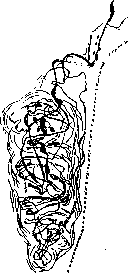
Figure 3. Meissner's corpuscle (after Dogel). These are elongated formations up to 40-180 microns in size. Their capsule consists of several rows of connective tissue lamellae. It merges with the Henle's and Schwann's sheaths of the medullated nerve fiber approaching the corpuscle. Inside the capsule are elongated cells (tactile) lying across it. Between these cells, the branches of the nerve fiber meander in a complex manner, having thickenings. According to the latest data (Boucke, Hering), the nerve fibers form delicate fibrillar networks inside the tactile cells. In addition to the medullated nerve fiber, non-medullated fibers also participate in the plexus inside the corpuscle (Timofeev, Dogel). More simply encapsulated nerve endings consist of a ball of nerve fibers covered by sheaths. These include Krause's end bulbs, found in connective tissue, and the genital corpuscles of Dogel and Martynov, located in the skin of the genital organs. The Vater-Pacini corpuscles (Vater, Pacini) differ in their regular ovoid shape, large size (4 mm in length), and complex capsule structure (see vol. XIII, art. 221, fig. 3). The capsule of the Vater-Pacini corpuscle consists of a large number (up to 60) properly arranged, layered connective tissue lamellae. Between these lamellae is tissue fluid. The capsule is called the outer bulb. The space inside the capsule is called the inner bulb; it is apparently also filled with fluid. Inside it is the actual nerve ending in the form of a flattened, ribbon-like axial cylinder passing along the axis of the corpuscle and ending in a button-like thickening or network. In addition to the thick medullated nerve fiber that forms the main plate or ball in the inner bulb, a thin medullated or non-medullated nerve fiber also participates in the formation of many Pacini corpuscles. This fiber loses its sheaths upon entering the inner bulb and wraps its branches around the main plate. This structure was first described by Timofeev and received the name of Timofeev's apparatus. According to data obtained by Yuryeva, Timofeev's apparatus is formed by sympathetic nerve fibers. Together with the nerve fiber, blood vessels enter the corpuscle, forming a capillary network in the basal part of the corpuscle and between the layers of the capsule. Vater-Pacini corpuscles are very widespread in the body, found in the loose connective tissue of the skin and in the connective tissue of many internal organs, blood vessels, and motor organs. Their function is not yet sufficiently clarified. Similar structures or a variety of Vater-Pacini corpuscles are Herbst's corpuscles (figure 4) in birds. They differ in smaller size, fewer lamellae in the outer bulb, and a number of "tactile cells" located in the inner bulb on either side of the nerve fiber. The same group includes the Golgi-Mazzoni corpuscles (Golgi, Mazzoni); they differ in small size, sometimes irregular shape, and less stratification of their outer bulb, and are found in the submucosal and subserosal tissues.
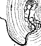
Figure 4. Herbst's corpuscle (after Dogel).
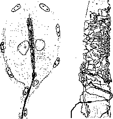
A special type of complex encapsulated nerve ending are Grandry's corpuscles (fig. 5), found in the skin of the beak and in the mucous membrane of the oral cavity in birds (ducks, geese); they are complexly constructed tactile cells surrounded by a capsule. Their size ranges from 10 to 50 microns. Usually they consist of two hemispherical, somewhat elongated cells joined by their flat sides. A medullated fiber approaches the corpuscle, loses its myelin, enters inside, and forms a reticular plate-like plexus between the cells - the tactile meniscus. The fibrils are in close union (Dogel, Boucke, Hering) with the intracytoplasmic network of the tactile cells. This forms a structure (corpuscula tactus), analogous to the periterminal network of motor plates (Boucke). If there are not two but more tactile cells, then tactile menisci are located between every two cells. Sometimes the cells are arranged in a stack, and sometimes single tactile cells are encountered, which then resemble Merkel's cells. The question of the origin of tactile cells from connective tissue or epithelium is not resolved. Hairs are richly supplied with nerve endings. In animals, there are especially many nerve endings in so-called sinus hairs. The nerve fibers approaching the hair branch off, forming a dense plexus surrounding the hair at the location of the sebaceous glands [see separate table (arts. 607-608), figure 6]. Other fibers extend
Figure 5. Grandry's corpuscle Figure 6. Neuromuscular spindle (after Bouke).
and form a wreath of thinning threads at the hair follicle, sometimes dividing dichotomously. These nerve plexuses are located on the outer epithelial sheath, into which thin branches penetrate shallowly. There are apparently no nerve endings in the papilla. Usually several medullated nerve fibers approach the hair, and by dividing into branches, they surround the connective tissue sheath of the hair somewhat below the level of the sebaceous glands. Thin branches penetrate to the outer part of the outer root sheath, where they form several circular spiral turns. Other branches, also lying on the periphery of the outer root sheath, are arranged parallel to the long axis of the hair and cross the previous ones at right angles. The resulting meshwork is called the 'hedgehog apparatus of the hair'. In a number of mammals, special nerve endings have been found in the so-called 'sinuous hairs'. The shaft of the sinuous hair, together with the root sheaths, is surrounded on all sides by cavities containing lymph and blood. Along the walls of these cavities and their connective tissue septa passes a huge number of nerve fibers. Thus, the slightest movement of the hair changes the pressure in the cavities and acts on the nerve receptors located in the connective tissue. In addition, sinuous hairs are supplied with a typical 'hedgehog apparatus'. Sensory nerve endings in striated muscles are called neuromuscular spindles (Figure 6). Among the muscle fibers, very thin cross-striated fibers rich in sarcoplasm stand out for their structure. One or more such fibers are covered over a considerable length by a special connective capsule in the form of a sheath. Between the capsule and the muscle fibers there is a considerable amount of lymph. Thick medullated nerve fibers, piercing the capsule, enter the lymphatic space and break up in it into a number of branches. The latter spiral around the muscle fibers, forming expansions, varicosities or plates and closely adhering to the sarcolemma. In addition to sensory nerve fibers, motor nerve fibers also approach this apparatus, forming typical motor plates in the muscle fibers. Experiments have established that neuromuscular spindles are organs of muscle sense. The nerve endings spirals are irritated both when the muscle fiber contracts and when it is stretched. Similar endings are also found on tendon fibers; these are so-called neuromuscular spindles. Nerve endings in individual organs. In arteries, there are in the tunica media thin plexuses of non-medullated fibers; in the t. adventitia-nerve endings of the free plexus type and nerve endings of the Vater-Pacinian corpuscle type. In veins, in the t. media-plexuses of non-medullated fibers and tree-like nerve endings. In large veins-Vater-Pacinian corpuscles. Capillaries are often accompanied by thin non-medullated fibers; actual nerve endings on them are rarely described. Shter the younger saw on the capillaries of the heart the finest intertwining branches, which he considers the terminal apparatus. He also described button-like endings on the capillaries of the meninges. In the heart, plexuses are described in the epicardium. In the myocardium, there are plexuses of thin nerve fibers, and between muscle fibers, meshworks are apparently sometimes formed. As for nerve endings in the heart muscle fibers themselves, it is definitely known that there are no endings analogous to the end plates of skeletal muscle here. Some authors (Cajal, Retzius, Smirnov, Mikhailov) describe button-like nerve endings on the fibers; Ranvier, Bouquet describe intracytoplasmic nerve endings. Some authors could not find such endings. In the endocardium, a very large variety of nerve endings are described, both free, in the form of bushes, tufts, etc., and encapsulated (Dogel, Mikhailov, Smirnov). In the pericardium, there are plexuses and encapsulated nerve endings of the Vater-Pacinian corpuscle type. In lymph glands and spleen, delicate nerve plexuses are found along the vessels and in the trabeculae. In the kidneys, nerve branches form plexuses in the walls of the renal pelvis; many nerve branches accompany the vessels; closely connected with them are nerve fibers spreading between and around the tubule cells, and perhaps also inside them. The mucous membrane of the respiratory tract is richly supplied with all types of sensory endings; here there are free nerve endings in the epithelium and subepithelial lying plexuses and tufts, both free and encapsulated. In the bronchi, a perichondral plexus is distinguished between the cartilage and the lung parenchyma, and a subchondral plexus in the submucous connective tissue between the cartilage and the muscular coat. From the latter, thin branches go, ending in a plexus near the glandular tissue and in the smooth muscle. In the parietal (Dogel) pleura, there are encapsulated bodies of the Vater-Pacinian-Golgi-Mazzoni type and free endings in the form of bushes. In the pulmonary pleura-free endings in the form of plexuses and branches with thickenings. In the submucous connective tissue of the tongue, there is a dense plexus formed by medullated and non-medullated fibers. From it, branches go into the papillae. Thin branches end in free endings in the epithelium and in the taste bulbs. In the dental pulp, there is a dense plexus with thicker branches in the center and thinner ones on the periphery. Nerve endings have not been found in the dentin. In the gums, all kinds of free and encapsulated nerve endings of the Vater-Pacinian, Merkel, Krause, etc. types are found. In all glands-salivary, gastric, intestinal, etc.-nerve endings have the appearance of plexuses around the lobules with button-like thickenings on the ends of the branches or clearly arranged on them (Figure ?). In the liver, only thin nerve fibers in the connective tissue are reliably known. The peritoneum has many free endings as well as encapsulated endings of the Vater-Pacinian corpuscle type and others. The meninges are abundantly supplied with nerve endings. Here there are plexuses and bush-like nerve endings. In the pia mater and in the tela choroidea, encapsulated nerve endings are described. After nerve section (Bouquet), as well as in case of intoxication (J. M. Villa-Verde), nerve endings undergo degeneration. This has been particularly studied on motor nerve endings in striated muscles. At first, the staining properties change; then the nerve endings break into lumps and clumps and then disappear completely. During regeneration of the corresponding nerve fiber, the nerve endings also regenerate (Tello, Bouquet). The new fiber penetrates through the sarcolemma and first forms thin delicate loops, then meshworks, and a new end plate is formed. Sensory nerve endings regenerate in the same order. The latest experimental works have shown that these nerve endings can undergo bizarre hypertrophy under the influence of external
Figure 7. Nerve plexus around glandular cells of the pancreas (according to Penza).
irritations, e.g., rays of radium, X-rays, ultraviolet rays (Lutershtein, Rahmanov, Shkarenko, Meissel).
Related articles
Mentioned in
Cite this page
“NERVE ENDINGS.” Soviet Medical Encyclopedia. English translation of Bolshaya Meditsinskaya Entsiklopediya, 1st ed. (Moscow, 1928–1936), ed. N. A. Semashko. https://sovietmedicalencyclopedia.pages.dev/article/nerve-endings/