Visual Pathways
Historical document, translated for reference. It reflects medical knowledge of the 1920s–30s and is not medical advice.
Summary
This article describes the anatomical structure and physiological function of the visual pathways, tracing the transmission of visual stimuli from the retina to the visual cortex. It details the peripheral and central neurons, the optic chiasm, and the primary visual centers, reflecting the neurological understanding of the early 20th century.
Encyclopedia article (1928–1936)
VISUAL PATHWAYS, CENTERS. The visual conduction pathways are nerve fibers connecting the visual sensory apparatus (the retina of the eye) with the visual center located in the cortex of the occipital lobe. The visual pathway is divided into two segments, or neurons—peripheral and central—which connect in the primary visual centers, where one neuron ends and another begins. The peripheral neuron includes the optic nerve (nervus opticus), the optic chiasm (chiasma nn. opticorum), and the optic tract (tractus opticus); the central neuron includes the primary visual centers (lateral geniculate body, pulvinar, and anterior quadrigeminal body), the optic radiation (radiatio optica), or Gratiolet's bundle, and the cerebral cortex on the inner surface of the occipital lobe, near the calcarine fissure. The optic nerve originates from the ganglion cells of the retina, which are located in its deep layers and, through bipolar cells, come into contact with the visual cells whose protoplasmic processes—rods and cones—receive visual stimulation. The axis-cylinder processes of the ganglion cells, gathering together, form the optic nerve, which penetrates the cranial cavity through the optic foramen and runs along the base of the brain to the midline, where it forms an incomplete decussation, or optic chiasm. All fibers entering into the composition of the optic nerve are divided into four groups: 1) the direct macular bundle; 2) the crossed macular bundle; 3) the direct peripheral bundle; and 4) the crossed peripheral bundle. The macular bundle begins in the macula lutea (blind spot), slightly outward and downward from the center, and consists of crossed and uncrossed fibers, as it partially decussates in the chiasm. Fibers originating from the outer parts of the retina form the direct, or uncrossed, peripheral bundle, while fibers originating from the inner half of the retina, together with a portion of the fibers of the macular bundle, pass to the opposite side, forming a decussation, and then join with the uncrossed fibers of the opposite side to form the optic tract (tractus opticus). The fibers of the optic nerve differ not only in direction but also in caliber; there are thin and thick fibers; the thick ones serve to transmit light stimulation to the visual centers, whereas the thin ones are reflex fibers and serve to transmit light stimulation to the nuclei of the third cranial nerve. In addition to centripetal fibers, centrifugal fibers also pass through the optic nerve, directed...
nm pm gag zv
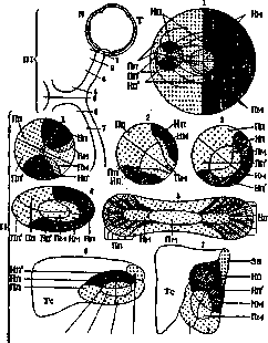
Pp Pp' Figure 1. Arrangement of visual fibers originating from the macula, and the inner and outer parts of the retina: I—in the optic nerve papilla, II—in the optic nerve, III—levels of sections: 1, 2, 3, and 4—sections through various parts of the optic nerve; 5—section through the retina; 6 and 6'—sections through the optic tract; Ze—pupillary fibers; N—nasal half of the retina; Nm—uncrossed macular fibers; Np and Np1—uncrossed peripheral fibers (upper and lower quadrant); Pm—crossed macular fibers; Pp and Pp1—crossed peripheral fibers (upper and lower quadrant); T—temporal half; Tc—tuber cinereum. (According to Henschen.) ...toward the retina; it is assumed that they originate in the superior colliculus and terminate in the granular layer of the retina; the significance of these fibers is not sufficiently studied (regulating, inhibitory action?). In the opinion of some authors, the above-described bundles occupy a distinct position both in the optic nerve and in the optic tract (Figure 1). After its formation, the optic tract is directed backward and outward, curves around the cerebral peduncle, and at its outer parts divides into three roots, which terminate in the lateral geniculate body, in the pulvinar, and in the anterior quadrigeminal bodies (Figure 2). In
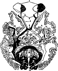
Figure 2. Visual conduction pathways: 1—visual fields; 2—retina; 3—optic nerve; 4—optic chiasm; 5—optic tract; 6—lateral geniculate body; 7—pulvinar; 8—anterior quadrigeminal body; 9—Wernicke's field; 10—optic radiation (Gratiolet's bundle); 11—cuneus (striate area); 12—calcarine fissure; 13—Sylvian aqueduct; 14—base of the cerebral peduncle; 15—red nucleus; 16—medial geniculate body; 17—inferior longitudinal fasciculus; 18—caudate nucleus; 19—corpus callosum (splenium).
humans and higher mammals, the main site of termination of the optic tract is the lateral geniculate body, and then the pulvinar (Figure 3); in the anterior quadrigeminal body, it is not visual fibers that terminate, but reflex ones. Apparently, all fibers of the optic tract terminate in the primary visual centers and do not pass into the optic radiation; thus, there is no direct connection between the retina and the cerebral cortex. Regarding the method of termination of the fibers of the optic tract, opinions differ; according to the data of some authors (Henschen, Wilbrand), visual fibers coming from specific places of the retina terminate in specific corresponding places of the lateral geniculate body; in other words, there is a projection of the retina onto the lateral geniculate body; from those cells near which the primary visual neuron terminates, the central neuron begins. Other authors (Monakow, Bernheimer) believe that upon entering the lateral geniculate body, all fibers are already mixed; they terminate near small intercalary cells and only through them communicate with the main cells that give rise to the central neuron; the fibers of the macular bundle also do not terminate in a specific place, but enter into connection with all cellular elements of the lateral geniculate body. The pulvinar has a structure analogous to the lateral geniculate body and the same relationship to the visual fibers (see Thalamus opticus). In the quadrigeminal body, the fibers of the optic tract terminate in the third layer of the anterior colliculus, but give numerous branches upward and downward; the ascending fibers come into contact with the cells of the superficial layer, while the descending ones descend into the middle and deep layers. The primary visual centers are connected to the cortex of the occipital lobe by centripetal and centrifugal fibers; these fibers are located in the white matter surrounding the posterior horn and in the so-called retrolenticular part of the internal capsule. The centripetal fibers coming from the geniculate body and the pulvinar first form a field
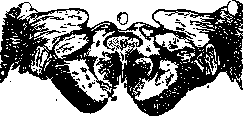
Figure 3. Section through the cerebral peduncle and the region of the geniculate bodies (site of termination of the optic tract): 1—optic tract; 2—pulvinar; 3—lateral geniculate body; 4—medial geniculate body; 5—anterior quadrigeminal body; 6—Wernicke's field; 7—base of the cerebral peduncle; 8—substantia nigra; 9—red nucleus; 10—Sylvian aqueduct; 11—oculomotor nerve nucleus; 12—oculomotor nerve. (According to Marburg.)
Wernicke's, which passes into the Gracilis bundle, and occupy the outer layer in the white matter of the occipital lobe, stratum sagitt. externum; while the centrifugal fibers, extending from the cortex of the occipital lobe to the quadrigeminal bodies and to the pulvinar, occupy the inner layer, strat. sagitt. internum. The fibers of the Gracilis bundle end in the occipital lobe, mainly on its inner surface, in both lips of the calcarine fissure (cuneus, gyr. lingualis); this area of the cortex has a special structure and is distinguished under the name of area striata (area 17 according to Brodmann); it is characterized by the following features: the presence of a myelin stripe visible to the naked eye, Vic d'Azyr's, in the lamina granularis interna (see Cerebral Cortex Architecture, Brain), the presence of large stellate cells, and in the lamina ganglionaris-single Meynert cells, the narrowness of the pyramidal layer and the absence of large pyramidal cells. In the cortex of the area striata, the visual fibers are related to the stripe of Vic d'Azyr and to the cells of the deep layers. Regarding the boundaries of the visual center in the occipital lobe, opinions differ. Some authors (Henschen) localize it exclusively on the inner surface, near the calcarine fissure; they assume that there is a projection of the retina throughout the entire visual pathway as well as in the cortex of the calcarine fissure, i.e., the upper homonymous quadrants of the retinas are projected onto the upper lip, and the lower ones onto the lower lip; the macular bundle ends at the bottom of this same sulcus. Other authors (Monakow, Brodmann) do not quite agree with these conclusions; not denying that the main visual center is near the calcarine fissure, they allow its extension to the adjacent gyri of the inner surface and even to the outer surface; they also deny the retinal projection and believe that even in the primary visual centers all fibers are mixed and in this form reach the cortex; they also do not agree with the strictly localized termination of the macular bundle, for which they allow the same wide termination as for the fibers of peripheral vision. Localizing color perception also in the calcarine fissure, some researchers recognize the possibility of special elements for it in the superficial layers of the cortex; other authors do not recognize special centers for different visual sensations, such as color perception, form, movement; the phenomena of dissociation in visual sensations in some visual disorders are explained by the fact that these different sensations are not all sufficiently finely differentiated, and therefore appear or disappear not simultaneously. Pathology. Diseases of different parts of the visual pathway are clinically expressed by various symptoms: in diseases of the retina and optic nerve, blindness will occur in the corresponding eye; in foci in the inner, crossed part of the chiasma, hemianopsia bitemporalis is observed, and in the uncrossed outer bundle-hemianopsia nasalis; disease of the optic tract, subcortical visual centers, Gracilis bundle and cortical center gives hemianopsia bilateralis homonyma with a hemianopic pupillary reaction when the visual pathway and primary centers are involved, and with a normal reaction in disease of one radiatio optica. With complete destruction of both visual centers, double hemianopia, or cortical blindness, results, sometimes with preservation of the central field of vision. Irritation of the visual pathways or centers can give visual or light hallucinations, which may be combined with phenomena of hemianopsia. Lesion of the outer surface of the left occipital lobe is accompanied by phenomena of psychic blindness.
E. Koponov»
Related articles
Mentioned in
Cite this page
“Visual Pathways.” Soviet Medical Encyclopedia. English translation of Bolshaya Meditsinskaya Entsiklopediya, 1st ed. (Moscow, 1928–1936), ed. N. A. Semashko. https://sovietmedicalencyclopedia.pages.dev/article/visual-pathways/