Visual Organs
Historical document, translated for reference. It reflects medical knowledge of the 1920s–30s and is not medical advice.
Summary
This article provides a historical overview of the comparative anatomy and embryological development of visual organs across the animal kingdom. It details the evolution from simple light-sensitive cells to complex, image-forming eyes, with a specific focus on the development of the vertebrate eye from the neural tube.
Encyclopedia article (1928–1936)
VISUAL ORGANS, organs of perception of light stimuli. In some animals, visual organs are not localized, and in them, the perception of light stimuli is possible even apart from special organs of vision. Often, the integuments themselves possess a general irritability in relation to light (lampreys). Properly speaking, organs of vision can be very simple, consisting in flatworms (Figure 1) of a sensory cell that continues into a nerve process and has a border of rod-like endings. The latter is covered in the form of a cap by a special pigment cell. Similar eyespots are also found


Figure 1. Eyespot of Tristomum molae. Figure 2. Pit-eye of Patella: 1-retina.

Figure 3. Eye of Helix: 1-pellucida ext.; 2-lens; 3-optic nerve.
in the spinal cord of the lancelet (Hesse). Light reaches these eyespots freely due to the transparency of the lancelet's tissues. More perfect organs are the eye pits (Figure 2) and vesicles of many higher worms and mollusks (Figure 3); in them, the sensory cells are equipped with rod-like outgrowths and form a continuous layer—the retina. The retina usually covers the posterior wall of the vesicle and is turned with its rods toward the light. In the anterior wall and inside the vesicle, light-refracting bodies develop—the lens and the vitreous body. In other cases, however, the lens develops in the integument, the retina is located in the anterior wall of the vesicle, and the latter bends inward, so that the vesicle acquires the shape of a goblet. In these cases, the rods are turned away from the light (inverted eyes of some mollusks and arachnids). The most perfect, image-forming vesicular eyes of the direct type are found in cephalopod mollusks (Figure 4), and of the inverted type—in vertebrates. 1 Figure 4. Cross-section through the eye of Sepia: 1-cornea; 2-iris; 3-ciliary body; 4-retina; 5-pigment zone; 6-gangl. opt.; 7-n. opticus; 8-head cartilage; 9-lymph gland; 10-eyeball; 11-vitreous body; 12 and 14-inner and outer segments of the lens; 13-sclera. In primitive vertebrates, however, unpaired eyes of the direct type (parietal and pineal) are still encountered, a rudiment of one of which is the suprabrain gland (epiphysis). The paired eyes of vertebrates (for details of structure, see Eye) differ from other sense organs by their development from the central nervous system. They develop from three sources: the retina with the pigment epithelium, the optic nerve, and the vitreous body originate from the primary brain
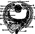
vesicle, the lens—from the ectoderm, the sclera, the basement membrane of the cornea, the choroid, and the iris develop from the mesenchyme. The eyes in the embryo are laid down in the anterior part of the neural plate in the form of paired pits, which then, with the formation of the neural tube, end up inside the latter, in the region of the anterior cerebral vesicle. At very early stages of development (in the chick on the 2nd day of incubation, in the human embryo in the 3rd week), the anterior cerebral vesicle gives two lateral outgrowths, which then constrict at the base and form primary optic vesicles, connecting to the brain by optic stalks (Figure 5). The lateral walls of the vesicles, facing the head ectoderm, are pressed inward, and the optic vesicles turn into double-layered optic cups. Simultaneously, the wall of the stalk also presses inward, forming a groove, and corresponding to the position of the groove in the wall of the cup, there is a notch—the choroid fissure (fissura chorioidea), which quite soon grows over (Figure 6). The inner, indented wall of the optic cup rapidly
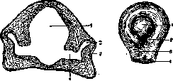
Figure 5. Primary optic vesicles and lens primordia of a 5 mm human embryo: 1-anterior cerebral vesicle; 2-primary optic vesicle; 3-thickening of the ectoderm (lens primordium); 4-optic nerve canal. (After Corning.)
Figure 6. Secondary optic cup and lens according to Manz's model: 1-lens; 2-fissura chorioidea; 3-groove on the lower edge of the optic nerve; 4-optic nerve canal. (After Corning.) The inner indented wall of the optic cup rapidly thickens and represents the primordium of the retina, the outer remains thin and forms the pigment epithelium, the stalk—the primordium of the optic nerve; the groove and fissure serve for the passage of the a. centralis retinae into the eye. Simultaneously with the formation of the cup, the head ectoderm (in the place where it lies adjacent to the optic vesicle) begins to thicken and deepen inward in the form of a pit—the primordium of the lens. The edges of the depression close, separate from the ectoderm, and the lens in the form of an epithelial vesicle ends up lying in the optic cup. Numerous experiments have proven that the formation of the lens is caused by a special stimulus emanating from the wall of the optic vesicle; the assumption that the optic cup is formed due to the pressure of the growing lens is now abandoned, since the formation of the cup proceeds even in the absence of the lens. The mesenchyme surrounding the optic cup condenses on its surface, marking the outer membranes of the eye, pushes the lens away from the ectoderm, and penetrates inside the cup, surrounding the lens; on the other side, the mesenchyme penetrates inside the cup
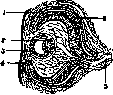
together with the a. centralis retinae, which is also directed toward the posterior wall of the lens (Figure 7). Thus, the laying down of the main constituent parts of the eye occurs, and their further development is connected with histological differentiation.
Figure 7. Eye of an 11.3 mm human embryo: 1-pigment epithelium of the retina; 2-posterior wall of the lens vesicle; 3-anterior wall of the lens vesicle; 4-edge of the optic cup; 5-arteria centralis retinae; 6-retina. (After Corning.) The development of the retina proceeds according to the brain type: embryonic cells differentiate into spongioblasts, giving rise to Müller's fibers and neuroglia, and neuroblasts. The latter form first ganglion cells, lying parallel to the inner edge in several rows, then bipolar cells and association cells; last of all, rod and cone cells develop; their processes—rods and cones—are formed only after birth. The pigment epithelium is formed from the outer leaf of the cup, which remains single-layered. Differentiation of the retina begins in the region of the central fovea and goes to the periphery, but does not reach the edges of the cup; in the area adjacent to the ciliary body and the iris, both leaves of the cup form a double-layered pigment epithelium, which is part of these formations. The stalk of the optic cup turns into the optic nerve when the neurites of the ganglion cells grow into it, heading to the brain. - The vitreous body also originates from the retinal membrane: its fibers grow out from the supporting elements of the retina, like glial ones. - Differentiation of the lens occurs in the second month of intrauterine life and boils down to the fact that the cuboidal epithelium on the posterior segment of the lens vesicle grows into long fibers; they fill the cavity of the vesicle and turn it into a narrow slit lying under the anterior, unchanged epithelium. The growth of the lens continues along its equator, where cell multiplication and their transition into fibers occur. The entire period of development, the lens is surrounded by a connective tissue capsule with blood vessels (tunica vasculosa lentis), the anterior section of which connects to the edge of the iris (membrana chorio-pupillaris); the capsule disappears before birth (Figure 8). - The choroid differentiates from the layer of mesenchyme closely adjacent to the cup; vessels appear in it very early, and pigment cells—in the 7th month. The anterior section gives rise to the ciliary body (in the 4th month) and the iris; the fibers of the zonulae Zinnii are formed by the epithelium of the cup, which is part of the ciliary body, and are analogous to the fibers of the vitreous body. - The sclera differs from the choroid by the development of dense bundles of connective tissue and is separated from it by the perichoroidal space—a lymph slit. In front, the sclera, leaving the edges of the cup, passes between the ectodermal epithelium and the lens, first lying tightly against it, and then separating by a slit (anterior chamber), lined with endothelium. In the 4th month, this anterior section of the sclera becomes transparent and forms the cornea. Since the optic cup is a part of the brain walls, it, like the latter, is surrounded by meninges: the "soft"—the choroid, and the "hard"—the sclera. The anterior part of the latter, in connection with the adjacent part of the integuments, gives a transparent cornea. As accessory organs of the eye, eye muscles develop—four straight and two oblique, to which in terrestrial vertebrates is added a muscle that retracts the eyeball into the orbit (develops from the external rectus). The sensory part of the eye—the retina—consists in vertebrates of a layer of long sensory cells (with rod-like endings turned away from the light), connected to a layer of bipolar nerve cells and finally a layer
"
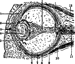
Figure 8. Eye of a 130 mm human embryo: 1-conjunctival sac; 2-tunica vasculosa lentis; 3-fusion of the eyelid edges; 4-lens; 5-cornea; 6-vitreous body; 7-retina; 8-pigment epithelium of the retina; 9-sclera; 10-m. rectus inf.; 11-optic nerve and a. centralis retinae; 12-m. rectus sup.; 13-optic nerve papilla; 14-canalis hyaloideus. (After Corning.)
of multipolar ganglion cells, covered with a layer of nerve fibers extending from them. The latter then enter the composition of the optic tract, developing in place of the eye stalk. Corresponding to its origin, the retina also includes supporting glial cells. In terrestrial vertebrates, the rod-shaped endings of the retina are clearly differentiated into two kinds of formations-thinner rods and swollen cones. The latter predominate initially, but in mammals the number of rods increases, especially in the peripheral parts of the retina. The number of sensory elements in the retina of higher vertebrates increases significantly. The main light-refracting body-the lens-in aquatic vertebrates has an almost spherical shape. Accommodation is achieved in fish by moving the lens inward by the action of a special muscle (campanula Halleri, fig. 9). In amphibians, another muscle (m. protractor lentis) pulls the lens forward. In reptiles and birds, a circular ciliary muscle develops in the vascular tunic, which by pressure on the lens changes its convexity (fig. 10). Finally, in mammals, the accommodative action of this muscle is achieved only indirectly-through a decrease in the tension of the lens capsule. The anterior edge of the eye cup is devoid of retina and together with the continuation of the vascular tunic forms the iris, provided with radial and circular muscle fibers of ectodermal origin. In reptiles and birds, these fibers, as well as the fibers of the ciliary muscle, are striated. The inner surface of the vascular tunic sometimes forms a shiny layer-the tapetum-consisting of flat cells with small crystals (tapetum cellulose of fish and many mammals, especially carnivores) or of thin elastic fibers (tapetum fibrosum of ungulates and cetaceans). This layer reflects light rays back onto the retina and causes the eyes to glow in the dark. In these cases, the pigment is reduced. The sclera of the vertebrate eye most often consists of cartilage and often contains bony plates in the anterior part (sclerotic ring of reptiles and birds, fig. 10). In mammals, the sclera is fibrous. Skin folds-eyelids-sometimes form a continuous ring (fish, chameleons), but are more often divided into upper and lower. In snakes (also
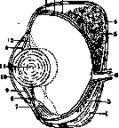
in geckos and amphisbaenas) the eyelids fuse above the eye and are completely transparent. ciliary body. There is often also a third eyelid lying deeper-the nictitating membrane, which draws the eye from the anterior (inner) corner, usually passively when the eye is retracted into the orbit (in reptiles and birds, however, there is a special t. nicticans). In the area of the eyelids, various glands develop that moisten the anterior wall of the eye. A group of large glands develops in the area of the lower eyelid at the anterior corner of the eye-the so-called gland of Harder, which moistens the nictitating membrane and is reduced in monkeys and humans. At the posterior corner of the eye lies the lacrimal gland, which in mammals moves under the upper eyelid. From the sebaceous glands along the edges of the eyelids in mammals, Meibomian glands also develop.-The compound eyes of insects and crustaceans consist of numerous ommatidia, jointly located on a common convex surface. Each ommatidium has the appearance of a narrow pyramid with a base formed by a crystalline lens, under which lies the so-called crystalline cone and finally a group of visual cells forming the "retinula", with a rod-shaped formation along its axis-the "rhabdom". Each ommatidium is isolated by pigment cells. In the compound eye, the image obtained is mosaic and direct.
velops in the area of the lower eyelid at the anterior corner of the eye-the so-called gland of Harder, moistening the nictitating membrane and reducing in monkeys and humans. At the posterior corner of the eye lies the lacrimal gland, which in mammals moves under the upper eyelid. From the sebaceous glands along the edges of the eyelids in mammals, Meibomian glands also develop.-The compound eyes of insects and crustaceans consist of numerous ommatidia, jointly located on a common convex surface. Each ommatidium has the appearance of a narrow pyramid with a base formed by a crystalline lens, under which lies the so-called crystalline cone and finally a group of visual cells forming the "retinula", with a rod-shaped formation along its axis-the "rhabdom". Each ommatidium is isolated by pigment cells. In the compound eye, the image obtained is mosaic and direct.

velops in the area of the lower eyelid at the anterior corner of the eye-the so-called gland of Harder, moistening the nictitating membrane and reducing in monkeys and humans. At the posterior corner of the eye lies the lacrimal gland, which in mammals moves under the upper eyelid. From the sebaceous glands along the edges of the eyelids in mammals, Meibomian glands also develop.-The compound eyes of insects and crustaceans consist of numerous ommatidia, jointly located on a common convex surface. Each ommatidium has the appearance of a narrow pyramid with a base formed by a crystalline lens, under which lies the so-called crystalline cone and finally a group of visual cells forming the "retinula", with a rod-shaped formation along its axis-the "rhabdom". Each ommatidium is isolated by pigment cells. In the compound eye, the image obtained is mosaic and direct.
velops in the area of the lower eyelid at the anterior corner of the eye-the so-called gland of Harder, moistening the nictitating membrane and reducing in monkeys and humans. At the posterior corner of the eye lies the lacrimal gland, which in mammals moves under the upper eyelid. From the sebaceous glands along the edges of the eyelids in mammals, Meibomian glands also develop.-The compound eyes of insects and crustaceans consist of numerous ommatidia, jointly located on a common convex surface. Each ommatidium has the appearance of a narrow pyramid with a base formed by a crystalline lens, under which lies the so-called crystalline cone and finally a group of visual cells forming the "retinula", with a rod-shaped formation along its axis-the "rhabdom". Each ommatidium is isolated by pigment cells. In the compound eye, the image obtained is mosaic and direct.
Related articles
Mentioned in
Cite this page
“Visual Organs.” Soviet Medical Encyclopedia. English translation of Bolshaya Meditsinskaya Entsiklopediya, 1st ed. (Moscow, 1928–1936), ed. N. A. Semashko. https://sovietmedicalencyclopedia.pages.dev/article/visual-organs/