Vitreous Body
Historical document, translated for reference. It reflects medical knowledge of the 1920s–30s and is not medical advice.
Summary
This article from the 1928–1936 Soviet Great Medical Encyclopedia provides a detailed anatomical description of the vitreous body, including its structure, attachments, and optical properties. It discusses the vitreous's relationship with the lens, retina, and ciliary body, and notes the absence of blood vessels and nerves within it.
Encyclopedia article (1928–1936)
VITREOUS BODY (corpus vitreum) fills the posterior part of the eyeball. It is located between the lens and the retina. The V. B. has the shape of a sphere flattened in the sagittal direction. Its lateral and posterior parts adhere to the retina, and the anterior edge is adjacent to the posterior half of the ciliary body. On the anterior surface of the V. B. there is a depression for placing the lens (fossa patellaris). The V. B. is relatively well attached in the region of the papilla, but its connection with the retina is less firm. A particularly strong connection exists at the ora serrata and in the region of the ciliary epithelium. The V. B. borders on the posterior chamber, having a weak connection there. This connection is further strengthened at the edge of the lens, where the hyaloideo-capsular ligament is located. The anterior part of the V. B. freely adheres to the posterior surface of the lens, as a result of which there is a slit—the posterior subcapsular space. Along the axis of the V. B. passes the central canal (canalis hyaloideus, s. canalis Cloqueti), which begins in front of the optic papilla and ends blindly behind the lens. This canal usually exists in the newborn's eye, but in an adult it turns into a wide lymph space (see figure). Keppe's question about the existence of a canal in the V. B. is decided in the negative sense. The V. B. possesses complete transparency, is colorless, and has a gelatinous consistency. The refractive index of the vitreous body is 1.334. The V. B. consists of a denser basis, a skeleton, the cells of which are filled with fluid. This basis becomes especially dense on the surface of the V. B., forming its cortex, the limiting layer. A posterior limiting layer adjacent to the internal limiting membrane of the retina and an anterior one are distinguished. The latter is located on the anterior surface of the V. B. and separates it from the posterior chamber and the lens. This layer creates the impression of a membrane (hyaloidea). The limiting layer is absent at the entrance to the central canal.
The basis of the V. B. is formed by delicate, thin, and soft fibrils. Part of the fibrils from the surface fan inward into the V. B., where they form a loose, often disintegrating nucleus of it (Salzmann). The V. B. contains neither blood vessels nor nerves nor its own fixed cells. Only wandering cells are encountered on the outer surface of the posterior limiting layer. Biomicroscopy with the slit lamp first made the V. B. visible to us. When examined by this method, it appears to consist of membranes hanging in the fluid, forming folds and having a fibrillar structure. In it one can note bright horizontal bands—illuminated folds of the membranes—and dim, weakly illuminated places. Of course, isolated
isolation is not possible.
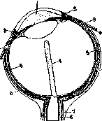
The fibers of the vitreous body at low magnification are not distinguishable, and their conglomerates are usually observed. With age, the spaces between the tissue fibers lose their regularity, strands disintegrate, and small white irregularities appear on them. The strands seem to be sprinkled with fine sugar, creating the so-called senile dusting of fibers. Vitreous body formation is of ectodermal origin. Initially, the cells of the lens primordium participate in the construction of the primary vitreous body, giving off processes. These processes break down into thin fibers, which connect with similar fibers emerging from the inner layer of the eye vesicle, thus creating a thin felt. With the formation of a capsule around the lens vesicle, the separation of the vitreous body from the lens occurs, while its connection with the retina primordium continues to exist. This connection is disrupted only after the appearance of a capsule on the retina, the future internal limiting membrane. The vitreous body is the refractive medium of the eye and maintains its spherical shape. The fluid of the vitreous body is identical in composition to the aqueous humor. It is poor in protein and contains crystalloids in the same percentage ratio as the fluid of the anterior chamber (see Eye). Pathological changes in the vitreous body, if they have an intense color and are located in the very anterior part of it, can still be observed using the method of oblique illumination. However, the latter recedes into the background when examining deeply located and weak changes, when it is necessary to use the ophthalmoscopic illumination method. Many details of the pathologically altered vitreous body can be elucidated by the method of examination with a slit lamp. Horizontal section of the right eye: 1 - anterior chamber; 2 - Schlemm's canal; 3 - its connection with the anterior ciliary veins; 4 - hyaloid canal; 5 - perichoroidal space; 6 - Tenon's space; 7 - suprachoroidal space; 8 - interchorial space; 9 - extensions of Tenon's capsule onto the tendons of the eye muscles. Congenital anomalies of the vitreous body occur in 2 forms: 1) persistent hyaloid artery and 2) congenital coloboma of the vitreous body. In the first case, we are talking about incomplete regression of the vitreous body arteries (a. hyaloidea). In this anomaly, a strand runs along the axis of the vitreous body from the posterior pole of the lens to the optic nerve papilla. More often, the undissolved embryonic artery has the appearance of a short flagellum, one end of which is attached either to the optic nerve papilla or to the posterior pole of the lens, and the other moves freely in the vitreous body. In the second case, we have a severe form of congenital anomaly of the vitreous body, the expression of which is an indentation in the lower-inner part. Nutrition of the vitreous body occurs by diffusion from the ciliary body. The flow of fluid in the vitreous body goes from front to back, in the direction of the optic nerve papilla. Being in such dependence on the ciliary body, the vitreous body is involved in pathological processes occurring in the former. The anatomical connection of the vitreous body with the retina entails a certain reaction on the part of the vitreous body in diseases of the retina and the choroid lying beneath it. The pathological state of the vitreous body is accompanied by a violation of its transparency, the appearance of opacities. These opacities acquire the appearance of flakes, threads, strands, detected by the ophthalmoscope against a bright red background - floccular opacities. In another type of opacities, we have a general dusty haziness of the red background (diffuse offuscatio). Such opacities are best examined using a flat mirror, behind which is placed a biconvex lens of 8.0D or 10.0D. Floccular opacities of the vitreous body are most often observed in severe myopia, diffuse ones in serous choroiditis. Liquefaction of the vitreous body (synchysis simplex) is usually observed together with its opacities, the latter acquiring mobility. When illuminated by the ophthalmoscope and with rapid movements of the eye, these opacities rise upward, settle downward, and appear in the center of the visual field. Sometimes liquefaction of the vitreous body takes a special form, in which illumination of the mirror of the examined eye reveals in the vitreous body a mass of glittering golden or silvery particles or small plates. These are cholesterol crystals, tyrosine, phosphates, fatty acid salts of calcium, sodium, etc. This form is called sparkling liquefaction of the vitreous body (synchysis scintillans, s. scintillatio corporis vitrei). Regarding cholesterol crystals, it is assumed that they precipitate from old exudates and hemorrhages. Cholesterolemia is considered a predisposing factor for this. Synchysis scintillans was obtained experimentally by introducing a soda solution into the vitreous body of a rabbit. Hemorrhage in the vitreous body (haemorrhagia in corpore vitreo) occurs as a result of trauma. It can arise on the basis of general diseases of the organism (tbc, malaria, hemophilia, metabolic disorders). Significant hemorrhages give the pupil a black appearance, and the red reflex from the fundus of the eye is absent during ophthalmoscopy. With small hemorrhages, red, brown-red, and coffee-colored masses are visible in the vitreous body. Special attention deserves the so-called recurrent juvenile hemorrhages in the vitreous body. This disease was first described by Graefe (v. Graefe). It is usually observed in youth. Among the causes, neuroses of the circulatory and digestive organs, reflex excitation of the vascular system under the influence of the sexual sphere, early local arteriosclerosis of the kidneys, and disorders of the internal secretion organs are indicated. In juvenile hemorrhages in the vitreous body, Axenfeld described tubercles in the adventitia of the retinal veins. He considers tuberculosis to be the main cause of this ailment. Müller explains the occurrence of recurrent juvenile hemorrhages in the vitreous body by the weakness of the capillaries. Being a vesselless organ, the vitreous body cannot serve as a focus for the occurrence of independent inflammatory processes, and therefore it is hardly possible to speak of its isolated disease (hyalitis). The inflammatory state of the vitreous body, in essence, is a consequence of the disease of the surrounding tissues. Penetrating injuries of the walls of the eyeball entail damage to the vitreous body and the introduction of infection into it. In this case, the development of microorganisms and the action of their toxins proceeds slowly and is initially accompanied by opacification of the vitreous body. The color of the opacification gradually changes from gray to yellow and yellow-green. Inflammation developing under such conditions leads to the formation of an abscess of the vitreous body. In some cases, an abscess develops around the introduced foreign body, occupying a small part of the vitreous body. In other cases, the entire cavity of the vitreous body becomes purulent. Abscesses of the vitreous body are surrounded by a layer of scar tissue or pus breaks out outward. Often, a purulent process in the vitreous body, accompanied by simultaneous and extensive suppuration of the retina and choroid, ends with panophthalmitis with a fatal outcome of atrophy of the eyeball. Deep dissection of secondary cataract may entail a violation of the integrity of the stroma of the vitreous body, which is accompanied by its opacification in the form of a grayish stripe running along the course of the injury. Sometimes such dissection entails prolapse of the vitreous body and its entrapment in the rupture of the capsule, a so-called vitreous body hernia develops. Penetrating injuries of the eyeball walls, operations on it can be accompanied by loss of the vitreous body. Loss of one third of it is tolerated by the eye relatively well, while large losses are dangerous for its fate: the eye becomes soft to the touch and furrows form on it corresponding to the four rectus muscles. Foreign bodies in the vitreous body - see Foreign bodies. Treatment of diseases of the vitreous body - general, causative (tbc, syphilis, rheumatism, gout); locally applied procedures: subconjunctival injections of sodium chloride, mercuric cyanide solutions; zur Nedden proposed for opacification of the vitreous body its aspiration using a syringe not more than 0.5 cm3. In purulent inflammation of the vitreous body, the following are indicated: intravenous injections of glucose, colargol, urotropin, aspiration according to zur Nedden, and even sclerotomy. Parasites of the vitreous body - cysticerci of Taenia solium - get into it from the retina or choroid. Ultimately, parasites lead to significant opacification of the vitreous body and the occurrence of inflammation of the vascular tract. Surgical removal of the parasite is indicated, the localization of which is established according to special schemes of Donders, Lenz, Levitsky, etc. (Donders, Lenz).
Related articles
Mentioned in
Cite this page
“Vitreous Body.” Soviet Medical Encyclopedia. English translation of Bolshaya Meditsinskaya Entsiklopediya, 1st ed. (Moscow, 1928–1936), ed. N. A. Semashko. https://sovietmedicalencyclopedia.pages.dev/article/vitreous-body/