Ciliary Body
Historical document, translated for reference. It reflects medical knowledge of the 1920s–30s and is not medical advice.
Summary
This article from the 1928-1936 Great Medical Encyclopedia describes the anatomy, structure, and histological features of the ciliary body of the eye.
Encyclopedia article (1928–1936)
CILIARY BODY (corpus ciliare) is the second section of the vascular tunic of the eye and is located between the iris and the choroid. It has the appearance of a belt about 5 mm wide in the upper and nasal parts and 6-7 mm in the lower and temporal parts. On a cross section, the ciliary body pre-
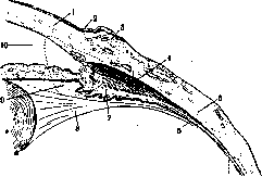
Figure 1. Anterior segment of the eye: 1-cornea, 2-limbus, 3-conjunctiva, 4-Müller's muscle, 5-sclera, 6-orbiculus ciliaris, 7-corona ciliaris, 8-Petit's canal, 9-posterior chamber, 10-anterior chamber.
sents a triangle, the apex of which lies at the place of its transition into the choroid, the outer side faces the sclera, the base looks toward the anterior and posterior chambers and the root of the iris, and the inner side is directed toward the lens and the vitreous body (Fig. 1). The anterior edge of the ciliary body, corresponding to the place of transition of the sclera into the cornea, is attached to the sclera behind Schlemm's canal; its posterior edge, corresponding to the anterior edge of the retina, passes into the choroid. The ciliary body closely adjoins the sclera, being attached to it by means of vessels and nerves, while the zonule of Zinn connects it with the lens. The ciliary body consists of two parts: the posterior, orbiculus ciliaris, s. pars plana corporis ciliaris, and the anterior, corona ciliaris. The first of these begins at the ora serrata of the retina, reaches the equator of the lens, and stretches for 3-4 mm. On it, no irregularities visible to the naked eye can be noted. The second part of the ciliary body is located from the equator of the lens to Schlemm's canal, occupies a space of 2 mm, and is covered with folds called ciliary processes (processus ciliares) [see separate plate (vol. VII, p. 275-276), Fig. 3]. The ciliary processes, up to 70 in number, are located on the inner surface of the ciliary body. Each ciliary process is a ridge whose length is 2 mm and height 0.8 mm. The tips of the processes do not come into contact with the lens. The tips of the processes, being not as strongly pigmented as the depressions between them, stand out on preparations in the form of whitish strips. The main mass of the ciliary body is formed by the ciliary, or accommodation, muscle (m. ciliaris), which is divided into several parts depending on the arrangement of the muscle bundles. In the outer layers adjacent to the sclera, the muscle bundles take a strictly meri-
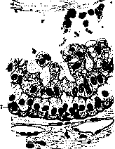
meridional direction, which is why 1 this part of the muscle has been assigned the name meridional. Its origin is formed by those smooth muscle fibers that can still be noted in the region of the choroid. The meridional part of the ciliary muscle ends at the scleral spur, passing into the plates of the scleral segment supporting the trabecular meshwork of the anterior angle. Figure 2. Orbiculus ciliaris close to corona ciliaris (transversal section): 1-anterior border layer of the vitreous body; 2-transverse sections of zonular fibers in the orbicular space; 3-membrana limitans interna ciliaris; 4-its folds, partly enclosing simple zonular fibers, and their bundles having a fan-shaped divergence toward the center of the eye. Both of these parts of the ciliary muscle
border layer of the vitreous
body; 2-transverse sections
of zonular fibers in the orbicular
space; 3-membrana limitans
interna ciliaris; 4-its folds,
partly enclosing simple zonular
fibers, and their bundles
having a fan-shaped divergence
toward the center of the eye. Both of these parts of the ciliary muscle
have a fan-shaped divergence toward the center of the eye. Both of these parts of the ciliary muscle
intercellular connective tissue; 9-elastic plate; 10-vascular layer
elastic plate; 10-vascular layer
plate (small meshes of reticuli H. Müller);
intercellular connective tissue;
ic layer;
of the ciliary body. They were discovered by Brücke and named after him. Finally, situated completely anteriorly and internally, near the root of the iris, the muscle fibers acquire a circular direction, forming the circular part of the ciliary muscle, discovered by Müller and named after him (Fig. 1, 4). The shape of the ciliary muscle depends on the degree of development of its circular part; in a hyperopic eye it is strongly developed, whereas in a myopic eye it is weakly developed (see Hyperopia, Fig. 1). Behind the muscular layer lies the vascular layer of the ciliary body (Fig. 2), serving as the direct continuation of the vascular layer of the choroid, and containing loose connective tissue, veins of various calibers, several small arteries, and a smaller number of chromatophores than in the choroid (Fig. 2, 10). The inner boundary of the vascular layer is formed by the elastic lamina (9), consisting of a network of elastic fibers. This lamina is followed by the interlamellar connective tissue (8). Still more internally, beyond the interlamellar connective tissue, lies the vitreous or cuticular lamina (7). Being a continuation of the lamina of the same name in the choroid, it has a reticulated structure; its meshes are filled with pigment epithelium. The next layer belongs to the epithelial covering of the ciliary body, the latter being divided into two sections: a) the pigment epithelium of the ciliary body (6) and b) the non-pigmented ciliary epithelium (5). The former represents the continuation of the pigment epithelium covering the posterior surface of the entire vascular tract. Embryologically, this epithelium is formed by the outer layer of the optic cup. It consists of a single row of pigmented cells, located with its outer surface on the vitreous membrane of the ciliary body and its inner surface on the next layer of non-pigmented epithelium. The latter, embryologically representing the undifferentiated part of the retina, also forms a single layer of cells. The protoplasm of the cells of this layer is devoid of pigment, and only near the root of the iris can the appearance of pigmented cells be noted. The inner surface of the ciliary body is covered by the internal limiting membrane of the ciliary body (membrana limitans interna ciliaris) (3). The ciliary processes also consist of loose connective tissue, penetrated by numerous blood vessels that form glomeruli resembling the glomeruli of the kidney. In general, the corona ciliaris is the part of the eyeball richest in blood vessels [see separate table (Vol. VII, pp. 275-276), Fig. 4]. The vascular system of the ciliary body is formed by the posterior and anterior ciliary arteries [see separate table (Vol. VII, pp. 275-276), Fig. 5]. Anterior to the circular muscle lies the circulus arteriosus iridis major, the branches of which supply blood to the ciliary processes [see separate color table (Vol. VII, pp. 303-304), Fig. 3]. In the processes, the arteries break up into numerous branchlets. Larger veins are formed from the thin venous branchlets. The latter, together with veins coming from the ciliary muscle, pass posteriorly and empty into the vortex veins. Partially, the venous blood from the ciliary muscle flows into the anterior ciliary veins, which pierce the sclera near the limbus of the cornea. The vessels of the uveal tract, in particular of the iris and the ciliary body, are connected to each other by numerous anastomoses, thanks to which circulatory disorders in this tract are rapidly equalized. The ciliary body is richly supplied with nerves. The latter originate from the trigeminal, oculomotor, and sympathetic nerves. Under the names of long ciliary nerves (from the nasociliary nerve) and short ciliary nerves (from the ciliary ganglion), they pierce the sclera near the optic nerve and run along the outer surface of the choroid to the ciliary body, in which they form dense networks, especially on the surface of the ciliary muscle [see separate table (Vol. VII, pp. 275-276), Fig. 1]. The nerve fibers of the ciliary body are characterized by the presence of ganglion cells. The short ciliary nerves carry sensory, motor, and sympathetic fibers, while the long ciliary nerves carry sensory fibers. The ciliary muscle is innervated by the oculomotor nerve. Due to the abundance of blood vessels and the numerous folds, the ciliary body plays an important role in the nutrition of the eye (the lens and probably the vitreous body). The ciliary muscle participates in the act of accommodation. The ciliary body is the main site of formation of the aqueous humor. Through the vessel walls and the ciliary epithelium, the aqueous humor is secreted into the posterior chamber, and then passes through the pupil into the anterior chamber as well. Its further pathway passes through the spaces of Fontana, the canal of Schlemm, and finally into the anterior ciliary veins. Thus, the process of formation of the aqueous humor is of a physical-chemical nature, proceeding by the type of ultrafiltration, in which only crystalloids pass freely into the aqueous humor and colloids are retained. This is the opinion held by the majority of ophthalmologists on the question of the source of the aqueous humor. Seidel regards the flow of fluid from the blood into the eye as the result of the active activity of the cells of the ciliary body, considering it an endocrine gland, albeit of a primitive character. Such a gland lacks chemical activity, as a result of which the synthesis of substances new to the blood occurs in true glands; in such a primitive gland there is only the osmotic work of suction of plasma from the capillaries, with the exception of colloids. According to Seidel's opinion, the movement of fluid in the ciliary body occurs by the type of fluid movement produced by a pump. The epithelium of the ciliary body possesses the biological property of retaining certain substances present in the blood of healthy or immunized animals (cytotoxins), or letting others pass through (agglutinins, precipitins). A pathological state of the ciliary epithelium disrupts this barrier function, and substances that were previously retained enter the aqueous humor. Eye irritation—puncture of the anterior chamber, subconjunctival injections, cauterization of the limbus—causes the formation of secondary aqueous humor, which is richer in protein, sodium, and potassium and has a lower chlorine content. The anatomical substrate of such a phenomenon is considered to be the so-called Greeff vesicles. These represent a vesiculiform detachment of the ciliary epithelium by fluid rich in protein and fibrin. Pathology of the ciliary body. Inflammation of the ciliary body (cyclitis) belongs to the most important afflictions of the eye, causing impairment and even loss of vision. The position of the ciliary body and the community of blood supply create all conditions for the easy transition of inflammation from it to the iris and choroid and vice versa. Therefore, pure forms of cyclitis are rarely observed; more often it is a matter of iridocyclitis or iridocyclochoroiditis. Objective signs of pure cyclitis: pericorneal injection, more or less pronounced depending on the strength of the process; tenderness upon pressure on the region of the ciliary body, with the patient's head involuntarily tilting backward; turbidity of the anterior part of the vitreous body; sometimes deposition of precipitates on the posterior surface of the cornea and the floor of the anterior chamber; the eye frequently becomes soft. Mandelstam considers the presence of blood and pus in the anterior chamber in the absence of changes on the part of the iris to be a particularly characteristic sign of cyclitis. Subjective signs of cyclitis: visual impairment due to turbidity of the vitreous body and severe pain, especially in acute cases. An example of pure cyclitis is the inflammation of the ciliary body in relapsing fever. Lozhechnikov observed cyclitis in 157 patients out of a material of 563 cases of ocular complications in relapsing fever. Estlander, however, considers pure cyclites in relapsing fever to be a rare exception. Svatikova believes that at first the process in ocular complications of relapsing fever is localized in the anterior segment of the vascular tract, causing slight turbidity of the vitreous body, then the ciliary body becomes involved, and still later the iris. In general, the etiology, clinical forms, and therapy of cyclites coincide with those of iritis (see). Among other pathological states of the ciliary body, mention must be made of its syphilis, which can proceed either in the form of diffuse inflammation or with the formation of granulomas. Theoretically dividing the latter into papules and gummas, practically ophthalmologists, following the suggestion of Evetsky, use the general term syphiloma of the ciliary body in such cases. The most constant signs of syphilomas of the ciliary body are iridocyclitis and a scleral swelling near the cornea, covered by strongly reddened conjunctiva. The swelling is immobile, of a dense elastic consistency, painful upon palpation, and has a rounded shape or the appearance of a ridge encompassing the cornea. The growth of the syphiloma into the sclera, the anterior chamber, and the vitreous body is possible. Intraocular pressure is usually reduced. Pathological-anatomical changes in syphilomas of the ciliary body boil down to the formation of more or less extensive foci of infiltration consisting of small round cells at the periphery and necrotic tissue in the center of the focus (Lieto Vollaro). In his preparations, Evetsky did not notice a focal structure in the syphiloma, which makes it possible to admit the existence of a syphiloma of a diffuse character alongside a syphiloma of the focal type. Treatment for it is general specific and local, as in iridocyclitis.
According to older observations (Filatov), despite treatment, blindness ensues in more than 62% of cases, whereas recent statistics bring the number of favorable outcomes up to 74% (Gastev), which is explained by the improvement in the treatment of syphilis at the present time (Pokrovsky). Tuberculosis of the ciliary body may occur simultaneously with tuberculosis of the iris and choroid. The diagnosis of this condition is established with difficulty and only when there is a large number of tubercular nodes bulging the sclera and even perforating it. - Atrophy of the ciliary body usually accompanies atrophy of the iris, but it is difficult to judge-atrophic changes during life. These changes may be assumed in long-lasting inflammatory processes of the uveal tract and the presence of atrophic phenomena in the iris. Histological examination of eyes enucleated for iridocyclitis reveals a decrease in the size of the ciliary body, atrophy of muscle fibers, vascular sclerosis, and proliferation of connective tissue. - Contusions of the eyeball in the region of the ciliary body entail hemorrhage into the ciliary muscle, which is clinically manifested by paralysis or weakening of accommodation. Penetrating wounds of the eye with their localization in the region of the ciliary body must be considered especially severe, resulting in a sluggishly proceeding iridocyclitis lasting for years and causing a complete nutritional disorder of the eye. Such a process is usually called sympathizing and is characterized, among other things, by a great tendency to the formation of adhesions. The described pathological condition of one eye is aggravated by the fact that it may entail sympathetic inflammation of the other eye (see Ophthalmia). Among the tumors of the ciliary body, sarcoma should be noted, which occurs quite rarely. Causing no disorders, it appears either as an elevation in the region of the ciliary body or as a black spot near the cornea. Increasing in size, the tumor grows into the anterior chamber, retaining the ability to grow also toward the choroid. Melanosarcomas are more common, have the ability to cause an increase in intraocular pressure early, and, located in the region of Schlemm's canal, perforate the sclera relatively early. - Inflammation of the ciliary body, as well as of the cornea and iris, and an increase in intraocular pressure are accompanied by pain, radiating most often to the forehead, less often to the ears or teeth of the upper jaw. This pain is usually called ciliary neuralgia. Especially severe pain occurs in iridocyclitis and acute glaucoma. In the latter, very often from the very beginning of the disease, severe trigeminal neuralgia develops, and only then are other manifestations of glaucoma joined to it. Frequently, attacks of ciliary pain recur daily at the same hours, especially in the evenings or at night, which gives them the character of neuralgia. As mentioned above, traumatic iridocyclitis can entail sympathetic inflammation of the eye (see Ophthalmia).
Related articles
Mentioned in
Cite this page
“Ciliary Body.” Soviet Medical Encyclopedia. English translation of Bolshaya Meditsinskaya Entsiklopediya, 1st ed. (Moscow, 1928–1936), ed. N. A. Semashko. https://sovietmedicalencyclopedia.pages.dev/article/ciliary-body/