Ovariotomy
Historical document, translated for reference. It reflects medical knowledge of the 1920s–30s and is not medical advice.
Summary
Ovariotomy is the general name for all operations involving the removal of an ovary. The article details the surgical techniques, historical development, and potential complications of this procedure as practiced in the 1930s.
Encyclopedia article (1928–1936)
Ovariotomy, general name for all operations involving the removal of an ovary. Removal of the ovary with preservation of at least a small portion of its tissue is called resection of the ovary. (According to Gubarev, it is more correct to say 'ovariectomy,' since almost always the entire ovary is removed, whereas the name O. indicates only its incision.) The first O. was performed in 1809 by McDowell in Kentucky. Subsequently, the development of technique and clarification of details were contributed by S. Wells, Koeberle, Olshausen, Schroder [Spencer Wells, Koeberle (1866), Olshausen, Schroder], Krassovsky, Reine, Slavyansky. In Russia, Severin Galenzovsky (Vilnius; 1827) was the first to attempt ovariotomy, but the operation was not completed due to extensive adhesions; the first successful O. was performed by Krassovsky in 1862 (St. Petersburg). O. can be performed by two routes—abdominal and vaginal. With
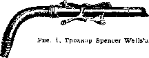
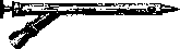
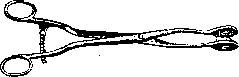
the abdominal method, the skin incision is made along the median line, paramedian, or transversely. Upon opening the abdominal cavity, the surgeon orients himself to the anatomical relationships. If the ovarian tumor is on a pedicle and without adhesions, then the operation is very simple: the tumor is easily extracted through the abdominal wound; beforehand, if necessary, the tumor is emptied using a trocar (figures 1 and 2), onto which a long sterilized rubber tube is placed. The emptying of the cyst is performed with the patient in a horizontal position. An assistant grasps the abdomen on both sides with both palms, compresses it, and thereby presses the cyst against the parietal leaf of the incised peritoneum. At this time, the operator inserts the trocar into the tumor, and the fluid flows out from the cyst through the lateral outlet of the trocar; the walls of the cyst begin to collapse, the surgeon quickly fixes them with two Nelaton forceps (figure 3), and the reduced tumor is removed from the abdominal cavity through a small incision. Multilocular cysts are
Figure 2. Trocar and tube.
punctured sequentially. Then the pedicle is located, ligated, and the tumor attached to it is excised together with the adjacent portion of the tube. When ligating, one should not use a simple ligature, but it is recommended to pierce the pedicle in the middle with a Deschamp needle with a double ligature—thus two ligatures, upper and lower, are obtained, which are tied in succession. In recent times, most surgeons operate without ligatures en masse (fig. 4); the pedicle of the tumor is fixed with a Kocher forceps, then it is cut through, the bleeding vessels are grasped with hemostatic forceps and ligated individually (spermatic artery, artery of the round ligament, and terminal branch of the uterine artery); subsequently, both layers of peritoneum covering
Figure 3. Nelaton's forceps.
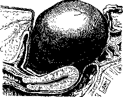
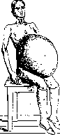
the pedicle are sutured together. This peritonization eliminates the possibility of adhesions between the intestines and the operative stump, which prevents the possibility of mechanical ileus. After treatment of the tumor pedicle, the toilet of the abdominal cavity is performed: any cyst contents that have accidentally entered the abdominal cavity are thoroughly removed (with gauze tampons), the bleeding sites are inspected again, and the abdominal wound is closed. At present, most surgeons consider it unnecessary to use for treatment of the cyst pedicle the instruments previously proposed [by Karavaev's clamp, angiotribe (Tuffier, Doyen)] or cauterization of the pedicle with carbolic acid; likewise, the so-called Staffordshire knot ligation (fig. 5) is no longer performed. Suture material—catgut. The most frequent complication in O. is the presence of adhesions with adjacent organs, more often with the intestines (fig. 6) (especially in so-called giant tumors) (fig. 7). In such cases, it is recommended to open the abdominal cavity near the umbilical ring, since here the peritoneum is most firmly fused with the underlying layers of the abdominal wall; a careful, layered incision of the abdominal wall excludes the possibility of detachment of the parietal peritoneum from the abdominal wall. If separation of intestinal adhesions presents great difficulties, it is better to leave a layer of the cyst wall on the intestine (Olshausen) than to worsen the prognosis of the operation by violating the integrity of the intestine in one or more places; the remaining inner layer of the cyst wall should be removed or cauterized with carbolic acid. In cases of extensive adhesions (rarely), one has to
Figure 6. Adhesion of cyst with intestine.
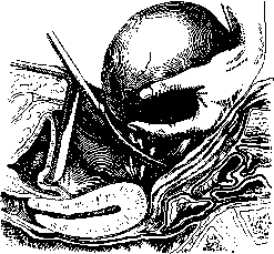
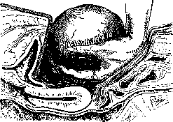
forego enucleation of the entire "tumor and use the so-called marsupialization (see). The omentum most often grows to the cyst; it is usually separated by blunt dissection, and bleeding sites should be ligated. If there is a suppurated or dermoid cyst, then the tumor must be removed entirely. If this cannot be accomplished, one must immediately perform a toilet, disinfect the contaminated areas (rivanol, ether); some in such cases drain the abdominal cavity. - A further complication is the twisting of the pedicle and cyst. The peculiarity of the operative technique in these cases is as follows: one should not untwist the pedicle, because by untwisting the thrombosed vessels are disrupted and embolism may occur. If the pedicle is elongated, it should be ligated en masse above the thrombi within it; if it is short, then a wedge-shaped excision of the pedicle together with a portion of the uterine angle is performed. After individually ligating the bleeding arteries, the edges of the wound are approximated with sutures. There are so-called interligamentary ovarian tumors, which have no pedicle at all and grow between the layers of the broad ligament. In addition to true interligamentary ovarian tumors, so-called pseudo-interligamentary cysts are also observed. There is no single technique for removing interligamentary tumors. The most accepted technique: the peritoneum is incised at the border of the implantation of the interligamentary cyst into the broad ligament, the serous covering is separated, and, holding the cyst wall, one proceeds to its enucleation (fig. 8). When enucleating the tumor, one must be extremely careful not to injure the ureter. After
Figure 7. Giant ovarian cyst.
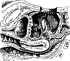
enucleation of the cyst and after stopping the bleeding, the resulting cavity is closed tightly with sutures. If it is not possible to completely remove the cyst wall due to its thinness or previous inflammation, then marsupialization is performed (fig. 9). Since statistics show that bilateral ovarian involvement occurs very frequently (51.8%, and according to Doderlein—90.9%), in cases of unquestionable malignancy of the tumor in one ovary, the other ovary should also be removed even in young subjects. O. can also be performed by the vaginal route—by incision of the anterior or posterior fornix. Posterior colpotomy was practiced by American surgeons
Figure 8. Enucleation of cyst.
(Battey, Atlee, Byford), in Russia—by Lyubovov (Kazan; 1892), and was significantly improved by Ott (Leningrad). Anterior colpotomy was practiced by German surgeons led by Duhrssen and Martin (Duhrssen, Martin). The vaginal route can be recommended for removing small, mobile cystic tumors, and the choice between anterior or posterior colpotomy depends on the anatomotopographic location of the tumor itself.
Figure 9. Marsupialization of cyst.
Performance of the operation - see Colpotomy. - The age of the patient is not a contraindication to O. The operation is successfully performed both at the very youngest age and in old age - in 80-year-old old women. In case of severe exhaustion of patients, O. is recommended to be performed under spinal anesthesia. In addition to impeccable surgical technique and strict adherence to asepsis, the prognosis depends especially on whether complications were present. According to the material of the gynecological clinic in Giessen, the mortality rate after O., excluding cancers and complicated cases, was 2%, and after ovariectomies performed for benign ovarian tumors, the mortality rate is only 0.7%. O. in complicated cases gives a much higher mortality rate (according to some statistics up to 15% and even 20%). Ovariectomy during pregnancy. Despite the fact that the childbearing function in ovarian tumors is reduced, the onset of pregnancy with them is not excluded: according to Martin, out of 1,732 deliveries, ovarian tumors were found in 20 cases (1.5%), according to Ols-hausen - in 3.9% of cases, according to the statistics of the Berlin obstetric clinic - in 0.1% of cases. Collective statistics show that out of 100 ovariectomies, 3 are during pregnancy; according to Yartsev's statistics, out of 2,000 ovariectomies - 68 ovariectomies complicated by pregnancy (3.4%); according to Lebedev's material, out of 412 ovariectomies - 28 cases (6.8%). Pregnancy can occur with both benign and malignant tumors. - The effect of ovarian tumors on the normal course of pregnancy and delivery depends on their anatomical location and size. Small cysts without adhesions, located in the small pelvis and having a long stalk, usually rise from the small pelvis to the large one as the pregnant uterus grows; but if the cyst lies interligamentary or is fixed in the small pelvis, then it is a mechanical obstacle during delivery. Pregnancy and delivery in the presence of an ovarian neoplasm can proceed normally, but complications can occur. A very serious complication during pregnancy, delivery, and in the postpartum period is twisting of the tumor stalk, rupture of the cyst due to compression, hemorrhage into the tumor, and finally suppuration. Recognition of an ovarian tumor in the first months of pregnancy presents no difficulty; it is much more difficult to diagnose a cyst at the end of pregnancy. - The question of indications for surgical intervention for ovarian tumors during pregnancy has not yet been finally resolved. Some authors object to performing O., based on the fact that in many cases of complications of pregnancy with ovarian tumors, delivery ends successfully, and after O., pregnancy is often interrupted (on average from 5% to 15%). In more recent data, this percentage is even lower. Therefore, the danger of interruption of pregnancy after operations is unfounded. It is better to operate in the first months of pregnancy despite the possibility of removal of the corpus luteum. The latter circumstance does not lead to interruption of pregnancy, as can be concluded from the observations of Essen-Moller, Ruge, and others. Even Veit pointed out that 'the prognosis of O. intra graviditatem is not only not worse than the prognosis of O. extra graviditatem, but even significantly more favorable'. This is because such operations are mostly performed on young patients whose bodies have greater resistance. Thus, as soon as an ovarian tumor is determined during pregnancy, its treatment should consist of surgical removal. Some recommend colpotomy, but most prefer laparotomy. In rarer cases, when the cyst is attached in the small pelvis or lies interligamentary and the pregnant uterus interferes with approaching the removal of the tumor, it is necessary to empty the uterus beforehand by means of the so-called small cesarean section.
Related articles
Mentioned in
Cite this page
“Ovariotomy.” Soviet Medical Encyclopedia. English translation of Bolshaya Meditsinskaya Entsiklopediya, 1st ed. (Moscow, 1928–1936), ed. N. A. Semashko. https://sovietmedicalencyclopedia.pages.dev/article/ovariotomy/