Colpotomy
Historical document, translated for reference. It reflects medical knowledge of the 1920s–30s and is not medical advice.
Summary
Colpotomy is a surgical procedure that provides access to the abdominal cavity through the vagina for diagnostic and therapeutic purposes. The article describes the history, techniques, indications, and complications of anterior and posterior colpotomy.
Encyclopedia article (1928–1936)
Colpotomy, colpotomy (from Greek kolpos-vagina and temno-I cut), or colpocoeliotomy, is a method of penetrating the abdominal cavity through the vaginal route in women for diagnostic and therapeutic surgical purposes. K. was first applied in 1822 by Sauter (Johann Nepomuk Sauter) in Germany for removal of cancerous uterus; then in 1857 Atlee opened the posterior vaginal fornix for removal of ovarian cysts. G. Thomas in New York in 1870 became the first to systematically apply the vaginal route for removal of ovarian cysts. Later in Germany Duhrssen (1894) and then A. Martin (1903) thoroughly developed this method and gave it a definite place in gynecology. In Russia, the vaginal route was first applied by Kiter and Elatic for removal of cancerous uterus. Two types of K. are distinguished: anterior K. (s. anterior) and posterior (s. posterior). The first consists in opening the abdominal cavity through the anterior fornix, while the second-through the posterior. Besides anterior and posterior K. in the narrow sense, one can also distinguish a special type of combined K.-sleeve resection of the uterus, in which anterior and posterior K. are performed and the uterus is removed.
Figure 1.
Figure 2. Thomas in New York in 1870 became the first to systematically apply the vaginal route for removal of ovarian cysts. Later in Germany Duhrssen (1894) and then A. Martin (1903) thoroughly developed this method and gave it a definite place in gynecology. In Russia, the vaginal route was first applied by Kiter and Elatic for removal of cancerous-
Figure 3.
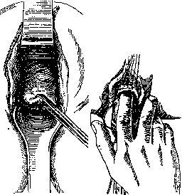
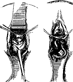
Figure 4. transformed uterus. Two types of K. are distinguished: anterior K. (s. anterior) and posterior (s. posterior). The first consists in opening the abdominal cavity through the anterior fornix, while the second-through the posterior. Besides anterior and posterior K. in the narrow sense, one can also distinguish a special type of combined K.-sleeve resection of the uterus, in which anterior and posterior K. are performed and the uterus is removed. Technique of K. To perform K., the woman is placed on the edge of the operating table with bent knee and hip joints and legs spread apart. The vagina is opened with specula, and the vaginal part of the uterus is grasped with bullet forceps or Muse-Duayen's tenaculum. When performing anterior K.-the vaginal part of the uterus is strongly lowered down, whereby the anterior vaginal wall is stretched and everted; then a penetrating incision through its entire thickness is made on the anterior vaginal wall-either a longitudinal median one, extending from the external urethral opening to the vaginal part of the uterus, or transverse, or arcuate. The latter two incisions, 5-6 cm long, are made in the area where the anterior fornix transitions to the vaginal part of the uterus (fig. 1). After the vaginal incision, the bladder is separated from the cervix partly by blunt dissection and partly with scissors, up to the uterovesical fold of peritoneum (fig. 2), which is then grasped with forceps and cut transversely with scissors, as a result of which the abdominal cavity is opened in the area of the anterior Douglas pouch (fig. 3). The pelvic cavity can be examined by bimanual examination. If adhesions are present, they are separated. Then, by inserting a plate speculum into the opened fornix, the necessary manipulations are performed-prolapse of the uterine body, ovaries and removal of existing tumors in them (fig. 4), etc. After these manipulations and hemostasis, the wound in the fornix is sutured (preferably with catgut), whereby the bladder edge of peritoneum is connected with the uterine one, and the edges of the vagina with each other. If the tissues were severely damaged, the wound is left partially open and a drain is inserted into it. A difficult moment of anterior K., especially for an operator inexperienced in vaginal operations, is the separation of the bladder from the cervix. Sometimes complications in the form of penetrating injury to the bladder may occur here. To avoid this, it is necessary in difficult cases to perform this step of the operation under the control of a catheter inserted into the bladder.
Posterior K. is technically easier. To perform it, a sufficiently wide but not long posterior speculum is inserted into the vagina; the vaginal part of the uterus with the help of bullet forceps or Muse-Duayen's tenaculum is pulled
Figure 5
COLSA
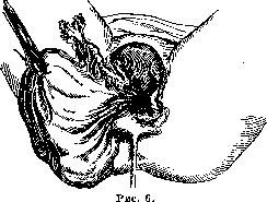
forward and upward. The posterior fornix, which has become accessible and stretched as a result, is opened with a longitudinal, or cruciate, or transverse incision made slightly below the site of attachment of the vagina to the vaginal part of the uterus (fig. 5). First, the vaginal wall is cut with a knife or scissors, then the peritoneum is opened. To stop bleeding and to facilitate the opening of the formed opening, 3 or 4 nodal sutures are placed on the incision in the posterior fornix, capturing the entire thickness of its wall. Then the necessary manipulations are performed, for which K. was undertaken, e.g., removal of ovarian cyst. The latter, depending on the location of the tumor, is done either without pulling or with pulling the uterus out (fig. 6). After completion of all
necessary manipulations and hemostasis, the wound in the fornix is sutured tightly. In the presence of purulent or bloody accumulation in the posterior Douglas pouch, opening of the posterior fornix is technically very easy. After incision of the vaginal wall, the bulging peritoneum is opened by blunt dissection, e.g., with forceps. After emptying the contents, a sufficiently thick rubber tube is inserted into the opening for drainage. In case of obliteration of the posterior Douglas or adhesion of thin intestines here, the performance of posterior K. becomes very difficult and sometimes even impossible. In these cases, injuries to the rectum and other, adhered sections of the intestine easily occur. The advantages of colpotomy as a method of gynecological laparotomies are 1) minimal trauma and operative shock, 2) good conditions for drainage, and 3) less danger of infection of the abdominal cavity. The disadvantages are the limited access to the abdominal cavity, the narrowness and crampedness of the operative field, and hence the impossibility of removing large tumors entirely, the difficulty of isolated hemostasis, peritonization, etc. In connection with this, colpotomy places greater demands on the technique and skill of the operating surgeon. To reduce some of the weak points of K., additional incisions of the perineum and vagina (Schuchardt's incision and median perineal incision of Ott) and vaginal Ott specula have been introduced, allowing almost the entire peritoneal cavity to be viewed. Pe- LAW
Anterior K. is performed for small (up to fist size), mobile or slightly adherent ovarian tumors located in the anterior fornix, for removal of small fibromyomas located in the anterior uterine wall, for correction of retroversion, retroflexion of the uterus and prolapse, for amputation of the uterine body according to Rieck, etc. Posterior K. is sometimes performed for diagnostic purposes. For therapeutic-surgical purposes, it is indicated for purulent and infected blood accumulations in the posterior Douglas. In these cases, it is an indispensable operation. Then it is done for ovarian tumors located in the posterior Douglas pouch, for uterine prolapse, for ectopic pregnancy, etc. Except in infected cases, ectopic pregnancy should be operated on as a rule by abdominal laparotomy. Contraindications for K. are abundant, dense adhesions of the uterus, ovaries and tubes, voluminous cystic, especially dense and malignant tumors of the genital sphere, suspected inflammation of the appendix, etc. Some ardent supporters of K., such as Duhrssen, Ott and others, set indications for them much wider and removed large tumors and even the appendix by this route; but in this case, due to the impossibility of completing the operation through the vagina, they sometimes had to switch to abdominal laparotomy or for technical reasons resort to extirpation of the uterus.-From this it is clear that compared to abdominal laparotomy, the scope of application of K. is limited; relatively simple operative cases fall under its jurisdiction. The majority of gynecologists perform it much less frequently than abdominal laparotomy: on average 8-30% (Gruzdev, Okinichits, Doderlein, Franz and others). The mortality rate for K. is lower than after abdominal laparotomies.

I. Horizontov. COLLES' LAW (Colles), a proposition published by the Irish physician Colles in 1837, according to which a mother showing no signs of syphilis, of a congenitally syphilitic infant who has infectious manifestations in the mouth that are dangerous to a healthy wet nurse, can breastfeed her child without risk of infection from him. This proposition was confirmed in 1840 by the Lyon physician Baumes (Francois Baumes) and entered the history of syphilology under the name of the Colles-Baumes law. This "law" formed the basis of the paternal (ex patre) theory of transmission of congenital syphilis, according to which a syphilitic virus mixed with the semen, supposedly without infecting the woman's sexual passages, infects either the germ plasm (ovulum) or the already developing embryo. The product of conception infected by one or the other method in turn either infects the mother or immunizes her, making her inaccessible to new infection with syphilis (the "Colles mother"). Subsequently, cases of "exceptions to the Colles-Baumes law" were even published, in which the mother nevertheless became infected from her congenitally syphilitic child. These latter cases made a particularly strong impression. Indeed, one such indisputable case would have been sufficient to prove the possibility of paternal transmission of congenital syphilis without the mother becoming infected. But as early as the 1860s, Norwegian authors Wilhelm Boeck and Ewre (W. Boeck, Oewre) noted that only a mother with syphilis can give birth to an infected child. Later, in 1898, Oedmansson, on the basis of studying a large amount of clinical material, came to similar conclusions. But the most categorical confirmation of this position was found in the classic work of Matzenauer. The latter, in 1903, on the basis of only clinical study of published cases, expressed the conviction that all "Colles mothers" have syphilis and that in the cases of exceptions to the Colles-Baumes law there is no new infection of the mother. In these latter cases, either a previously healthy mother became infected with syphilis from her infant who was born healthy but became infected after birth, or as a result of constant irritation during lactation, chancriform papules appeared on her nipples. Furthermore, these cases can be considered as a result of superinfection due to the constant rubbing of the child's spirochetes into the nipples. Since the time of Matzenauer's work, cases of exceptions to the Colles-Baumes law have been published less and less frequently, and in the last 10 years none have been published at all (Rietschel). One can therefore assert that the "exceptions" in question do not exist in nature at all. The correctness of the views expressed by Matzenauer regarding "Colles mothers" is confirmed by the already sufficiently accumulated clinical and laboratory data. The brilliant discoveries of the early 20th century fully confirmed the bold, for that time, conclusions of Matzenauer, based on a subtle analysis of clinical facts. However, the possibility, though rare, of transmission of congenital syphilis only by the father, without the participation of the mother, is still recognized by some authors, among whom it is necessary to mention the names of such major scientists as Finger, Hochsinger, Almkvist; the latter, in a detailed work (1928), cites 5 cases that seem to testify to the possibility of a healthy mother giving birth to a child with syphilis. However, the cases cited by Almkvist are not without flaw: all the mothers were not examined sufficiently thoroughly (for example, none of them had a spinal fluid examination). Furthermore, Almkvist does not explain the fact that none of these "healthy" mothers became infected with syphilis from their children who had active manifestations of congenital syphilis, and nowhere does he express his opinion on the "immunity" of these mothers. Moreover, all these and similar observations cannot refute the basic fact that to this day it has not been possible to detect either the pale spirochete nor pathological changes in the placenta before the fifth month of pregnancy. And this circumstance stands in clear contradiction with the hypothesis of infection of the egg (ovulum) by a spirochete, whether unfertilized or already fertilized (Meshchersky). Thus, even this work by Almkvist cannot be considered sufficiently convincing. The positions of Oedmanson-Matzenauer remain in force. The more thoroughly "Colles mothers" are studied, the more often they have to be transferred from the ranks of healthy and immune to the category of diseased. Rare cases in which syphilitic infection cannot be established in such a mother testify only to the insufficient sensitivity of modern diagnostic methods, to the extreme diversity of the course of syphilis, and not to their health. Since the fact of spirochete carriers (Nullerbi Kolle) has been established, the "Colles mother" does not represent anything exceptional in the pathology of syphilis (Sobolev). In any case, for practical purposes, it should be remembered that the birth of a diseased child by a diseased mother is such a frequent clinical fact that the birth of a diseased child not only gives the physician the right but also imposes on him the duty to subject the mother of such a child, whatever the results of her examination, to vigorous specific treatment both for the purpose of her personal prevention and for the prevention of her future offspring.
Related articles
Mentioned in
Cite this page
“Colpotomy.” Soviet Medical Encyclopedia. English translation of Bolshaya Meditsinskaya Entsiklopediya, 1st ed. (Moscow, 1928–1936), ed. N. A. Semashko. https://sovietmedicalencyclopedia.pages.dev/article/colpotomy/