Colpopoiesis
Historical document, translated for reference. It reflects medical knowledge of the 1920s–30s and is not medical advice.
Summary
This article from the 1928-1936 Soviet Great Medical Encyclopedia discusses the history and surgical techniques of colpopoiesis, the creation of an artificial vagina for congenital absence or atresia of the organ. It reviews early methods using skin flaps, mucous membranes, and peritoneum, as well as the pioneering contributions of Russian surgeons such as Snegirev and Popov using intestinal segments.
Encyclopedia article (1928–1936)
COLPOPOESIS (colpopoesis), the creation of an artificial vagina. Even ancient physicians comparatively often observed the complete or partial absence of internal genital organs. Among these defects, the partial or complete absence of the vagina must have particularly attracted attention. This defect inevitably made itself known by the absence of menstruation at an age when menstruation should have already appeared, and, in the presence of a uterus capable of secreting menstrual blood, by the formation of blood tumors from accumulating blood (haematometra, haematocolpos, haematosalpinx). In the absence of the uterus or in its rudimentary state, blood tumors are not formed, and a woman often learns of her defect only after marriage and consultation with a physician. In rare cases, the absence of the vagina is a consequence of the obliteration of its lumen under the influence of various inflammatory processes, especially in early childhood. The operation aiming to create an artificial vagina in place of the absent one, in order to provide an outflow for menstrual blood when a menstruating uterus is present, to eliminate the formation of blood tumors, and also to enable a woman, even one devoid of a menstruating uterus, to live a normal sex life, has its own history. In the event of blood tumors, the formation of an artificial passage in place of the absent vagina is usually a life-saving operation in order to deliver the woman from a gradually increasing blood tumor, which often threatens rupture and severe complications, as well as to stop often severe pains, especially intense during periods corresponding to menstrual days. Physicians of the ancient world were already familiar with these sufferings (Hippocrates, Aristotle, Pliny, Celsus) and, to relieve women of them, had to resort to punctures of such tumors. With the development of surgery, physicians tried to improve these punctures by replacing them with the creation of an artificial passage at the site where the vagina should be located. However, all these attempts in the pre-antiseptic period usually ended very poorly: the emptied cavity easily became infected from the wound opening, and patients often died from septic infection. In addition, even in cases that ended comparatively successfully, the artificially made passage quickly closed, and the entire effect of the operation was nullified. Thus, despite the fact that the operation proposed by Dupuytren in 1817 made it possible to create a voluminous artificial vagina, it still did not lead to the desired results, since this passage also quickly closed through scarring. The conquests of the antiseptic era made it possible to begin a stubborn struggle for the preservation of the artificially formed vaginal passage. Attempts were made not only to create an artificial passage, but also to epithelialize it by transplanting skin or mucous membrane onto the inner surface of the newly created vagina. The so-called plastic method appeared. In this case, for the transplantation, the nearest areas of skin of the lips, perineum, or thighs were used as material, which remained connected by means of a thin pedicle to the place from which such a flap was taken (autoplasty). Free transplantation of skin flaps was also performed, in which no connection of the transplanted areas with the mother soil was left (autotransplantation). Finally, attempts were made to use skin or mucous membrane for transplantation from other patients (heteroplasty) and even from animals. However, all these methods only improved the results of Dupuytren's operation, only temporarily delaying the subsequent process of scarring of the artificially made passage. As a rule, the ultimate result was the obliteration of such a vaginal passage, even an epithelialized one, and the result of the operation was reduced to zero. It should be noted that Russian physicians occupy a particularly honorable place in the creation of an artificial vagina. Thus, Hepner (St. Petersburg) was the first to conceive the brilliant idea of using a skin flap, taken from the patient's labia majora and buttocks, transplanted into the vagina formed by Dupuytren's method. Shalita (Kiev) was the first to propose transplanting islands of mucous membrane, cut with scissors from the inner surface of the patient's labia majora and minora, into the vagina. Much was done for the development and winning the right of citizenship of this operation by Rein (Kiev), and especially Snegirev (Moscow) and Popov (Leningrad). In the pre-antiseptic period, the operation to create an artificial vaginal passage was performed for only one purpose: to eliminate the formation of blood tumors and create conditions under which the blood secreted during menstruation would find a free exit outward. With the development of antisepsis, when the results of this operation began to improve significantly, reports gradually began to appear about cases of the colpopoiesis operation performed with the aim of giving a woman the opportunity to live a normal sexual life. The necessity of such indications was also dictated by the circumstance that the majority of married women with the absence of the vagina still lived a sexual life, but coitus was performed either into an artificial depression formed under its influence in the perineal region or through the widened urethra. Such a "broad" application of the operation caused a whole explosion of indignation and heated debates in the medical world and in scientific societies. Should a physician, by creating an artificial vagina, pander to the desire of a woman and her husband to have a sexual life when the main goal of sexual intercourse—the possibility of pregnancy—is absent? A whole series of opponents of this operation appeared, striving to prove all its sterility, barrenness, and immorality. However, along with this, passionate defenders of this operation also appeared, proving that "the surgeon has the right and must intervene with active assistance to restore the missing organ, even if not entirely perfect. These operations are not only fully indicated, but they must be regarded as a noble duty of the surgeon" (Rein). At the present time, with the exception of isolated individuals, there are few surgeons who deny the important significance of this operation, especially since its necessity, as it turned out in a large number of cases, is dictated by a number of mental factors. As noted, the method of transplantation did not yield stable results despite persistent measures to delay the process of epithelial displacement and its replacement by connective tissue. Various bodies made of glass, ebonite, metal, and other materials were introduced into the newly formed vagina. These bodies were given a spherical, cylindrical, or some other shape; they were introduced for months, and still the newly formed vagina slowly obliterated. In 1897, Ott for the first time applied the method of using the peritoneum in order to avoid the desolation of the vagina. After the formation of an artificial passage and the opening of the peritoneum deep within the formed channel, he brought down the edges of this peritoneum to the level of the skin incision, to which he sewed the edges of the peritoneum. Even earlier, Pfannenstiel formed a similar passage and, having reached the cervix of the existing uterus, brought it down and sewed the edges of the skin incision to it. But both of these methods did not receive wide distribution because they were significantly inferior in their results to methods that appeared almost simultaneously with them, the idea of which was to use a tubular organ—a section of the small or large intestine—for the formation of the vaginal tube. The first attempts in this area were made by Gersuny. However, they did not yield the desired results. In Russia, this idea was used in an extremely original form by Snegirev, who, despite the fiercest attacks, laid a serious and deeply thought-out beginning for the modern colpopoiesis operation. Snegirev's idea is extremely simple. He separates the lowest section of the rectum from the one located higher up. Having performed a resection of the coccyx, he sews the edge of the upper section of the intestine into the region of the resected coccyx and thus creates an artificial anus (anus praeternaturalis); the lower segment of the intestine serves as the vagina. To widen the anal opening and bring it closer to the pudendal lips, Snegirev makes an incision from the side of the anus along the midline of the perineum to the external opening of the urethra and sews the incised mucous membrane of the rectum into this wound. Thus, a large, oblong-shaped introitus is obtained, in the formation of which the anal opening also takes part. Snegirev's operation was modified and significantly improved by Popov. He freed it from the unpleasant aspects of the artificial anus, left the anal opening in its place, but formed the vagina from the lower section of the rectum. Popov achieved this by, like Snegirev, detaching the lower section of the rectum from the sacrum, separating this section of the intestine from below from the anal opening, and from above from the upper section of the same intestine; he brought down the upper section of the intestine to the anal opening and fixed it there with interrupted sutures, and moved the lower, isolated section of the rectum to the place of the vaginal tube formed by him and fixed it.
Somewhat later than Popov, the German gynecologist Schubert proposed his own method, which does not essentially differ from Popov's operation (Fig. 1). The difference lies only in the method of approach to the rectum. To separate the rectum and isolate its lower segment, Schubert used the method already proposed by Snegirev
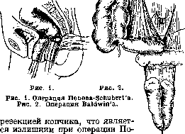
Popov. In any case, at the base
of
both operations lies one and the same idea of Snegirev, but Popov carries it out with less trauma and the same end results. With the development of abdominal surgery, the exclusively vaginal route for the formation of an artificial vagina was replaced by an operation associated with opening the abdominal cavity through the anterior abdominal wall. This became necessary in order to use the lower sections of the small or large intestines for the formation of the vagina. For the first time, such an operation was proposed and performed by the American surgeon Baldwin (Baldwin; Fig. 2). The idea of this operation, detailed by Stoeckel, is extremely simple: an access is made, as always, at the site of the absent vagina (partly with sharp instruments, partly by the blunt route); a curved forceps is introduced into this access, after which the patient is positioned for laparotomy, the abdominal cavity is opened along the midline, a section of the small intestine is found as close as possible to the cecum, its most mobile loop 25 cm long is isolated, leaving the isolated section on the mesentery and trying not to damage the mesenteric vessels nourishing the isolated section. The patency of the intestine is restored by anastomosis, and the isolated section of the intestine, having pierced the mesentery in the middle with a thick ligature, is pulled by this ligature using the inserted forceps into the artificial passage described above (Fig. 2). After this, it only remains to suture the lower edge of the loop of the resected section of the small intestine into the skin wound of the introitus, open the intestine here, and the artificial double vagina is ready. Of course, the abdominal wall is sutured tightly. This operation, when properly performed and while preserving the integrity of the mesenteric vessels, yields excellent results both in terms of primary operative outcomes and long-term results. At present, the vast majority of gynecologists use either the Popov-Schubert operation or the Baldwin operation. A heated debate over the advantages of one or the other method is attempted to be resolved by references to the already quite extensive material of both operations accumulating at present. The Popov-Schubert method is particularly widespread in the USSR and Germany, while the Baldwin operation is performed both in these countries and everywhere. Among Russian gynecologists recently, the Baldwin operation, judging by increasingly frequent reports, is also beginning to acquire many supporters. Both the method of forming an artificial vagina from the rectum and the Baldwin operation provide a good and permanent vagina, fully suitable not only for sexual life, but even for childbirth, as indicated by Wagner's case.
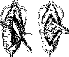
Figure 1.
Figure 2. the influence of intra-abdominal pressure and the filled urinary bladder on the sutured vaginal wall is also reduced, which promotes healing and facilitates it. When separating the flap, which can be either thickened or thinned, it must be remembered that the urethra, urinary bladder, or ureters can be injured during this process, and therefore the separation must be performed in the layer of cellular tissue; the tip of the knife is recommended to be directed toward the tissue being removed, i.e., the vaginal wall. If the cellular tissue is loose, the separation succeeds by the blunt route. Upon removal of the flap, the posterior surface of the urinary bladder is exposed. Before
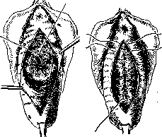
Figure 3.
Figure 4. the operation, it is better to empty the urinary bladder to avoid injuring it. Bleeding vessels are circum-sutured. If the resurfaced area turns out to be uneven, it should be trimmed with curved scissors. The edges of the wound are sutured; to avoid the formation of pockets, purse-string sutures are applied to the cellular tissue. The suture material used by most authors is catgut, iodine-catgut; the suture can be interrupted, or continuous, or interrupted-continuous (Vydrin). The latter consists in the fact that the thread is passed twice through the loop, thereby achieving that the continuous suture appears to consist of interconnected interrupted sutures; with this suture, each individual section of it is completely independent in terms of integrity from another section, and there is not the mass of knots that would exist when applying only interrupted sutures. If after the removal of the flap the urinary bladder strongly protrudes, i.e., there is a cystocele, then it is necessary either to apply one or concentrically several purse-string sutures according to Schroeder, Gershuni (Fig. 3) to the wall of the urinary bladder or a sagittal suture to the anterior retinaculum uteri according to Bumm and Martin (Figs. 4 and 5). Posterior colporrhaphy is performed in the same way, but since in prolapse of the posterior vaginal wall there is mostly a strongly gaping genital cleft and an old perineal rupture, in these cases Simon-Hegar colpoperineorrhaphy is performed. The technique is as follows: having determined the required degree of elevation of the perineum according to the case, bullet forceps or two threads are applied to the extreme points, which are tied at the ends, and thus loops are obtained with which assistants stretch the perineum and the posterior vaginal wall. The almost straight line obtained at the border of the vaginal mucosa and the perineal skin of COLPOTOMY
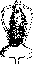
Figure 5.
is the base of the triangle, the apex of which is located on the posterior vaginal wall, more or less high depending on the degree of prolapse of the latter; a Kocher forceps is applied to the apex of the triangle. Assistants, pulling on the threads applied to the extreme points of the base of the triangle and on the Kocher forceps applied to the apex of the triangle, spread out the area of the posterior vaginal wall to be removed. A transverse incision is made at the border of the mucosa of the posterior vaginal wall and the perineal skin between the threads, the posterior vaginal wall is separated by sharp (Fig. 8) and blunt routes from the rectum to the upper point of the triangle; the entire separated flap is removed. Lawson Tait proposed not removing the flap, but suturing its edges in a sagittal direction and leaving it (flap plastic surgery). Then, to restore the rectovaginal septum, the muscles and fasciae that make up the tendinous center of the perineum in front of the rectum are sutured. So that during THIS
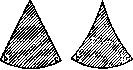
rectum with the index finger of the left hand wearing a rubber glove. After applying three or four interrupted a
a
sutures made of iodine-catgut
to the separated or ruptured muscles and fasciae of the rectovaginal septum, a continuous-interrupted suture made of iodine-catgut is applied from top to bottom on the edges of the triangle of the posterior vaginal wall in a sagittal direction until the extreme points of the base of the triangle on which the loops are applied are connected. This concludes the actual posterior colporrhaphy. Then, interrupted silk stitches or Michel clips are applied to the perineal skin (Figs. 9-10). Upon completion of the operation, the patient is put to bed and remains in the supine position until d
d Figure 7. Left—figure of resurfacing according to Hegar. Right—corrected Hegar figure of resurfacing. the morning of the next day; then she is turned on her side and allowed to independently change position, but not sit up. A light diet is prescribed. Giving opium is not recommended. On the fourth day, the patient receives a laxative, mostly Magnesia sulfurica. Care
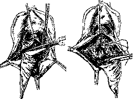
Figure 8.
Figure 9.
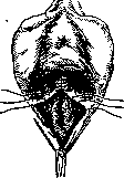
Figure 10.
of the wound consists of irrigating it several times a day (mandatory after each urination) with a lysoform solution. On the 5th or 6th day, the sutures or Michel clips are removed from the perineum. On the 12th day, the patient gets up, and sitting in bed is not allowed before this. On the 14th
^ day, the patient is discharged. Mention should also be made of the operation proposed first by Neugebauer in Warsaw and some time later by Le Fort in Paris for major uterine prolapses. The operation consists in cutting out flaps of equal shape facing each other from the anterior and posterior walls of the vagina, and the resurfaced surfaces thus obtained are sutured together; as a result of this operation, a longitudinal septum is obtained in the vagina. This operation has not gained wide distribution. Colporrhaphy can be performed under general or local anesthesia. Anterior colporrhaphy is performed without any anesthesia (Vydrin), while colpoperineoplasty is performed under local anesthesia: a 1/2% solution of novocaine with three drops of Sol. Adrenalini hydrochlorici (1:1.000) per gram of solution.
M. Vydrin
Related articles
Cite this page
“Colpopoiesis.” Soviet Medical Encyclopedia. English translation of Bolshaya Meditsinskaya Entsiklopediya, 1st ed. (Moscow, 1928–1936), ed. N. A. Semashko. https://sovietmedicalencyclopedia.pages.dev/article/colpopoiesis/