Hysterotomy
Historical document, translated for reference. It reflects medical knowledge of the 1920s–30s and is not medical advice.
Summary
This article describes hysterotomy, a surgical incision of the uterine wall, historically used for uterine examination and removal of growths. It details the technique, including the use of forceps and sutures, and its application in obstetrics, particularly for vaginal cesarean section.
Encyclopedia article (1928–1936)
HYSTEROTOMIA (vaginal), an operation for incising the anterior wall of the uterus; it was proposed by Defontain for digital examination of the uterine cavity, removal of polyps, enucleation of submucous myomas, etc. The cervix is exposed with a speculum; the cervix is grasped with Muesse forceps; a transverse incision is made at the edge of the vaginal part of the uterus, through the mucous membrane of the anterior fornix, and the bladder is displaced upward. One blade of scissors is introduced into the cervical canal, which is pulled down, and the anterior wall of the uterus is incised down to the peritoneal fold. The operation is performed extraperitoneally. After the operation, catgut sutures are applied to the uterine wall and the vaginal mucosa. Hysterotomy found wide application in obstetrics, where under the name of "vaginal cesarean section" it was proposed by Duhrssen. Initially, Duhrssen incised both the anterior and posterior walls of the cervix. Now it is considered quite sufficient to incise only the anterior wall. Additional incision of the posterior wall is performed only in exceptional cases (when it is difficult to pull the cervix down). Kronig's proposal to perform vaginal cesarean section by means of only posterior hysterotomy, with opening of the abdominal cavity, did not gain popularity. The operation begins with a longitudinal incision, a little away from the urethral opening, to the external edge of the cervix; a transverse incision can be added to this incision, stepping back from the edge of the cervix by 1-1/4 cm. Some limit themselves to a transverse arcuate incision of the mucosa (see Figure 1). After this, the bladder is separated by an acute or blunt method (see Figure 2), which is usually performed very easily due to the looseness of the tissue. After separating the bladder, it is displaced upward with a speculum and the peritoneum is separated from the uterus as far as possible. Then the anterior wall of the cervix is incised. Grasping the edges of the incision with Muesse forceps and pulling the cervix down, the anterior wall of the uterus is incised as high as possible, down to the peritoneal fold (see Figure 3). The fetal sac is opened, and a footling version is performed or forceps are applied; with a dead fetus, perforation is performed. The last part has to be delivered by the Crede method or manually. The edges of the uterine incisions are sutured with catgut sutures, not including the uterine mucosa (see Figure 4). When suturing the vaginal wound in its lower part, some insert a strip of gauze, which is removed after 2-3 days. With careful and cautious separation of the bladder, injury to it can be easily avoided: if the peritoneum is opened, it is carefully closed with separate sutures; a sufficiently long uterine incision prevents its rupture during delivery, and a T-shaped or arcuate incision of the mucosa prevents vaginal ruptures. The cause of bleeding during ante partum operation may be excessive deviation from the midline during separation of the bladder. Significant bleeding is also possible in placenta previa, in which the tissues of the lower segment become brittle and easily bleed. Various additions to the technique—preliminary dilation of the cervix for good drainage of secretions in the postpartum period, preliminary introduction of metreurynter and incision of the uterus under it, Shukhardovsky incision, midline episiotomy—did not gain wide popularity and are applied only for special indications. According to Selitsky's data, the mortality rate from the operation itself is zero. Sufficiently numerous observations have shown that vaginal cesarean section does not affect menstruation and does not influence subsequent4 births.
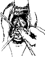
Figure 2. Longitudinal incision of the mucous membrane of the fornix; separation of the bladder by a blunt method.
vaginal incision

Figure 3. Separated bladder pulled upward with a speculum; incision of the uterus along the midline.
in view of the looseness of the tissue is usually performed very easily. After separating the bladder, it is displaced upward with a speculum and the peritoneum is separated from the uterus as far as possible. Then the anterior wall of the cervix is incised. Grasping the edges of the incision with Muesse forceps and pulling the cervix down, the anterior wall of the uterus is incised as high as possible, down to the peritoneal fold (see Figure 3). The fetal sac is opened, and a footling version is performed or forceps are applied; with a dead fetus, perforation is performed. The last part has to be delivered by the Crede method or manually. The edges of the uterine incisions are sutured with catgut sutures, not including the uterine mucosa (see Figure 4). When suturing the vaginal wound in its lower part, some insert a strip of gauze, which is removed after 2-3 days. With careful and cautious separation of the bladder, injury to it can be easily avoided: if the peritoneum is opened, it is carefully closed with separate sutures; a sufficiently long uterine incision prevents its rupture during delivery, and a T-shaped or arcuate incision of the mucosa prevents vaginal ruptures. The cause of bleeding during ante partum operation may be excessive deviation from the midline during separation of the bladder. Significant bleeding is also possible in placenta previa, in which the tissues of the lower segment become brittle and easily bleed. Various additions to the technique—preliminary dilation of the cervix for good drainage of secretions in the postpartum period, preliminary introduction of metreurynter and incision of the uterus under it, Shukhardovsky incision, midline episiotomy—did not gain wide popularity and are applied only for special indications. According to Selitsky's data, the mortality rate from the operation itself is zero. Sufficiently numerous observations have shown that vaginal cesarean section does not affect menstruation and does not influence subsequent4 births.

Figure i. Layer-by-layer suturing of the operative wound. Sutures on the uterus do not include the mucous membrane.
az7 (Selitsky). A necessary condition for performing vaginal cesarean section is the absence of obstacles from the pelvic bone and free access to the cervix. Indications for performing vaginal cesarean section can be life-threatening conditions of the pregnant woman or parturient (eclampsia, heart, lung, kidney diseases, etc.), premature separation of the placenta, prolapse of the umbilical cord, incorrect fetal position—transverse, oblique. With an obstruction for delivery from the side of the cervix (scars), as well as with central or low attachment of the placenta, due to dangerous for life bleeding, preference should be given to classic cesarean section. Vaginal cesarean section is indicated for septic infection of the pregnant uterus with a living or dead fetus. Due to the ease of the operation technique, hysterotomy found wide application for terminating pregnancy, starting from the 3rd-7th month. The advantage of hysterotomy besides that lies in the conditions of greater asepsis compared with other methods of terminating pregnancy and in the elimination of conditions for accidental damage to the uterus (cervical ruptures, uterine perforation, etc.). Hysterotomy is the most reliable, gentle, and safe method of artificial abortion in late pregnancy. In order to avoid deformation of the vaginal part of the cervix, which does not always fuse after its incision, some authors propose starting the incision of the uterus above the external os or, by opening the peritoneum of the anterior Douglas space, incising the body of the uterus itself—Dutzmann's operation, sectio caesarea vaginalis vera (Kakushkin).
Related articles
Mentioned in
Cite this page
“Hysterotomy.” Soviet Medical Encyclopedia. English translation of Bolshaya Meditsinskaya Entsiklopediya, 1st ed. (Moscow, 1928–1936), ed. N. A. Semashko. https://sovietmedicalencyclopedia.pages.dev/article/hysterotomy/