Obstetric Forceps
Historical document, translated for reference. It reflects medical knowledge of the 1920s–30s and is not medical advice.
Summary
Obstetric forceps are an instrument used during childbirth to replace lacking traction force, functioning as an extension of the obstetrician's hands. The article details their mechanical effects, indications, contraindications, and principles of application.
Encyclopedia article (1928–1936)
OBSTETRIC FORCEPS, an instrument that replaces the lacking or absent traction force (vis a tergo) during childbirth. In this sense, they serve as an extension of the obstetrician's hands ('iron hands' of the obstetrician). (For the history of the instrument's invention, see Obstetric Operations; for the design of forceps and their various models, see Obstetric Instrumentation.) When applying forceps, one must rely only on the mechanical effect. The so-called dynamic action of forceps (their ability to cause uterine contractions when introduced), which was previously discussed, if it has any significance at all, is only secondary. The main action is reduced to purely mechanical moments: a) compression of the head, b) its straightening, and c) extraction. Of these, the first two also have secondary importance, and the compression of the head, which is inevitable when applying forceps, should be minimal and in any case should not exceed that which occurs during natural normal first configuration of the head. Otherwise, the bones, vessels, and nerves of the child's head will inevitably suffer. Most obstetricians at present believe that forceps should not be used to correct various abnormal presentations and positions of the head (forceps atypical in function), as they give a high percentage of injuries. Indications for the operation. Previously, indications were based on personal discretion. At present, more or less definite indications have been developed for forceps. Applying forceps without indications, the obstetrician bears a certain responsibility for the professional error made with all its consequences. The developed indications for forceps are as follows: forceps are applied in cases where rapid termination of labor is necessary in the interests of the mother, the child, or both together. Specifically, this will be eclampsia, premature detachment of the placenta, prolapse of the umbilical cord, beginning asphyxia of the child, all diseases of the mother that complicate the expulsion period (heart defects, nephritis), primipara with high degree, etc. Weakness of labor activity itself should not serve as an indication for applying forceps. In such cases, in a clinical setting, one should wait for indications either from the mother's side or from the child's side (asphyxia). The practical physician, especially under district conditions, still has to consider weakness of labor activity as one of the indications for applying forceps. In this case, obstetric practice has developed the following rule: with weakness of labor activity, forceps can be applied if the expulsion period in a primipara lasts more than 5-6 hours, and in a multipara more than 2-3. Contraindications follow from the conditions under which forceps can be applied: 1. For applying forceps, it is necessary that the true conjugate be not less than 8-8.5 cm. 2. Forceps should be applied only with complete opening of the uterine os. So-called 'intrauterine forceps' of Nevyrtz (with opening of 2-3 fingers) should definitely be considered an error against the rules of operative obstetrics. 3. Forceps should, as a rule, be applied only with cephalic presentations, and the head should not be too large (hydrocephalus) or too small (forceps cannot be applied to a head of less than 7 months' age). 4. The head must be immobile. Forceps on a mobile head are contraindicated. 5. The fetal bladder must be ruptured and the membranes tucked behind the greatest circumference of the head. 6. The child must be alive. 7. Forceps cannot be applied with threatening and with occurred rupture of the uterus, as well as with the posterior face presentation (chin to the back) and with the so-called Litzmann's insertion of the head. General principles of applying forceps. When applying forceps, it is first necessary to clearly and precisely know the mechanism of the act of labor (see table) and always remember the following three basic rules: 1. Forceps must grasp the greatest periphery of the head, i.e., in other words, the tops of their blades must always go beyond the parietal tubers. If this rule is not observed, slipping of the forceps may occur due to improper fixation of the head. 2. Forceps should always be applied so that the tops of their blades look toward the conducting point (see table); the concavity of the pelvic curvature of the instrument should always be turned toward the pubis. 3. Forceps should be closed so that the conducting point always lies in the plane of the head curvature of the instrument, i.e., by positioning the locking parts of the instrument in one plane, one should connect its handles in such a way that the blades grasp the proper periphery of the head. Forceps can be closed horizontally (handles should look directly at the obstetrician), with handles raised upward, and with handles lowered downward. In all these cases, the conducting point must necessarily lie in the plane of the forceps; otherwise, if during closure it lies outside the plane of the head curvature of the instrument, the forceps can easily slip. When applying forceps, the following possibilities arise: with occipital presentations, forceps can be applied a) typically and b) atypically. Typically, forceps are applied to the head that has completely completed the mechanism of the act of labor (necessary data for forceps). Conducting point Size through which the head passes through the Bulwark ring Point of fixation, resp. rotation (hipomochlion) I. Occipital presentations: a) anterior view b) posterior view c) anterior-parietal presentation d) middle and low (deep) position of the head II. Face presentations: a) anterior view b) frontal presentation c) middle and low (deep) position of the face Small fontanelle Large fontanelle Small oblique Medium oblique Straight Large font. In artificial delivery according to the type of anterior view (see I, a). Suboccipital fossa Border of the hairy part of the forehead Nose bridge should conduct labor Chin Vertical Root of the nose Medium between large oblique and straight In artificial delivery, labor should be conducted according to the type of anterior view (see II, a). Sublingual region Upper jaw has performed internal rotation (rotation). Forceps in such cases are applied to the transverse (biparietal) size of the head and in the transverse size of the pelvis (see figure 1).
Figure 1. Typical forceps in occipital presentation (according to Halban-Seitz).
In occipital presentations, forceps can be applied a) typically and b) atypically. Typically, forceps are applied to the head that has completely completed the mechanism of the act of labor (necessary data for forceps). Conducting point Size, through which the head passes through the Bulwark ring Point of fixation, resp. rotation (hipomochlion) I. Occipital presentations: a) anterior view b) posterior view c) anterior-parietal presentation d) middle and low (deep) position of the head II. Face presentations: a) anterior view b) frontal presentation c) middle and low (deep) position of the face Small fontanelle Large fontanelle Small oblique Medium oblique Straight Large font. In artificial delivery according to the type of anterior view (see I, a). Suboccipital fossa Border of the hairy part of the forehead Nose bridge should conduct labor Chin Vertical Root of the nose Medium between large oblique and straight In artificial delivery, labor should be conducted according to the type of anterior view (see II, a). Sublingual region Upper jaw has performed internal rotation (rotation). Forceps in such cases are applied to the transverse (biparietal) size of the head and in the transverse size of the pelvis (see figure 1).
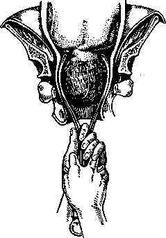
Figure 1. Typical forceps in occipital presentation (according to Halban-Seitz).
Some call such forceps outlet forceps (the head is usually, but not always, in the outlet of the pelvis), but it is more correct to call them typical. As the same figure shows, with typical forceps the head is grasped in the suboccipitobregmatic plane (the forceps lie somewhat in front of the ears). This is the so-called 'ideal grip' of Farabeuf (Fara-beuf), named thus because with it all three of the above rules for applying forceps are ideally fulfilled. Strictly speaking, forceps are adapted only for application to the head that is in the direct diameter of the pelvic outlet (that is, having completed rotation completely). Forceps that have to be applied to a head that has not yet completed rotation can be called atypical. They are usually called cavity forceps, since with them the head is indeed more often in the cavity of the pelvis. Such atypical forceps have to be applied: 1) to a head that has only partially completed internal rotation (the sagittal suture is in one of the oblique diameters of the pelvis), 2) in so-called middle and low, resp. deep, transverse positions of the head, and 3) in so-called high forceps. When applying atypical forceps (excluding high ones), one should be guided by one rule common to all: atypical forceps must be applied in that oblique diameter of the pelvis toward which the guiding point looks (in occipital presentations, -the anterior fontanelle). When applying atypical forceps to a head that has not yet completed full rotation (the sagittal suture is in the right oblique diameter), -the guiding point (anterior fontanelle) is on the left, therefore the forceps are applied, according to the stated rule, in the left oblique diameter: the left blade lies on the left and behind, the right - above, in front. The forceps grasp the head in the region of the ears. With atypical forceps, therefore, there are two possibilities: a) on the right the guiding point - in the right oblique diameter the forceps are applied and the right blade must be below; b) on the left the guiding point - in the left oblique diameter the forceps are applied, the left blade will lie below. -With middle and low transverse positions of the head, forceps have to be applied even more atypically. To apply forceps correctly here, in Farabeuf's ideal grip, they would have to be applied to the transverse diameter of the head, but in the direct diameter of the pelvis. Ordinary forceps (with two curves) cannot be applied this way, as the pelvic curvature prevents it. For this special forceps [Lazarevich's, Kjelland's (Kjel-land)]; ordinary forceps, with middle and low transverse position, have to be applied, like any atypical forceps, according to the general rule: in that oblique diameter toward which the guiding point looks]. The guiding point in this case should be considered the anterior fontanelle, since only by bringing it under the symphysis pubis is correct rotation of the head obtained (anterior view). The forceps grasp on one side the parietal eminence, and on the other the suboccipitobregmatic area. This grasp cannot be called good, but for ordinary forceps it will be the only possible one under these conditions. With facial presentations, forceps are applied according to the same rules as with occipital presentations. Here also there are a) typical forceps, which are applied in the transverse diameter of the pelvis on either side of the face that has completed rotation (the facial line is in the direct diameter of the pelvis), and b) atypical ones, which are applied according to the same rule as atypical forceps in occipital presentations (in that oblique diameter toward which the guiding point looks, that is, the chin). In both cases the forceps grasp the face in the region of the ears. The technique of the operation in occipital presentation. The patient is prepared for the operation in the usual way (enema, ordinary toilet of the external genitalia, etc.). The operation can be done, in the absence of an operating table, on a cross-bed. Anesthesia is given, in the interests of the child, only in severe cases. It is necessary, in any case, besides the forceps, to prepare instruments for stopping possible bleeding from a torn cervix (mirrors, clamps, needle holder, needles, material for sutures), as well as everything necessary for craniotomy. Before applying the forceps, it is necessary to perform the most careful internal examination and to determine exactly where the guiding point is located, to orient oneself as to the position of the sagittal suture, the degree of opening of the uterine os, and so on. In extreme cases, it is necessary to perform such an examination even by inserting the whole hand into the vagina (under anesthesia). The technique of the operation consists of five moments, which in relation to typical forceps in occipital presentations will be as follows: the first moment of the operation - application of the blades. The left blade is always applied first.
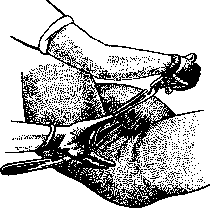
Figure 2. Introduction of the right blade from the side of the opposite paxoBorocrn6a (noHaIban-Seitz).
left blade. It is introduced into the left half of the pelvis under the control of two fingers of the right hand, which are placed between the head and the vaginal wall. The blades must necessarily go beyond the parietal eminences in order to be positioned in the suboccipitobregmatic area. The second blade is introduced in the same manner. When introducing the blades, they should always be brought from the side of the opposite inguinal fold (see Figure 2), with the blade, or rather the shank, descending and describing an arc to that horizontal plane at which it should stop. When introducing the blades, certain difficulties may be encountered. Sometimes after their application the blades are not in parallel planes. This is very easily corrected if one places the thumbs on the Bush hooks and, pressing on them, straightens the blades. In some cases the blades are applied in such a way that one of them is placed deeper in the birth canal (turns out to be longer) than the other. In such cases one should only pull up that blade which turns out to be longer. The second moment, closing the blades, is performed in the following way according to the general rule: the guiding point (anterior fontanelle) should be in the plane of the forceps, i.e., in other words, with typical occipital forceps, the forceps must be closed in such a way that their shanks point directly at the obstetrician (horizontal closure). The third moment - trial traction. The forceps are grasped with the right hand, the index finger of the left hand is applied to the head, with the other fingers of this hand necessarily lying on the shank of the forceps (only with this hand position can one not only see but also feel the slipping of the forceps). The fourth moment - the actual tractions, which should be similar in nature to contractions. Their direction must strictly correspond to the position of the head: the higher the head stands, the more backward the direction of the tractions should be. With the head in the pelvic outlet, the shanks of the forceps during tractions should point directly at the obstetrician. No rocking, rotating, pendulum-like or other movements should be made during tractions. if eight to ten tractions are unsuccessful, further application of forceps should be abandoned. The fifth moment - delivery of the head. The head is delivered either in the forceps or the forceps are removed first. The forceps are removed in the same way as they were applied, that is, the left blade describes an arc and is drawn to the right inguinal fold, while the right one - back. Some recommend, in the interests of protecting the perineum, to remove the forceps before delivery of the head, at the moment when the fixation point appears under the symphysis pubis (with typical occipital forceps - the suboccipital fossa). So that the head cannot go back after removal of the forceps, the maneuvers of Olshausen and Ritgen (Olshausen, Ritgen; see Obstetric Manual Maneuvers) have been proposed, which do not always, however, achieve their purpose. Delivery of the child's body is performed according to general rules. -In exactly the same way forceps are applied to the head that has completed rotation, but has done so incorrectly, with the spine and occiput backward (forceps in the posterior view of occipital presentation and in anterior parietal presentation). It is clear that here there will be a different guiding point (posterior fontanelle) and a different fixation point (the border of the hairy part of the forehead in posterior views and the bridge of the nose in anterior parietal presentation). In other respects
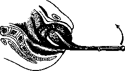
Figure 3. Forceps in the posterior view of occipital presentation (according to Halban-Seitz).
The forceps are applied in full accordance with the technical moments just described. A small but essential detail will be the direction of traction: the latter should be performed in such a way that the handles of the forceps always face the obstetrician. With any other direction of traction, extension of the head inevitably occurs, which is undesirable (see Figure 3). - Atypical forceps for occipital presentations should, as a general rule, be applied in the oblique diameter toward which the small fontanelle (guiding point) is facing. The technical peculiarities will only concern the first moment (introduction of the blades) and the fourth (traction). When introducing the blades, the left blade is always introduced first, as with typical forceps, and it is introduced from the opposite inguinal fold, etc. As for the second blade, it is also first introduced from behind, but then it is raised together with the fingers of the controlling hand (the handle of the forceps is lowered at this time) to the parietal eminence (the blade wanders). In the first position, this wandering is performed by the right blade, and in the second position, by the left blade. In the fourth moment of the operation, the character, direction, strength, etc. of the traction are determined by the same rules as with typical forceps. The question in this case only concerns how to perform the traction so that the head completes rotation. It usually tends to turn on its own into the anteroposterior diameter of the pelvis. The obstetrician can gradually, over a series of tractions, facilitate such a rotation (in the first position, perform traction while turning the head from left to right, and in the second position, vice versa). - Atypical forceps for low, resp. deep, transverse positions of the head are a difficult and responsible operation. Cases of the forceps slipping off are often observed due to insufficient and unsatisfactory grasp of the head. Forceps of the ordinary type must be applied here, like any atypical forceps, in the oblique diameter of the pelvis, in accordance with the guiding point (small fontanelle). Among the technical peculiarities in this case, mention should be made of changing the position of the forceps. At the moment when the sagittal suture, after several tractions, moves from the transverse position to an oblique position, the forceps must be removed and reapplied as atypical forceps are applied to a head that has not completed rotation.
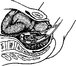
The technique of applying forceps in face presentations essentially differs in no way from the technique in occipital presentations, but here there will be a different mechanism of labor, a different guiding point (chin), and a different point of fixation (sublingual region), etc. In addition, when applying forceps, there is one essential peculiarity which must be kept in mind and which concerns the moment of closing the forceps: the forceps should always be closed with the handles raised upward, since in these presentations the guiding point (chin) is always at the pubic symphysis. If cases are observed where the chin is placed behind (the so-called posterior variety of face presentation), then labor in such cases is impossible, and consequently the use of forceps is contraindicated. Taking into account this peculiarity of the technique of applying forceps in face presentations, it is necessary when applying the forceps to ensure that the left blade introduced first is held by the assistant with the handle turned upward (see Figure 4). Otherwise, the technique of applying forceps in face presentations presents no peculiarities.
High forceps. Some obstetricians call this the forceps applied to a still mobile head, i.e., when it is above the plane passing through the terminal line. According to the considerations presented above, forceps should not be applied to such a mobile head. Others consider high forceps to be those that must be applied to a head standing at the inlet of the pelvis with the so-called small segment (sagittal suture in the transverse diameter, promontory freely reached, but the plane passing through the innominate line is already occupied by some segment of the head). With such a position of the head, applying forceps is also not recommended, since essentially it is still mobile here. Most obstetricians, however, call high forceps those that are applied to a head standing at the inlet of the pelvis with the large segment (high transverse position of the head, where without pushing the head, it is already impossible to reach the ischial spines). For applying high forceps, special instruments are used - Tarnier's and Breus's forceps (Tarnier, Breus). All these instruments, as well as ordinary type forceps but with longer handles, must be applied in the transverse diameter of the pelvis, no matter how the head is positioned (in oblique or transverse diameters). The grasp of the head under these conditions will be unsatisfactory, and most importantly, unsafe, especially in the transverse position of the sagittal suture (in such cases, one blade must be applied to the face and the other to the occiput (see Figure 5). In Tarnier's and Breus's forceps, the head is given the opportunity to perform its mechanism more or less correctly thanks to the devices present in these forceps. The same can be achieved approximately with ordinary forceps (with long handles), if only one uses the technique first recommended by Osiander. During traction, the left hand should be placed on the lock part of the instrument and, by pressing it energetically backward, thereby allow the head to freely bypass the obstacle present near the symphysis. When the head has passed through the inlet of the pelvis and descended into the cavity, the forceps must be repositioned according to the general rules for atypical forceps.
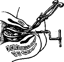
More convenient for extracting a high-lying head are the Russian Lazarevich forceps (straight, parallel) and the new Killian forceps. They can be applied in the anteroposterior diameter of the inlet and on the transverse diameter of the head, which is undoubtedly more correct and advantageous for the head. High forceps are currently the subject of great disagreement among obstetricians, and the majority of the latter deny their usefulness.
Kielland's Forceps. Attempts to create an absolutely perfect model of obstetric forceps have not ceased to the present day. At present, there are already over 300 modifications of this important instrument, and despite this, new and new models continue to be proposed. Among the most recent models belong the forceps of the Norwegian obstetrician Kielland, which have also already been modified. Kielland's forceps were proposed by him as an instrument that can be successfully used in the most various complications of the act of labor. These forceps are constructed on the type of Lazarevich's forceps. The great advantage of Kielland's forceps lies in the fact that they can be applied to the transverse diameter (biparietal) of the head, however and wherever the latter may be positioned. An essential feature of the technique for applying Kielland's forceps is the application of the first blade, which, typically according to Kielland, is applied in such a way that when introduced into the uterus, its concavity is directed toward the pelvis and only then it makes a 180° turn, adapting to the corresponding parietal eminence. The second blade is applied in the same way as with ordinary forceps. Kielland's forceps have caused great controversy in the specialized literature. In any case, these forceps represent a certain step forward in operative obstetrics. The greatest danger in the use of Kielland's forceps lies in the above-mentioned rotation of the blade, already introduced into the uterus. Several cases of rupture of the lower segment of the uterus, as well as injuries to the bladder, have been described. In any case, these forceps cannot yet be recommended for widespread use. - Forceps for the next head are currently used by few. They are recommended to be applied in those cases when extraction of the fetus by the breech fails to deliver the subsequent head. In such cases, forceps have to be applied to the transverse diameter of the head and in the transverse diameter of the pelvis, and they are always applied from under the child's body (the latter must be raised upward). With such extraction of the head, there is a great danger of causing irreparable damage, on the one hand, to the soft and bony parts of the pelvis, and on the other hand, to the cranial box and its contents in the child. - Forceps in breech presentations. In obstetric practice, obstetric forceps have been used for a long time in these presentations. Here, complications are often encountered in which every physician more than once will regret that he does not have at his disposal such an instrument as forceps for the head. Such complications are also observed in manual assistance, which has to be rendered in breech presentations and in extraction of the fetus by the breech end. The operation of extracting the breeches appears particularly difficult in those cases when the latter are already impacted in the pelvis. The various methods of extraction recommended in this case - extraction by the inguinal flexure simultaneously with compression of the fetus according to Kristeller, extraction with a blunt hook, with a loop, etc. - often give no results even in the most experienced hands. Special breech forceps were invented, but they also do not achieve the purpose. In the end, ordinary obstetric forceps were proposed for extracting the breeches. In transverse or oblique positions of the breeches, the forceps are applied so that the terminal part of the blades lies over the trochanter major on the iliac crest of both sides. When the breeches are in the anteroposterior diameter of the pelvis, it is recommended (Gauss) to apply one blade to the sacrum and the other to the posterior surface of the thighs. It is still more convenient to use Lazarevich's or Kielland's straight forceps for extracting the breeches. Prognosis with forceps. It was stated above that forceps are by no means a safe instrument for the mother and for the child. If one counts all the injuries that have to be observed in the mother in the soft parts of the birth passages after forceps (ruptures of the cervix, vagina, vulva, perineum, rarer cases of rupture of the bladder, urethra), as well as injuries in the region of the symphysis, sacroiliac joint, coccyx, in the region of the sciatic plexus, etc., then the figure obtained is 50% (Hans). Undoubtedly, not all these injuries should be attributed to the forceps, but the indicated figure nevertheless speaks sufficiently clearly in favor of the fact that forceps cannot be regarded as a safe operation. The morbidity of parturients after forceps is observed, approximately, in 17% of applications, and the mortality rate is 0.18-0.58% (Hans, Lesewitz). In the child, serious injuries are also observed after forceps in the form of facial nerve paralysis (4-5%), depressions of the skull, hemorrhages into the cranial cavity, etc. The mortality rate of children after application of forceps is given by various authors within limits from 7 to 27%.
M. Malinovsky. In obstetric practice, a large number of obstetric instruments proposed at different times have long been applied—both for operative intervention in pathological births, and for the examination of pregnant women. Below are listed the most important obstetric instruments, with their design and most commonly used models (the illustrations are on three plates). Instruments for the examination of pregnant women and parturients. The obstetric stethoscope—for listening to the movements and fetal heartbeat. It differs from the ordinary one in the width of the end placed on the abdomen (see Figure 1).—The pelvimeter, for measuring the external and internal dimensions of the pelvis, fetus and its head. It resembles a compass with curved branches. The degree of opening of the compass is marked on a scale attached to one of the legs of the compass. The most commonly used model is Martin's pelvimeter (see Figure 2).—Breusky's compass, a very convenient model of pelvimeter, specially designed for measuring the external dimensions of the pelvic outlet, is constructed from two straight intersecting branches (see Figure 3).—The Bylicki-Gauss measuring instrument, for the direct measurement of the true conjugate, consists of a handle and two branches. One branch is rigidly attached at a right angle to the handle, the other is movable—sliding inside the first branch, and at the end facing the obstetrician, it carries a scale and a ring. The distal end of the fixed branch is bent upward and should, during measurement, touch the inner surface of the symphysis. The distal end of the movable branch is bent and looks forward, and during measurement, it touches the promontorium. The degree of separation of the branches is marked on the scale (see Figure 4).—The measuring centimeter tape is intended for measuring the abdominal circumference, the height of the fundus of the uterus, and other dimensions. The most convenient model is the tape measure (see Figure 5). Instruments for dilating the cervix.—Hegar's dilators are metallic bougies that sequentially increase in diameter. Their sequential insertion into the cervix causes its gradual dilation. They are used mainly in the first half of pregnancy. There are two varieties of dilators—cylindrical and conical. A significant advantage in terms of gentle dilation is possessed by sets of bougies that have a diameter difference of not 1 mm, but 0.5 mm (see Figures 6 and 7).—The metreurynter, a rubber balloon intended for dilating the cervix. Inserted in a folded state (in the form of a cigar) behind the internal opening of the cervix, the balloon expands when filled with fluid, which is poured through a rubber tube firmly attached to the balloon. Metreurynters vary in size and shape. The most common shapes are pear-shaped, violin-shaped, and cone-shaped (see Figures 8 and 9). The Champetier de Ribes meteurynter is a cone-shaped balloon (see Figure 10).—The colpeurynter is a balloon intended for insertion into the vagina.—The Bossi dilator, an instrument consisting of a series of metal rods; in its folded state, it resembles a conical bougie. At one end, the metal branches are fixed to a screw, and when this screw is rotated, their distal ends spread apart. The dilator is inserted in a folded state into the cervix, which, when the screw is rotated, is spread apart by the branches to the sides. Due to the extremely rough dilation of the cervix, the instrument is now almost never used (see Figure 11).—The obstetric bougie (see Figure 12), elastic, long, 0.5 cm in diameter, is intended for inducing premature labor (it is now extremely rarely used in practice). Instruments for extracting an undamaged fetus. Forceps, represent an instrument for extracting the head (in rarer cases—the buttocks). Forceps consist of three parts: blades, lock, and handles. According to the type of lock, forceps are divided into three types: French, German, and English. In French forceps, the lock consists of a pin and a screw thread. The connection of the two branches of the forceps is achieved by placing the hole of the right branch of the forceps over the pin located on the left branch, and as the pin is gradually screwed in, it presses one branch against the other (junctura per axin). In German-type forceps, the pin is fixed and carries a button; on the other branch (right) there is a corresponding notch into which, when closing, the pin of the left branch enters (the lock of Würzburg). In English forceps, the lock is a simple depression on each branch, and when the instrument is closed, one depression enters the other, thus fixing the forceps branches immovably (junctura contabulationem). The blades of the forceps have two curves—pelvic and cephalic. In some forceps, there is no pelvic curve (straight forceps). Forceps with three curves have been proposed. The following modifications have practical significance.—Simpson's forceps, the most commonly used model of forceps with an English lock (see Figure 13).—Naegele's forceps, one of the common models of German forceps (see Figure 14).—Lazarevich's forceps; these forceps have the following two features: the forceps branches do not cross (parallel) and lack a pelvic curve. Closing is accomplished with the help of a pin located on the inner side of the right handle of the forceps, which freely enters a hole present on the left handle (see Figure 15).—Kjelland's forceps, a light, elegant instrument with almost no pelvic curve. The lock consists of a small rectangular plate bent at a right angle (sliding lock, bayonet-like); embracing the other blade, such a lock allows the blades to shift longitudinally and excludes any other shifts. The part of the blade located between the lock and the terminal part is round. On the handle, there are two buttons that serve to indicate the direction of rotation of the blade after its insertion. The forceps are intended for application to a head that is high in the transverse diameter, with the application of the blades to the head in the biparietal diameter. With the help of these forceps, rotational movement can be imparted to the head (see Figure 16).—Tarnier's forceps, with axial traction. Their main difference from ordinary forceps is that to the posterior part of the blades, movable branches are attached, which in turn are movably connected by a handle to a crossbar. These branches together with such a handle form a device with which traction is performed. In addition to the ordinary French lock, for stable closure of the forceps, there is a special screw lock located closer to the handle. The advantage of these forceps lies in the possibility, with the help of the traction device, to guide the head during traction strictly along the axis of the pelvis. Another advantage is that the head retains full mobility during traction due to the movable attachment of the cross handle to the rest of the traction device. The traction apparatus can be removed, and in this case, Tarnier's forceps become ordinary simple forceps (see Figure 17).—Breus's forceps—another model of forceps with movable blades, intended for extracting a high head. They are not entirely correctly classified as axial forceps. Axial traction in Breus's forceps takes a back seat, and the main point is the mobility of the blades, which facilitates the movement of the head along the axis of the pelvis. The instrument is ordinary Simpson's forceps, only with a special addition that allows the terminal part of the blades to move independently. The design and purpose of such an additional device are self-evident from the illustration (see Figure 18). Traction in Breus's forceps is performed as in ordinary forceps, i.e., by the handle.—The obstetric hook for extracting the fetus by the inguinal flexure. Kiistner's hook, the most convenient modification of the obstetric hook (see Figure 19).—Bunge's instrument for passing a loop into the inguinal flexure when extracting the fetus by the breech. It consists of a curved, hollow probe, which is attached to an elongated, firmly fixed handle. In this probe slides a strong gutta-percha loop, to the end of which a strong silk thread is attached. The hollow probe has a longitudinal slit of such a size that the gutta-percha loop can be inserted and removed from the groove with slight effort. After the probe is passed behind the inguinal flexure, the silk thread is grasped from the perineal side of the fetus, which holds the distal end of the gutta-percha loop. When the hollow probe is raised upward, the gutta-percha loop slips out of the groove and remains lying alone in the inguinal flexure. After the grooved probe is removed, the obstetrician has both ends of the loop, by which traction of the fetus is performed (see Figure 20). Instruments for disrupting the integrity of the fetal head. The perforator—for piercing the cranial vault. There are three varieties: scissors-shaped, spear-shaped, and trephine-shaped. The advantage of scissors-shaped and spear-shaped perforators is the ease and convenience of performing perforation.
The advantage of trephine-like perforators is obtaining a smooth hole without protrusions and fragments. - The scissor-shaped perforator by Naegele consists of a flat point made of two halves that spread apart like scissors when the instrument's handles are compressed. This point, when spread apart, destroys the skull (see figure 21). - The spear-shaped perforator by Blot resembles a spear longitudinally split into two halves. When pressure is applied to the handle, both halves separate and, through their movement, destroy the cranial bones (see figure 22). - The trephine-shaped perforator by Martin consists of a sleeve through which a rod passes, ending on one side with a handle and on the other with a toothed crown. The toothed crown constitutes the cutting part of the instrument. When the handle is turned to the right, the toothed crown extends forward and, with further movement, cuts out a round hole in the cranial bone. When the handle is turned in the opposite direction, the cutting crown retracts into the sleeve. In the center of the crown, there is a corkscrew-shaped hole intended for secure fixation of the instrument during perforation (see figure 23). - The Phenomenov perforator, resembling a drill, is a very convenient model. - A catheter for destroying and washing out the brain of a perforated head - a wide metal catheter that fits onto the rubber tube of an irrigator. Instruments for extracting the perforated head of the fetus. The Breusky craniotrib consists of ordinary but more massive forceps, with the difference that on their handles there is a screw lock with which powerful compression of the blades is produced, and thereby - vigorous flattening of the head (see figure 24). Extraction is performed with ordinary forceps.
Related articles
Mentioned in
Cite this page
“Obstetric Forceps.” Soviet Medical Encyclopedia. English translation of Bolshaya Meditsinskaya Entsiklopediya, 1st ed. (Moscow, 1928–1936), ed. N. A. Semashko. https://sovietmedicalencyclopedia.pages.dev/article/obstetric-forceps/